Understanding Needle Biopsy Procedures: What to Expect and How to Prepare
Needle biopsy is a minimally invasive way to obtain tissue for laboratory diagnosis. This article summarises the main biopsy types, how imaging guidance improves accuracy, practical preparation, risks, and recovery so patients and referrers know what to expect.
Key Takeaways
- Needle biopsies provide tissue samples with minimal invasiveness.
- Fine Needle Aspiration (FNA) and Core Needle Biopsy (CNB) are the primary techniques.
- Ultrasound (or CT) guidance improves sampling accuracy and safety.
- Follow clinical instructions, arrange transport if sedated, and wear comfortable clothing.
- Risks are usually minor: bleeding, infection and temporary soreness.
- Post-procedure care focuses on wound care, symptom monitoring and simple analgesia.
Types of Needle Biopsy
Clinicians choose the technique based on lesion type and diagnostic needs.
- Fine Needle Aspiration (FNA): Uses a thin needle to sample cells or fluid; quick and less invasive.
- Core Needle Biopsy (CNB): Uses a larger needle to collect a tissue core for greater histological detail.
Comparative evidence guides selection for targets such as breast lesions and other soft-tissue masses.
FNA vs. CNB: Comparing Needle Biopsy Types for Breast Lesion Diagnosis
In Japan, fine needle aspiration biopsy (FNA) of the breast has long been recognized as a useful diagnostic tool, and has been used in many institutions because it provides a rapid, accurate and cost-effective evaluation. However, the use of core needle biopsy (CNB) is increasing, and vacuum assisted biopsy devices have been developed to produce larger specimens for analysis. CNB is useful because the frequency of inadequate specimens is lower than in FNA, and it requires a less invasive procedure than open biopsy.
Core needle biopsy (CNB) as a diagnostic method for breast lesions: comparison with fine needle aspiration cytology (FNA), 2004
Specialised techniques, such as the modified Bergström needle for muscle sampling, are used in specific clinical or research settings.
Understanding the Bergström Needle Biopsy Technique for Muscle Samples
The percutaneous biopsy technique enables researchers and clinicians to collect skeletal muscle tissue samples. The technique is safe and highly effective. This video describes the percutaneous biopsy technique using a modified Bergström needle to obtain skeletal muscle tissue samples from the vastus lateralis of human subjects. The Bergström needle consists of an outer cannula with a small opening (‘window’) at the side of the tip and an inner trocar with a cutting blade at the distal end. Under local anesthesia and aseptic conditions, the needle is advanced into the skeletal muscle through an incision in the skin, subcutaneous tissue, and fascia. Next, suction is applied to the inner trocar, the outer trocar is pulled back, skeletal muscle tissue is drawn into the window of the outer cannula by the suction, and the inner trocar is rapidly closed, thus cutting or clipping the skeletal muscle tissue sample.
Human skeletal muscle biopsy procedures using the modified Bergström technique, RA Shanely, 2014
Knowing the chosen method helps patients understand what happens during the procedure.
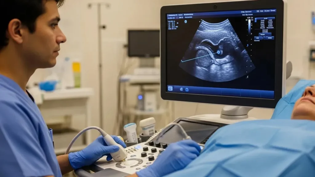
Role of Ultrasound Guidance
Ultrasound provides real-time imaging to target lesions, improving sample adequacy and reducing injury to nearby structures. CT or other imaging may be used when ultrasound is unsuitable.
Essential Preparation Steps
Good preparation reduces complications and improves the appointment flow. Follow instructions from your clinical team.
- Follow healthcare instructions (including medication adjustments).
- Transport: Arrange someone to drive you home if sedation is used.
- Clothing: Wear loose, accessible clothing for the biopsy site.
What Are the Essential Preparation Steps Before the Biopsy?
Confirm any fasting instructions, discuss medications (especially blood thinners), and notify the team of allergies or bleeding disorders. Follow specific provider guidance to reduce procedural risk.
How Can Patients Address Common Concerns About the Procedure?
Discuss the steps, anaesthesia and pain control with your clinician. Sharing medical history and asking questions reduces uncertainty and can improve the experience.
Managing Pre-Biopsy Anxiety and Pain: Patient Experience Factors
Worry about the procedure was the only variable found to be significantly correlated with both biopsy-related outcomes (pain and physical discomfort). From a clinical perspective, this item could be used as a brief screening tool to identify patients who might be at risk for poorer biopsy experiences and who might benefit from brief interventions to reduce pre-biopsy worry.
Pre-biopsy psychological factors predict patient biopsy experience, SJ Miller, 2014
Simple coping techniques (controlled breathing, a support person) and clinician reassurance often reduce anxiety and pain perception.
Common Concerns
Pain is usually controlled with local anaesthesia; most soreness is short-lived. Result turnaround varies by biopsy type and laboratory workload—ask your referrer for an expected timeframe.
Risks and Safety Considerations
Needle biopsies carry low but specific risks: bleeding, infection and transient soreness. Clinicians minimise risk with imaging guidance and sterile technique.
- Bleeding: Usually minor and self-limited; higher risk with clotting disorders.
- Infection: Small risk, reduced by aseptic technique.
- Discomfort: Mild soreness or bruising is common.
What Potential Complications Should Patients Be Aware Of?
Serious complications are uncommon but can occur in patients with risk factors. Seek prompt care for excessive bleeding, signs of infection, or severe pain.
How Do Expert Radiologists Minimize Procedure Risks?
- Imaging guidance (ultrasound/CT) for precise needle placement.
- Sterile technique and standard protocols to prevent infection.
These measures increase safety and the chance of obtaining diagnostically useful tissue.
Follow-Up Care
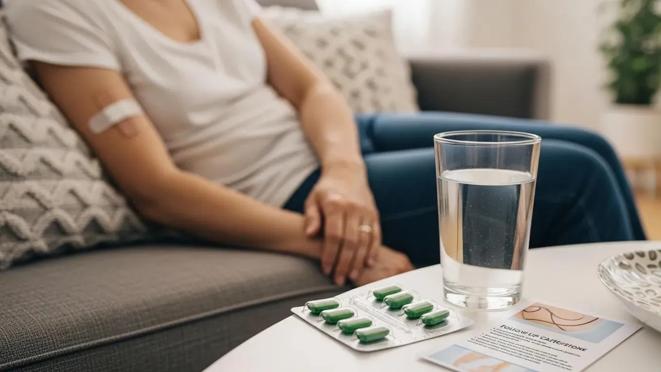
After the biopsy, keep the site clean, watch for infection signs, and use simple analgesics for soreness. Follow any specific aftercare instructions given by your team.
- Monitor for infection: increasing redness, swelling, discharge or fever warrant contact with your provider.
- Manage discomfort: paracetamol or ibuprofen (as advised) typically suffices.
What Is the Recovery Process and Results Timeline After a Needle Biopsy?
Most people recover within a few days. Mild soreness or bruising is expected; significant pain or worsening symptoms should prompt medical review.
What Should Patients Expect During Recovery and Aftercare?
Expect mild local symptoms and follow aftercare instructions to aid healing. If symptoms deviate from expectations, contact your healthcare team.
How Long Does It Take to Receive and Understand Biopsy Results?
Results usually arrive within several days to a week, depending on pathology processes. Your clinician will discuss findings and next steps when reports are available.
Understanding the procedure, preparation and aftercare can reduce anxiety and improve outcomes. For diagnostic imaging and ultrasound-guided biopsies, Life Imaging provides comprehensive services and support.
Frequently Asked Questions
What is the difference between Fine Needle Aspiration (FNA) and Core Needle Biopsy (CNB)?
FNA samples cells with a thin needle and is less invasive; CNB removes a tissue core for more structural detail. Clinicians choose the method that best answers the diagnostic question.
How can patients manage anxiety before a needle biopsy?
Discuss anaesthesia and pain control with your clinician, use relaxation techniques, and bring a support person if helpful.
What should patients do if they experience complications after a needle biopsy?
Contact your healthcare provider for excessive bleeding, signs of infection, or severe pain. Early assessment ensures timely treatment.
Are there any dietary restrictions before a needle biopsy?
Follow provider instructions. If sedation is planned, fasting for a set period is commonly required.
What types of imaging are used during needle biopsies?
Ultrasound is common for real-time needle guidance; CT or other modalities are used when appropriate for the lesion location.
How can patients prepare for potential discomfort during the procedure?
Discuss pain management options with your clinician. Local anaesthesia is routine, and simple analgesics are usually effective afterwards.
Conclusion
Concise, practical information about needle biopsy techniques, preparation and aftercare helps patients and referrers make informed decisions. For personalised advice, consult your healthcare provider.