
Erina Central Coast Medical Imaging & Radiology Services — Comprehensive Diagnostic Care
At Erina Central Coast Medical Imaging we combine essential diagnostic imaging services to help clinicians and patients get timely, accurate answers. This guide outlines the tests we offer, how cardiac and bone-health imaging work, and what patients and referrers should expect from referral through to the final report.
People searching for “Erina MRI,” “Erina ultrasound” or “Central Coast DEXA” usually want clear prep steps, realistic expectations about comfort and safety, and straightforward billing information. This page delivers that: concise modality explanations, practical preparation checklists, technology notes, bulk-billing guidance and a clear referrer workflow so results can be acted on efficiently. Below you’ll find a short services summary and quick anchors to sections on available services, cardiac imaging, DEXA scans, patient comfort and accuracy, the patient journey, and how referrers can access our services.
Central Coast Imaging, Central Coast Radiology, Central Coast Cardiac Imaging, Central Coast Ultrasound, Central Coast DEXA, Central Coast Imaging, Central Coast MRI
What Medical Imaging Services Are Available in Erina Central Coast?
We provide a broad range of imaging tools to visualise internal anatomy, assess function and guide treatment. Each modality works differently—MRI uses magnetic fields, CT and DEXA use X-rays, and ultrasound uses sound waves—and each has strengths, such as soft-tissue characterisation, bone density measurement or evaluation of blood flow. Knowing what a test shows and why it’s ordered helps patients and referrers choose the right investigation and prepare correctly. Below is a compact list with short explanations to help decision-making and referral planning.
- MRI: Cross‑sectional scans using magnetic resonance to show soft tissues, brain, spine and joints with excellent contrast.
- CT: Fast X‑ray based scans for bone, lung, abdomen and vascular assessment, available with low‑dose protocols.
- Ultrasound: Real‑time imaging for pregnancy, vascular, musculoskeletal and abdominal assessments without ionising radiation.
- DEXA: Dual‑energy X‑ray absorptiometry for bone mineral density and body composition measurements.
- X‑ray & Mammography: Projection imaging for bones, chest and breast screening or diagnostic studies.
These modalities often work together — combining structural detail, functional assessment and safe repeatability — to guide personalised management and follow‑up.
The sections that follow compare common services, list practical preparation steps, and outline typical indications so patients and referrers can decide what test to pursue next.
Which MRI Scans Does Erina Central Coast Offer?
Our MRI service routinely covers neurological, musculoskeletal, spinal and targeted body studies that use high soft‑tissue contrast to detect pathology. MRI differentiates tissue composition to identify brain lesions, disc disease, tendon and ligament injuries and joint cartilage problems. For sports and musculoskeletal injuries we look for tendon tears, stress injuries and cartilage defects; for neurological concerns we assess stroke, inflammation and tumours. Scan times vary with the sequences requested — from moderate to longer studies — and we provide cushions, ear protection and constant contact with the technologist to support comfort. Knowing the scanner field strength and planned sequences helps set expectations for image detail and the patient experience before considering ultrasound or CT alternatives.
What Types of Ultrasound Scans Are Provided on the Central Coast?
Our ultrasound services include obstetric (including advanced 3D/4D when appropriate), gynaecologic, vascular, musculoskeletal, abdominal and paediatric studies. Ultrasound uses reflected high‑frequency sound to create live images, allowing assessment of organ structure, fetal growth, blood flow with Doppler and guidance for procedures — all without ionising radiation. 3D/4D obstetric modes provide volumetric views useful for growth checks and anomaly screening; vascular duplex studies evaluate flow and stenosis in peripheral and carotid arteries. Image quality depends on sonographer skill and patient factors (body habitus, bowel gas, positioning); occasionally CT or MRI is recommended for further characterisation. After ultrasound, clinicians can decide whether cross‑sectional imaging is needed to complete the diagnostic pathway.
Below is a quick comparison to help match clinical questions to the most appropriate modality and preparation.
Different imaging modalities address specific clinical needs and require distinct preparation steps.
This comparison helps you pick the right initial test by matching the clinical question to each modality’s strengths and preparation needs.
How Does Cardiac Imaging in Erina Central Coast Support Heart Health?

Our cardiac imaging program includes non‑invasive studies that assess coronary anatomy, heart function and calcified plaque to guide risk assessment and treatment. Tests such as CT coronary angiography (CTCA), calcium scoring, echocardiography and stress testing each provide different but complementary information: CT‑based studies visualise coronary anatomy and plaques, while echocardiography evaluates valve function and myocardial performance in real time. Calcium scoring quantifies calcified atherosclerotic burden, CTCA images luminal narrowing with contrast, and echo assesses wall motion and valve integrity. Choosing the right test and following prep instructions ensures safety and diagnostic accuracy so clinicians can personalise risk stratification and treatment plans.
The table below summarises when each cardiac test is typically used and what it adds.
What Are the Benefits of CT Coronary Angiogram and Calcium Scoring?
CTCA and calcium scoring offer non‑invasive ways to detect coronary artery disease and assess atherosclerotic burden. CTCA visualises coronary lumen and plaque features, often avoiding invasive catheter angiography in low‑to‑intermediate risk patients. Calcium scoring gives a numeric estimate of calcified plaque that correlates with long‑term risk and helps guide preventive therapy. Both tests can clarify chest pain causes and reduce unnecessary invasive procedures; radiation exposure is managed with low‑dose protocols when possible. Patients needing contrast for CTCA will have renal function checked and receive clear pretest instructions — these safeguards improve diagnostic yield while reducing risk.
How Do Echocardiograms and Stress Tests Work at Erina Central Coast?
Echocardiography uses ultrasound to create moving images of the heart’s chambers, valves and wall motion, giving immediate functional information without radiation. Stress echocardiography combines exercise or medication‑induced stress with imaging to reveal inducible ischaemia by comparing rest and stress wall motion, complementing anatomical CT assessments. These operator‑dependent studies provide quick, clinically useful data that we integrate with CT or other tests to refine diagnosis and management, particularly for suspected heart failure or valve disease. Resting echoes are generally short; stress studies take longer. Our staff will advise on clothing, medication adjustments and whether exercise or pharmacologic stress is appropriate.
What Should Patients Know About DEXA Scans and Bone Health on the Central Coast?
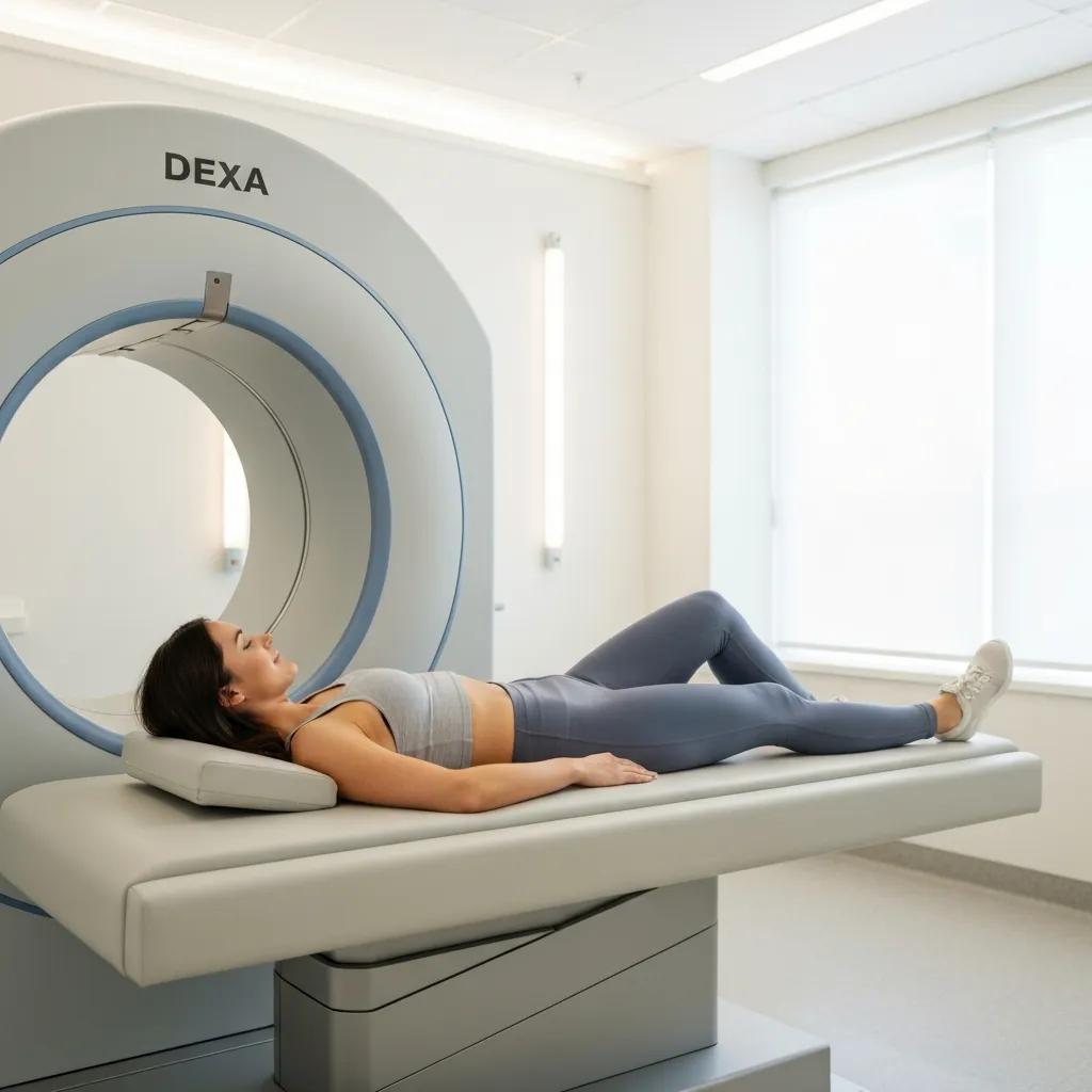
DEXA scans measure bone mineral density to diagnose osteoporosis, estimate fracture risk and monitor treatment using low‑dose X‑ray absorption differences between bone and soft tissue. The test reports T‑scores and Z‑scores that guide decisions about medications, supplementation and lifestyle measures to reduce fracture risk. DEXA is sensitive to small changes in bone density, quick to perform and generally comfortable. The table below outlines common DEXA types, what they measure, who they’re for and typical billing notes so you can plan your appointment.
Research has also explored advanced analyses (such as Hip Structure Analysis) that extract geometric strength information from DXA scans, highlighting that conventional DXA metrics do not capture every aspect of bone strength.
Advanced Bone Strength Analysis Using DXA Scans
The Hip Structure Analysis (HSA) approach was developed to derive geometric strength estimates from archived hip DXA scans used in large research cohorts. Evidence shows that some strength characteristics are not fully reflected by standard DXA measures, so clinicians and researchers are increasingly interested in methods that assess bone strength more directly. While DXA can contribute useful geometric data, it was not originally designed as a complete measure of structural strength.
Extending DXA beyond bone mineral density: understanding hip structure analysis, 2007
How Does a Bone Density Test Diagnose Osteoporosis?
Bone density testing diagnoses osteoporosis by comparing a patient’s BMD to a young adult reference (T‑score) and to age‑matched peers (Z‑score). A T‑score of −2.5 or lower typically defines osteoporosis; scores between −1.0 and −2.5 indicate osteopenia. These thresholds correspond to increasing fracture risk and guide decisions about medications, fall prevention and further investigation. Serial DEXA tests let clinicians monitor treatment response and decide when specialist referral or additional imaging is needed. After abnormal results we work with your GP or specialist to outline risk‑reduction steps, treatment options and follow‑up plans.
What Preparation Is Needed for a DEXA Scan in Erina?
Preparing for a DEXA scan is straightforward: wear loose, comfortable clothing without metal fastenings, avoid taking calcium supplements immediately beforehand if instructed, and bring any previous bone density reports. The scan is painless and takes only a few minutes per site while you lie still on the table as the low‑dose X‑ray arm passes over the region. Tell the imaging team about recent fractures, metal implants or pregnancy so we can adapt the protocol if needed. Clear prep reduces the chance of repeat scans and improves comparability with prior studies.
How Do Erina Central Coast Radiology Services Ensure Patient Comfort and Accuracy?
We prioritise patient comfort and diagnostic accuracy through up‑to‑date equipment, experienced staff and robust quality assurance across image acquisition, reporting and follow‑up. Modern scanners and optimised protocols shorten scan times, lower radiation dose and improve image quality. Our patient‑centred practices — clear pre‑test instructions, in‑scan communication and simple anxiety‑reduction measures — help most patients complete their studies comfortably. Quality assurance includes radiologist oversight, standardised reports and reliable turnaround times so results support timely clinical decisions.
What Advanced Technologies Are Used in Erina’s Imaging Center?
We use advanced imaging technologies — higher field‑strength MRI, low‑dose CT protocols, modern digital mammography and high‑resolution ultrasound — to improve diagnostic precision while keeping scans quicker and safer. Higher field MRI increases signal and contrast for subtle soft‑tissue findings; low‑dose CT reduces radiation without compromising key information. Software improvements, including AI‑assisted reconstruction and automated measurements, speed image processing and support radiologist accuracy by flagging relevant findings. Paired with experienced sonographers and radiologists, these tools enable earlier detection and clearer problem definition.
How Does Bulk Billing Work for Medical Imaging Services in Erina?
Bulk billing means Medicare can cover the cost of eligible imaging services when specific clinical criteria are met, so the patient has no out‑of‑pocket fee in those cases. Eligibility depends on the clinical indication, MBS item rules and a valid referral from a treating practitioner; common bulk‑billed examples include urgent or hospital‑referred studies and specified screening tests. To confirm eligibility, check the referral, review Medicare rules and contact the imaging provider about bulk‑billing slots and booking options. Doing so can reduce financial barriers and improve access to necessary diagnostics. Central Coast Imaging, Central Coast Radiology, Central Coast Cardiac Imaging, Central Coast Ultrasound, Central Coast DEXA, Central Coast Imaging, Central Coast MRI
What Is the Patient Journey for Imaging Services in Erina Central Coast?
The patient journey runs from referral and triage through booking, preparation, scanning, reporting and clinical follow‑up. After your referrer sends a referral, we triage urgency, provide pre‑test instructions and schedule the appointment. Clear prep reduces delays and repeat imaging. On the day, staff will guide you through registration and the scan, ensuring safety and comfort while acquiring diagnostic images. Radiologists then report the study and deliver results to the referring clinician within standard turnaround times, enabling timely treatment decisions or specialist referrals.
The numbered checklists below summarise modality‑specific preparation and what to expect for results and follow‑up.
- Pre‑appointment checklist for MRI, CT and Ultrasound:
Bring your referral and photo ID. Tell staff about any implants or pregnancy.
Follow fasting or bladder instructions given for your specific test.
Remove metal and wearable electronics for MRI; disclose any allergies if contrast CT is planned. - On‑the‑day checklist for comfort and safety:
Wear comfortable clothing and arrive early to complete registration.
Tell us if you have claustrophobia — we can discuss open MRI options or support measures.
Staff will confirm your details and explain how long the scan is expected to take.
These steps reduce anxiety and avoid avoidable delays so tests proceed smoothly from referral to result.
How Should Patients Prepare for MRI, CT, and Ultrasound Scans?
Preparation varies by test but follows clear principles to ensure image quality and safety: MRI requires removal of metal and full disclosure about implants; CT may require fasting and renal function checks when contrast is used; ultrasound prep depends on the target area (fasting for some abdominal studies, a full bladder for pelvic scans). If you have claustrophobia or anxiety, ask about open MRI alternatives, relaxation techniques or in‑scan communication options. Bringing prior imaging and a brief medical history helps radiologists compare studies and reach an accurate diagnosis. Following these steps prevents delays and reduces the need for repeat scans.
How Are Imaging Results Delivered and Explained to Patients?
Radiologists interpret images and send standardised reports to the referring clinician; urgent findings are communicated directly to the referrer to speed patient care. Turnaround times vary by modality and urgency — some reports are ready within hours, others within a few days — so check with your referrer about when you’ll hear back and arrange follow‑up to discuss implications. Many centres, including ours, offer secure online access to images and reports where appropriate. When results are abnormal or urgent, the radiologist and clinical team coordinate to ensure timely patient notification and clear next steps.
How Can Referring Doctors Access and Utilize Erina Central Coast Imaging Services?
Referrers can access our services by following standard referral guidelines: clearly state the clinical question, include relevant history and prior imaging, and specify the requested study so we can triage and choose the most appropriate protocol. Clear referrals that note urgency and clinical indicators reduce delays and improve appropriateness. Referrers should also be familiar with our portal for images and reports and the process to request urgent imaging; these tools support rapid collaboration and better patient outcomes. The short list below summarises commonly requested services.
Services referrers commonly request include:
- Central Coast MRI for neurological, spinal and musculoskeletal questions.
- Central Coast Ultrasound for obstetric, vascular and MSK assessments.
- Central Coast DEXA for bone mineral density testing.
- Central Coast Cardiac Imaging for CTCA, calcium scoring and echocardiography.
Including a clear clinical question and indicating urgency when needed helps ensure timely scheduling and the right imaging choice for optimal patient care.
What Are the Referral Guidelines for Imaging Procedures?
Good referrals include patient identifiers, a concise clinical question, relevant medical history, prior imaging and the requested modality or specific study so we can triage and book appropriately. For some tests, MBS item rules or local criteria affect bulk‑billing eligibility or prioritisation, and acute symptoms should be flagged for fast‑tracking. Clear indications — for example suspected fracture, progressive neurological deficit or suspected malignancy — help imaging services allocate resources and select the right protocol. If you’re unsure, contact our clinical liaison to discuss complex cases and reduce unnecessary imaging.
How Can Referrers Access Patient Images and Reports Online?
Referrers usually access images and reports via secure web portals or PACS viewers that provide DICOM images and PDF reports for review, measurement and integration into electronic medical records. Portal access typically requires user registration and authentication; images become available within standard turnaround times after reporting, and urgent cases are shared faster through direct channels. From the portal you can download reports, compare studies and request additional views or second opinions; IT and clinical support are available for access issues. Familiarity with portal workflows improves multidisciplinary collaboration and speeds patient management based on imaging findings.
This page maps our modalities, preparation steps, cardiac and DEXA specifics, technology benefits, billing pathways and referrer processes so patients and clinicians on the Central Coast can move from referral to treatment with clarity and confidence.
Frequently Asked Questions
What should I expect during my first imaging appointment at Erina Central Coast?
On your first visit you’ll be welcomed by our reception team and asked to provide your referral and ID. The technologist will take a brief history, explain the procedure and estimate how long the scan will take. We offer comfort measures such as cushions and in‑scan communication to help you through the exam. Arriving a little early gives you time to complete any paperwork and ask questions.
Are there any risks associated with medical imaging procedures?
Most imaging tests are safe when used appropriately. X‑rays and CT scans involve ionising radiation, so we balance diagnostic benefit against exposure and use low‑dose protocols where possible. MRI and ultrasound do not use ionising radiation. If you have concerns — about radiation, contrast agents or pregnancy — speak with your referrer or our staff so we can explain risks and benefits for your situation.
How can I access my imaging results after the procedure?
Radiologists review and report studies, and the report is sent to your referring clinician who will discuss the findings with you. Many centres, including ours, also provide secure online portals where patients can view images and reports once they’re released. You may need to register for portal access; if you’re unsure when results will be available, check with your referrer.
What should I do if I have claustrophobia and need an MRI?
Tell your referrer and our booking staff if you have claustrophobia. We can discuss options such as open or wider‑bore MRI scanners, relaxation strategies and in‑scan communication. In some cases light sedation may be appropriate; we’ll explain the process and requirements during booking so you arrive prepared.
What are the typical turnaround times for imaging results?
Turnaround times depend on the study and urgency. Routine X‑rays or ultrasounds may be reported within hours to a couple of days; MRIs and CTs often take longer, typically up to several days for detailed reporting. Urgent cases are fast‑tracked and communicated directly to the referrer as soon as possible.
Can I bring someone with me to my imaging appointment?
Yes — having a friend or family member for support is usually fine. Some areas (for example the MRI scan room) have safety restrictions, so check the centre’s visitor policy when you book. A companion can help with transport and post‑scan arrangements, especially if sedation is used.
Conclusion
Erina Central Coast Medical Imaging provides a full range of diagnostic services designed to help patients and referrers make informed health decisions. Understanding each modality, its preparation and what to expect can make the imaging pathway clearer and less stressful. If you’re ready to take the next step, explore our services or contact us to schedule an appointment — we’re committed to comfortable care and reliable results to support your health journey.