Understanding Ultrasound Scans: Clear, practical information about imaging and patient care
An ultrasound scan is a safe, non‑invasive test that uses high‑frequency sound waves to produce live images of structures inside the body—without ionising radiation. This guide explains how sonography works, the common types of scans your doctor may request, and simple steps to prepare so your appointment runs smoothly and the results are accurate. Many patients arrive unsure about safety, preparation or what the report will mean for their care; this article answers those questions with clear explanations and practical checklists. You’ll find an overview of transducer technology, the main ultrasound categories (obstetric, vascular, musculoskeletal and more), how ultrasound compares with CT and MRI, and what to expect during and after your scan. We also outline preparation by scan type, the examination and reporting process, and why Life Medical Imaging Central Coast is a trusted local provider. Throughout, common search terms like ultrasound scans, sonography, ultrasound safety and ultrasound preparation are used to help you find reliable, evidence‑informed answers and support conversations with your referring doctor.
What Is an Ultrasound Scan and How Does Sonography Work?
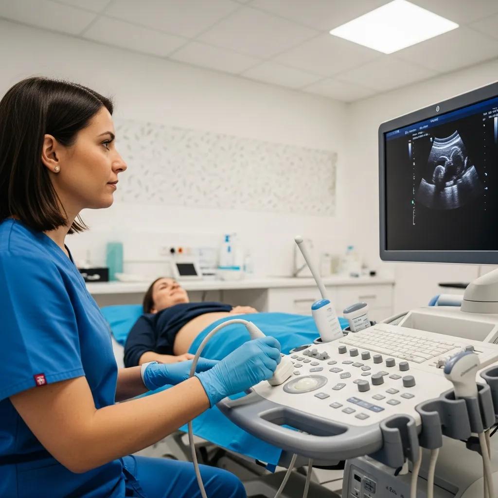
An ultrasound scan is a diagnostic procedure that uses a handheld probe, called a transducer, to send and receive sound waves and create real‑time images of tissues and organs—without exposing you to ionising radiation. The transducer turns electrical pulses into sound, then listens for echoes reflected by tissue boundaries; the machine converts echo timing and strength into greyscale images. Because images are live, sonographers can assess movement, blood flow and organ function as they happen, which is why ultrasound is useful for pregnancy checks, vascular studies and image‑guided procedures. Image quality depends on probe frequency, the patient’s body habitus and the operator’s technique, so experienced sonographers adjust settings to give the best balance of resolution and depth for each clinical question.
What Are Ultrasound Scans and Their Diagnostic Uses?
Ultrasound scans are used to diagnose and monitor many conditions by visualising organs, blood vessels and soft tissues in real time. Typical uses include pregnancy assessment, investigation of abdominal pain, looking for gallstones and evaluating vascular problems. For example, obstetric ultrasound dates pregnancy and tracks fetal growth; abdominal scans can reveal gallstones, liver changes or kidney enlargement. Doppler ultrasound measures blood flow and helps detect deep vein thrombosis or carotid disease, while musculoskeletal scans assess tendons, muscles and joints for tears or fluid. Referrers choose ultrasound when soft‑tissue detail, dynamic assessment or avoiding radiation is important—matching the scan to the clinical question ensures useful results.
How Does the Ultrasound Transducer Use Sound Waves to Create Images?
The transducer works like a tiny speaker and microphone: it sends short pulses of high‑frequency sound into the body and then listens for the returning echoes. The scanner translates echo timing and strength into the image you see. Higher‑frequency probes give finer detail for shallow structures such as the thyroid or breast, while lower‑frequency probes penetrate deeper for abdominal and pelvic imaging; sonographers select probes based on the area being examined. Advanced modes such as Doppler analyse flow, colour mapping shows direction and velocity, and 3D/4D obstetric techniques reconstruct volumetric fetal anatomy. Operator technique—probe angle, pressure and patient positioning—affects image quality, which is why skilled sonographers and specialist reporting help achieve accurate diagnoses.
Are Ultrasound Scans Safe? Understanding Ultrasound Scan Safety and Benefits
Ultrasound scans are regarded as safe because they use non‑ionising sound waves rather than X‑rays. Clinical guidelines support their routine use in pregnancy and in many diagnostic settings when medically indicated. Professional bodies recommend sensible use of diagnostic ultrasound and following safety principles; at diagnostic levels there is no evidence of long‑term harm. Practical benefits include immediate bedside assessment, portability and lower cost than many other imaging options. Limitations include operator dependence and reduced image quality when a patient is very large or when bowel gas obscures the view. Knowing both the strengths and limits of ultrasound helps clinicians select the right test and helps patients understand why ultrasound may be the preferred option.
Different imaging methods vary in radiation exposure and clinical purpose; the table below summarises radiation use and practical risk for commonly used modalities so you can see ultrasound’s safety advantage at a glance.
This comparison highlights that ultrasound is a radiation‑free option suitable for many indications—particularly when safety and bedside assessment are priorities.
Below are ultrasound’s main benefits and practical limits, summarised so patients can discuss imaging choices with their clinician.
Ultrasound offers several patient‑centred advantages:
- No ionising radiation: safe for repeated use and pregnancy monitoring.
- Real‑time, dynamic imaging: allows assessment of motion and blood flow as they happen.
- Portable and fast: many exams can be done at the bedside or completed quickly in clinic.
These strengths make ultrasound a first‑line tool for many problems, while CT or MRI may be chosen when more detailed cross‑sectional views are required.
Why Are Ultrasound Scans Considered Radiation-Free and Safe?
Ultrasound is radiation‑free because it uses mechanical sound waves rather than ionising photons. These waves pass through tissue but do not ionise atoms or damage DNA the way X‑rays can. Regulatory and professional guidance promote ALARA principles—use the lowest useful acoustic output for the shortest practical time—and following these protocols minimises theoretical risk. In pregnancy, ultrasound is the preferred routine modality because it balances diagnostic benefit with safety, and targeted scans follow established guidelines to answer specific clinical questions. Knowing ultrasound is non‑ionising reassures patients that diagnostic imaging can usually be done with minimal risk when clinically justified.
For detailed regulatory and technical information, see the official ultrasound safety guidelines.
What Are the Key Benefits of Ultrasound Imaging Compared to Other Scans?
Ultrasound provides clear clinical benefits—safety, cost‑effectiveness and live imaging—while its main limitations are operator dependence and reduced penetration through air or bone. Compared with CT, ultrasound avoids ionising radiation and is more portable; compared with MRI, ultrasound is quicker and often more accessible but may offer less soft‑tissue contrast for some conditions. Understanding these trade‑offs helps referrers choose the best test for each patient—for example, ultrasound for gallbladder problems or Doppler for vascular flow, and CT/MRI when detailed cross‑sectional anatomy is needed. This targeted approach gives patients faster, safer assessments tailored to their clinical needs.
What Types of Ultrasound Scans Are Offered and What Do They Diagnose?
Ultrasound covers a range of specialised scans—general abdominal/pelvic, vascular (Doppler), musculoskeletal, obstetric, gynaecological, breast and advanced 3D/4D obstetric imaging—each designed to image particular anatomy and answer specific clinical questions. General abdominal scans look at the liver, gallbladder, spleen, pancreas and kidneys for stones, masses or inflammation; pelvic scans assess the bladder, uterus and ovaries. Vascular (Doppler) studies measure blood flow to detect blockages or clots, while musculoskeletal ultrasound evaluates tendons, ligaments and joints for tears or inflammation. Obstetric and gynaecological scans support pregnancy care, fertility assessment and pelvic pathology; 3D/4D imaging adds volumetric detail for fetal anatomy and parent‑facing images.
The table below compares common scan types, the body area examined and typical diagnostic uses to help you identify which scan may match your referral.
This comparison shows how each ultrasound subtype matches clinical questions and supports discussion with your referring clinician.
Life Medical Imaging Central Coast provides a full range of these ultrasound services across several Central Coast locations, including general, vascular, musculoskeletal, obstetric, gynaecological, breast and 3D/4D obstetric imaging. As an independent, NATA‑accredited radiology clinic with sub‑specialist Women’s and Cardiac Imaging, we invest in current technology, experienced clinical staff and simple appointment options by phone or online. Central Coast patients therefore have access to specialist reporting and the convenience of local clinics when referred for ultrasound.
For a detailed overview of specific diagnostic applications, explore the different types of ultrasound scans available.
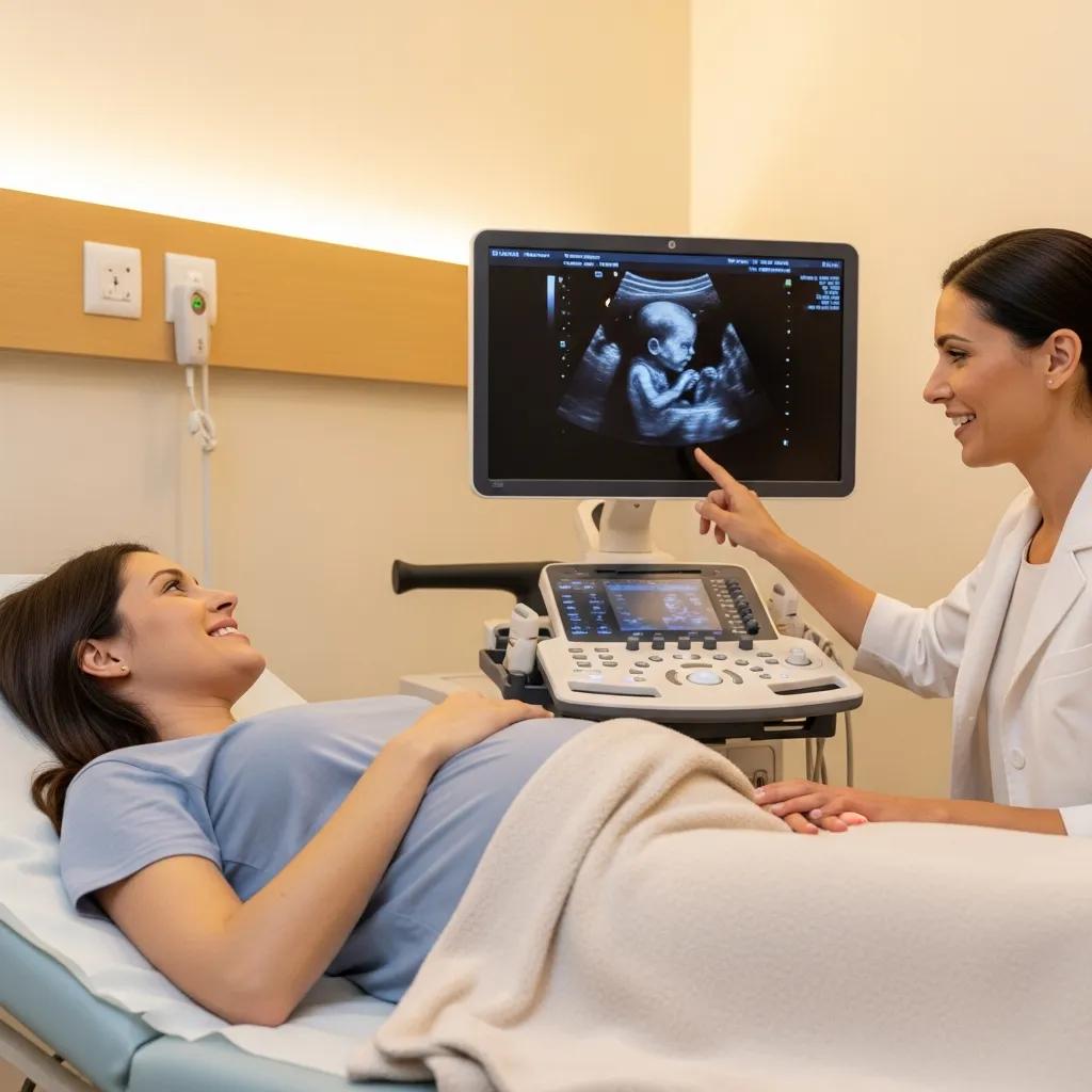
Advanced 3D/4D Ultrasound in Prenatal Diagnosis
3D and 4D ultrasound techniques are now practical for routine clinical use. Unlike traditional 2D scans, 3D modes provide volumetric views that can reveal anatomical features not seen in a single 2D plane, helping clinicians identify normal and abnormal fetal structures. 4D imaging adds real‑time motion, which aids assessment of fetal movements and cardiac function when used with specialised acquisitions such as STIC‑Colour. For counselling, rendered volumes help parents understand findings and may reassure in low‑risk cases. Storing volumes (not just 2D images) also allows later review of equivocal findings and supports teaching. This review demonstrates current 3D/4D capabilities and display options for clinicians who do not routinely perform these exams.
How Should Patients Prepare for an Ultrasound Scan?
Preparation depends on the type of scan because image quality is affected by organ distension, bowel gas and bladder fullness. Proper preparation reduces the need for repeats and improves diagnostic accuracy. Generally, upper abdominal scans require fasting to limit bowel gas and improve views of the gallbladder and pancreas, while pelvic and obstetric scans usually ask for a full bladder to act as an acoustic window. Wear clothing that gives easy access to the area being scanned and bring any previous imaging or referral notes to help the sonographer focus on the clinical question. Clinic‑specific instructions may vary, so confirm preparation details when you book to avoid delays.
Below is a concise table mapping common scan types to preparation steps and why they matter.
This table helps you match your appointment type to clear, timed steps and reduces the chance of needing to reschedule.
Practical tips to improve comfort and the success of your scan:
- Wear loose, two‑piece clothing so the sonographer can access your abdomen or pelvis easily.
- Bring your referral paperwork and any previous imaging reports if available.
- Tell the clinic in advance about mobility needs or if you would like a chaperone present.
These simple actions make appointments smoother and let sonographers focus on getting diagnostic‑quality images.
At Life Medical Imaging Central Coast we give location‑specific preparation details when you book so you know exact fasting times or bladder requirements for the clinic you choose. Our online and phone booking channels provide tailored instructions by appointment type and site, which helps for multi‑location services across the Central Coast.
For a comprehensive guide on how to prepare, see our ultrasound preparation page.
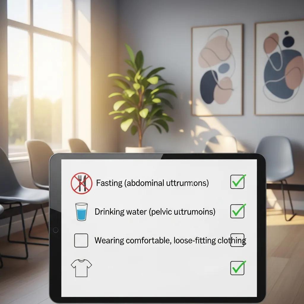
What Are the Common Preparation Steps for Different Ultrasound Types?
Common preparation patterns are predictable: fasting for upper abdominal scans, a full bladder for pelvic and obstetric scans, and comfortable, accessible clothing for musculoskeletal exams. For abdominal imaging, fasting reduces bowel gas and helps visualise the gallbladder; for pelvic exams, drinking water about an hour before the appointment creates a helpful acoustic window. Musculoskeletal scans usually need minimal prep but wearing short sleeves or shorts makes access faster. Always confirm these steps with your imaging provider at booking to avoid delays and improve diagnostic yield.
How Can Patients Ensure a Comfortable and Successful Ultrasound Appointment?
To ensure comfort and a successful scan, arrive with any referral paperwork, wear clothing that allows easy access to the scanned area, and tell staff about mobility or anxiety concerns when you book or arrive. If you prefer a chaperone, request this at booking so staff can arrange it. If transport or parking is an issue, ask the clinic for access details—many Central Coast sites provide guidance when you call. Clear communication and simple preparation reduce stress and support the best possible imaging outcome.
What Happens During and After Your Ultrasound Scan?
During the scan a sonographer will position you on the couch, apply a water‑based gel to the skin and move the transducer over the area while capturing images. The test is usually comfortable and non‑invasive; you may feel mild pressure as the sonographer works to get clear views. You may be asked to change position, hold your breath or move a limb to help acquire the necessary images. Most studies take between 10 and 45 minutes depending on complexity. A radiologist interprets the images and issues a formal report to your referring doctor; urgent findings are communicated promptly under clinic protocols. Knowing the typical workflow helps reduce anxiety and sets expectations for next steps after imaging.
The step‑by‑step sequence below summarises what to expect from arrival to report distribution.
- Registration and check‑in: confirmation of your referral and identity.
- Preparation in the exam room: clothing adjustment and gel application.
- Image acquisition: the sonographer records static and dynamic views.
- Reporting: a radiologist reviews images and sends a report to the referrer.
This straightforward sequence explains the clinical workflow and likely follow‑up after the scan.
What Can Patients Expect During the Ultrasound Examination?
Expect a comfortable exam where the sonographer applies warm gel and moves a handheld probe over the skin to capture images. Most scans are painless, though you might feel mild pressure. The sonographer will explain what they’re doing, take measurements and ask brief questions about your symptoms or history to correlate with the images. If an urgent issue is seen, the sonographer or radiologist will escalate it to the referring clinician for timely management. You may be asked to wait briefly after the scan while images are checked for completeness before you leave.
How Are Ultrasound Results Interpreted and Communicated?
After images are acquired, a radiologist interprets them and prepares a structured report that is sent to your referring doctor, who will then discuss findings and next steps with you. Turnaround times vary by urgency but are commonly a few days for routine studies; critical or urgent findings are communicated immediately. Reports outline findings, offer diagnostic impressions and sometimes recommend further imaging or specialist referral. If you have questions about the report, discuss them with your referring clinician, who will place the imaging results within the context of your overall care.
Why Choose Life Medical Imaging Central Coast for Your Ultrasound Scan?
Life Medical Imaging Central Coast is an independent, NATA‑accredited radiology service offering a broad range of ultrasound options, supported by sub‑specialist Women’s and Cardiac Imaging, modern equipment and experienced clinical staff. NATA accreditation reflects our commitment to consistent quality and calibration standards, while sub‑specialist reporting improves accuracy for complex referrals. With multiple Central Coast locations and straightforward booking by phone or online, we aim to make local access to specialist imaging easy for patients and referring clinicians. These trust signals help ensure reliable imaging with specialist interpretation when it matters most.
Key provider strengths that matter to patients and referrers:
- NATA accreditation for quality and consistency across services.
- Sub‑specialist Women’s and Cardiac Imaging for expert reporting.
- Modern ultrasound equipment and experienced sonographers for accurate studies.
- Multiple Central Coast locations with convenient booking options.
This combination of accreditation, specialist capability and local access supports timely imaging and confident clinical decision‑making for patients on the Central Coast.
What Makes Life Medical Imaging Central Coast a Trusted Ultrasound Provider?
Life Medical Imaging Central Coast blends independent practice with NATA accreditation, sub‑specialist imaging services and current ultrasound technology to deliver accurate diagnostic reports and patient‑focused care. NATA accreditation confirms adherence to strict quality and calibration standards, while sub‑specialist Women’s and Cardiac Imaging ensures targeted expertise for complex referrals. Skilled sonographers and supportive staff focus on patient comfort and optimise imaging protocols to answer the referring clinician’s question. Our multiple Central Coast locations improve access for local patients and support continuity of care between community referrers and imaging services.
To learn more about our services and commitment to patient care, visit the Life Medical Imaging Central Coast website.
How Can Patients Book an Ultrasound Appointment on the Central Coast?
You can book an ultrasound appointment online or by calling the clinic to arrange a suitable time and receive location‑specific preparation instructions. Multiple Central Coast sites let you choose the most convenient clinic when booking. When arranging your appointment, tell us about any mobility needs, request a chaperone if you prefer one, and confirm fasting or bladder requirements so the visit is efficient and diagnostic quality is maximised. Referring doctors should include clear clinical indications on referrals to ensure the correct ultrasound protocol and speed up specialist reporting when needed. Booking is designed to be straightforward, with staff ready to advise what to bring and how to prepare for your scan.
After your ultrasound, follow up with your referring doctor to review the radiologist’s report and agree next steps; if urgent treatment or further imaging is required, your clinician will advise promptly. Life Medical Imaging Central Coast supports patients through the imaging process with clear instructions at booking and specialist reporting to help timely clinical decisions.
Frequently Asked Questions
What should I expect during my ultrasound appointment?
At your appointment you’ll lie comfortably on an exam couch while a sonographer applies warm, water‑based gel and moves a handheld probe over the area being examined. The scan is generally painless, though you may feel mild pressure. You might be asked to change position or hold your breath to get clearer images. Most scans take between 10 and 45 minutes depending on the study.
How long does it take to receive ultrasound results?
Images are reviewed by a radiologist who prepares a report for your referring doctor. Turnaround times vary; routine, non‑urgent reports are usually available within a few days. If the scan shows an urgent problem, the clinic will ensure your referring doctor is notified promptly. Always follow up with your referrer to discuss results and the next steps.
Are there any risks associated with ultrasound scans?
Ultrasound is considered very safe because it uses non‑ionising sound waves rather than the ionising radiation used in X‑rays and CT scans. Current guidelines support routine clinical use, including in pregnancy. Known risks at diagnostic settings are minimal, and clinicians follow the ALARA principle—”As Low As Reasonably Achievable”—to minimise exposure. If you have concerns, speak to your healthcare provider.
Can I eat or drink before my ultrasound?
Preparation depends on the scan type. For abdominal ultrasounds you’ll usually be asked to fast for 6–8 hours to reduce bowel gas and improve visibility of the gallbladder and pancreas. For pelvic and obstetric scans you’ll often be asked to drink water before the appointment to ensure a full bladder. Confirm specific instructions with your imaging provider when booking.
What types of ultrasound scans are available?
Common ultrasound types include general abdominal, vascular (Doppler), musculoskeletal, obstetric, gynaecological and breast scans. Each targets different body regions and clinical questions—organ health, blood flow or fetal development, for example. Knowing the scan type helps you prepare and discuss concerns with your clinician.
How can I ensure a successful ultrasound experience?
To get the best result, bring your referral paperwork and any previous imaging, wear clothing that gives easy access to the area being scanned, and tell staff about mobility or anxiety needs when you book. Request a chaperone in advance if you prefer one. Following preparation instructions, such as fasting or drinking water, also helps improve image quality.
Conclusion
Understanding ultrasound scans helps you make informed choices about diagnostic imaging. Ultrasound is a safe, non‑invasive tool that provides real‑time images without ionising radiation, and simple preparation steps help ensure a comfortable, useful exam. For more information about ultrasound services and to book an appointment, visit Life Medical Imaging Central Coast or contact your referring doctor. Take the next step in your care by arranging your scan today.