
Your Complete Guide to Medical Imaging Exams and Diagnostic Radiology
Medical imaging studies are specialised radiology exams that create pictures of the body’s internal structures to help clinicians diagnose, stage and manage illness. Common modalities include CT, MRI, ultrasound, X‑ray and DEXA — each highlights different anatomy (and sometimes physiology) so care teams can make clearer decisions and plan targeted treatment. Many people want straightforward guidance about what each test shows, how to prepare, what to expect during the scan and how safety is managed. This guide explains the main imaging types, practical preparation tips for CT and ultrasound, the typical patient journey before and after scans, which conditions suit which tests, and the risks and safety measures taken by radiology teams. You’ll also find simple booking steps for the Central Coast so you can move from understanding to scheduling with confidence.
What Are Medical Imaging Studies and Diagnostic Radiology Procedures?
Medical imaging studies are healthcare exams that produce images of organs, bones, tissues and blood vessels to detect disease and guide treatment. They use targeted energy — ionising radiation for CT/X‑ray or sound waves for ultrasound — to form images; each modality emphasises different tissue characteristics and clinical advantages. The main benefits are improved diagnostic accuracy, faster triage in urgent care and precise guidance for procedures, all of which reduce uncertainty for patients and referrers. Knowing how the modalities differ helps clinicians and patients choose the most appropriate test for a given symptom or condition.
Below is a concise summary of the common modalities, their typical uses and basic preparation needs.
Which Types of Medical Imaging Studies Are Commonly Performed?
Typical studies include general CT, Cardiac CT and CT angiography for vascular assessment, ultrasound for obstetrics and vascular work, digital X‑ray for bones and chest, and DEXA for bone density and body composition. Each test fits particular clinical needs: CT is excellent for acute abdominal pain and complex fractures; cardiac CT angiography assesses coronary anatomy; ultrasound evaluates pregnancy and blood flow; X‑ray quickly demonstrates fractures; and DEXA measures bone loss or composition. Interventional procedures — spinal or joint injections, biopsies and aspirations — combine imaging guidance with therapeutic or diagnostic techniques to improve accuracy and reduce complications. Paediatric imaging uses smaller, tailored protocols to limit radiation exposure, while specialised services such as dental imaging and musculoskeletal ultrasound provide focused assessments.
This overview leads naturally to how imaging findings influence diagnosis and everyday clinical decision‑making.
Medical imaging is continually evolving — new techniques and applications keep improving diagnosis and patient outcomes.
Recent Trends in Medical Imaging Modalities and Their Applications in Disease Diagnosis
Medical imaging is central to modern healthcare, supporting diagnosis, treatment planning and ongoing monitoring. Non‑invasive tools such as X‑ray, PET, CT, MRI and ultrasound allow clinicians to see internal structures without surgery. Advances in medical image processing (MIP) have strengthened disease prediction, detection and analysis, and MIP datasets are increasingly used in machine learning and deep learning models to build intelligent decision‑support tools. Because human interpretation can be time‑consuming and fallible, these technologies aim to improve accuracy and efficiency. This review describes the working principles, benefits and limits of diverse imaging modalities, surveys recent methodological advances and highlights emerging trends and future directions in the field.
Recent trend in medical imaging modalities and their applications in disease diagnosis: a review, B Abhisheka, 2024
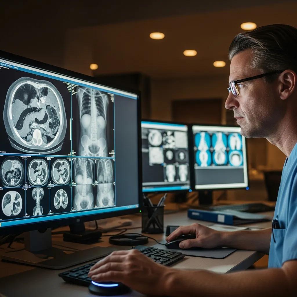
How Do Medical Imaging Studies Aid in Diagnosis?
Imaging turns symptoms and clinical signs into visual evidence that locates disease, measures severity and clarifies relationships between structures. For example, CT can detect internal bleeding after trauma, ultrasound can show ovarian cysts or fetal wellbeing, and X‑ray confirms fractures — each finding directly influences surgical, medical and rehabilitation choices. Radiologists integrate image findings into structured reports that highlight urgent results, suggest follow‑up and note study limitations; referring clinicians use these reports to plan next steps. Modern imaging also supports cancer staging, treatment response monitoring and guidance for minimally invasive procedures, making it an essential part of ongoing patient care.
Accurate interpretation depends on choosing the right protocol and providing clinical context, so the next section explains how to prepare to ensure safe, high‑quality images.
How Do I Prepare for Different Medical Imaging Examinations?

Preparation varies by modality and the clinical question. Correct preparation improves image quality and reduces the chance of a repeat scan. Key general points: follow fasting instructions for contrast studies, tell staff about allergies and medications (particularly diabetes medicines), and bring any prior imaging or referral notes. Specific preparation steps for CT and ultrasound depend on the exact study and are described below to help you arrive ready. Good preparation also improves safety and diagnostic yield, which speeds up clinical decision‑making.
Arriving prepared reduces anxiety and delays — please follow the CT checklist and the ultrasound guidance we provide with your booking.
What Are the Preparation Steps for CT Scan Appointments?
For a CT scan, confirm whether IV contrast is planned, follow any fasting instructions, check medication guidance for diabetes and wear comfortable clothing without metal. If contrast is required, tell staff about any previous contrast reactions or kidney disease; you may be asked about medicines such as metformin and given specific advice. Arrive early to complete registration and screening forms; during the scan you’ll lie on the motorised table and may be asked to hold your breath for short periods. Preparing well shortens the appointment and ensures the radiologist receives the images needed for an accurate report.
Next we cover ultrasound preparation, which differs by the body area being examined.
Consistent protocols across sites help ensure the same quality of imaging and reporting for patients and clinicians.
Standardising CT and MR Protocols for Consistent Medical Imaging Experiences
CT and MR studies are performed across multiple sites and scanners. To deliver consistent ordering, acquisition and image appearance, many services reduce the number of protocols and adopt indication‑driven standard templates. This process typically includes creating standard protocol templates, a change‑request workflow and governance oversight. Rapid improvement events can streamline protocols — for example, reducing a large set of CT or MR protocols into a smaller, standardised group — and ongoing work aims to standardise reporting output. The result is a more consistent experience for radiologists, patients and referring providers.
CT and MR protocol standardization across a large health system: providing a consistent radiologist, patient, and referring provider experience, 2017
How Should Patients Prepare for Ultrasound Studies on the Central Coast?
Ultrasound prep depends on the area being scanned. Abdominal ultrasounds often require fasting for 6–8 hours to reduce bowel gas; pelvic or early pregnancy scans commonly need a full bladder to improve the acoustic window. Wear loose clothing and bring your referral and any prior imaging; remove jewellery and be ready to expose only the area being examined. For obstetric appointments, bring pregnancy documents and consider whether a partner or support person is permitted. Paediatric or mobility‑limited patients may need additional assistance. Following the specific instructions on your appointment notice helps the sonographer capture clear, real‑time images and shortens scan time.
Proper preparation leads to faster, more accurate exams — and a smoother visit for you.
What to Expect During and After Diagnostic Radiology Exams
Most patients follow a clear pathway: arrival and check‑in, clinical screening and consent if needed, the imaging procedure, then reporting and follow‑up with the referring clinician. Radiology teams explain the scan, answer questions and carry out safety checks (for example, screening for allergies or pregnancy) before starting. After many scans there is minimal downtime and you can resume normal activities; some interventional procedures require short observation and specific aftercare instructions. Knowing the typical journey reduces anxiety and helps you prepare useful questions for clinicians and radiographers.
Below are brief descriptions of what to expect for CT (including Cardiac CT angiography), ultrasound and X‑ray exams — sensations, timings and procedural steps.
What Happens During a CT Scan or Cardiac CT Angiography?
During a CT you lie on a motorised table that passes through the scanner gantry while X‑ray detectors capture cross‑sectional images. Scans are usually painless — you may hear brief machine noise and feel slight table movement. If IV contrast is given, you might notice a warm flush or metallic taste; staff monitor you for immediate reactions and will have checked your allergy history and kidney function beforehand. Cardiac CT angiography often requires heart‑rate control and ECG gating to synchronise images with the cardiac cycle, and breath‑hold instructions are used to reduce motion artefact. After the scan, patients who received contrast are observed briefly and images are sent to a radiologist for reporting; turnaround depends on clinical urgency.
Understanding the CT process helps set expectations for ultrasound and X‑ray, which are generally quicker and less invasive.
How Are Ultrasound and X‑ray Studies Conducted?
Ultrasound uses a handheld transducer that emits and receives sound waves while a sonographer moves it over the skin with gel to improve contact; exams are real‑time and often allow immediate verbal feedback. X‑rays use a short burst of ionising radiation to capture static projections; they are fast — usually completed in minutes — and staff apply shielding and low‑dose techniques where appropriate. Both modalities usually require minimal recovery time and you can often leave immediately after imaging; the team will advise if follow‑up or additional tests are likely. Bring your referral, ID and prior imaging, and expect an estimated timeline for when your referring clinician will discuss results.
Next we outline which conditions are best investigated with each imaging type.
Which Medical Conditions Are Diagnosed Through Imaging Studies?
Imaging is essential across many clinical areas because it localises disease, defines severity and often suggests likely causes that guide treatment. Common examples: cardiovascular disease assessed with cardiac CT and vascular ultrasound; fractures and arthritis evaluated with X‑ray and CT; cancers investigated with CT and targeted ultrasound for biopsy planning. Neurological emergencies such as stroke and haemorrhage rely on CT and MRI for rapid assessment. Choosing the right modality increases diagnostic yield and reduces unnecessary tests, which benefits patients and referrers alike.
The table below maps frequent conditions to recommended imaging tests and typical findings so you understand why a specific study is chosen.
How Is Imaging Used for Cardiovascular and Neurological Disorders?
For cardiovascular disease, cardiac CT and CT angiography show coronary anatomy, measure calcified plaque and non‑invasively assess stenosis; vascular ultrasound evaluates blood flow and detects peripheral stenosis or aneurysm. In neurological emergencies, CT rapidly identifies haemorrhage or large infarction, while MRI provides detailed characterisation of soft‑tissue lesions, small infarcts and demyelinating disease. These imaging results inform urgent interventions (for example, thrombolysis or endovascular treatment) and elective care such as revascularisation planning. Imaging also supports chronic disease monitoring and helps guide secondary prevention.
Knowing how cardiovascular and neuroimaging contribute to care clarifies when further diagnostic or interventional steps are recommended.
What Role Does Imaging Play in Detecting Cancer and Musculoskeletal Issues?
Imaging is central to cancer diagnosis and care: CT and ultrasound localise lesions, describe their size and relation to surrounding structures, and guide image‑guided biopsies for tissue diagnosis and staging. For musculoskeletal problems, X‑ray detects fractures quickly, CT details complex bony anatomy, ultrasound identifies tendon tears and effusions and guides joint injections, while MRI is used when detailed soft‑tissue or cartilage assessment is required. Results from these studies determine referrals to oncology, orthopaedics or allied health and help plan surgery, systemic therapy or rehabilitation. Imaging shortens the pathway from suspicion to definitive treatment by offering precise anatomical — and sometimes functional — detail.
After learning which tests suit which conditions, many patients ask how to book locally; the next section explains booking steps on the Central Coast.
How Can Patients Book and Access Radiology Services on the Central Coast?
To book radiology services you generally need a referral from your treating clinician, a choice of appropriate modality and an appointment time, and to prepare for the study. Referral details vary by exam — referrers should state the clinical question and relevant history to guide protocol selection. Many providers accept online or phone bookings and have pathways for urgent or paediatric referrals. On the day, bring the referral, identification and any prior imaging. If you need help with bookings or enquiries, contact the clinic directly for guidance.
- Obtain a referral from your GP or specialist that describes the clinical question and requested test.
- Pick a convenient appointment time and site, and tell staff about urgency, paediatric needs or mobility issues.
- Follow the specific preparation instructions sent with your booking and bring the referral and any prior imaging.
These steps give the radiology team the clinical context needed to select the right protocol, reduce delays and improve diagnostic quality.
What Are the Steps to Book a CT Scan or Ultrasound Study?
Booking a CT or ultrasound follows a simple workflow: the clinician issues a referral, the patient or referrer contacts the imaging provider to schedule, and the clinic supplies pre‑scan instructions and screening forms. For CT scans with contrast, pre‑scan checks include allergy and kidney function screening and specific medication advice; ultrasound bookings include bladder or fasting instructions tailored to the exam. Urgent requests are triaged by clinical need and clinics commonly prioritise acute referrals from emergency departments or outpatient services. Arrive with your referral and any prior studies so the reporting radiologist can compare images and provide an accurate report.
Following these booking steps reduces the risk of rescheduling and speeds up clinical decision‑making for both urgent and routine care.
Where Are Life Medical Imaging Central Coast Locations and Facilities?
Life Medical Imaging Central Coast operates across multiple locations, offering diagnostics from general CT and Cardiac CT to DEXA and interventional procedures. Our clinics hold NATA accreditation and provide sub‑specialist expertise in Women’s and Cardiac Imaging, supported by skilled clinical and support staff using modern equipment in comfortable settings. Choose the site most convenient for you and check clinic‑specific details about parking, accessibility and arrival times when booking. For current location information and appointment availability, contact the clinic using the details provided with your referral.
What Are the Risks, Benefits, and Safety Measures of Medical Imaging Studies?
Medical imaging delivers clear benefits — accurate diagnosis, non‑invasive assessment and procedural guidance — while carrying modality‑specific risks, such as ionising radiation with CT and X‑ray or contrast reactions with iodinated agents. Radiology teams reduce risks through dose‑reduction protocols, choosing ultrasound where appropriate (no ionising radiation), allergy screening and sterile technique for interventional procedures. Accreditation, staff qualifications and adherence to professional guidelines further improve safety and consistency. Presenting balanced information on benefits and risks helps patients and referrers choose the study that best answers the clinical question.
The table below summarises benefits, risks and common safety measures across major modality groups to help quick comparison.
When studies are clinically justified and safety protocols are followed, benefits generally outweigh risks.
What Are the Common Risks and Benefits of CT Scans and X‑rays?
CT and X‑ray provide rapid, high‑value diagnostic information for fractures, internal injury and many disease processes, enabling timely interventions and tailored treatment plans. The main risk is exposure to ionising radiation, but modern scanners and protocols substantially reduce dose while keeping image quality. Clinicians always weigh diagnostic benefit against radiation risk when ordering tests. Contrast agents increase diagnostic accuracy for vascular and soft‑tissue evaluation but can cause allergic reactions or affect kidney function in some patients; pre‑scan screening and hydration strategies help reduce these risks. Careful patient selection, dose‑reduction practices and clear communication about benefits and risks maximise safety and diagnostic value.
The next section explains how radiologists and teams manage safety for ultrasound and interventional procedures, which carry different risk profiles.
How Do Radiologists Ensure Patient Safety During Ultrasound and Interventional Procedures?
Ultrasound uses non‑ionising sound waves and is inherently safe; sonographers and radiologists maintain quality through standardised protocols, experienced operators and real‑time checks to ensure diagnostic completeness.
Interventional procedures combine imaging guidance with sterile technique, local anaesthesia and monitoring to minimise infection and bleeding risks. Radiologists obtain informed consent, explain aftercare and advise on warning signs. Teams use checklists, keep equipment sterile and provide clear discharge instructions, including activity restrictions and contact details for concerns. These layered safety measures, together with staff expertise and accreditation, protect patients and support positive outcomes.
If you need further information about our imaging services or help with booking, please contact the clinic — our team is here to assist.
If you need further information about our imaging services or help with booking, please contact the clinic — our team is here to assist.
Frequently Asked Questions
What should I do if I have a fear of medical imaging procedures?
Feeling anxious about imaging is common. Tell the radiology team about your concerns before the appointment — they can explain the process, offer reassurance and discuss ways to make you more comfortable. Some centres allow a support person to be present. Techniques such as slow breathing, grounding exercises or having someone with you can help. Our staff are trained to support your comfort and safety throughout the exam.
Are there any alternatives to ionising radiation imaging techniques?
Yes. Ultrasound and MRI do not use ionising radiation. Ultrasound uses sound waves for real‑time imaging and is ideal for pregnancy and many soft‑tissue assessments. MRI uses magnetic fields and radio waves to produce detailed soft‑tissue images and is especially useful for neurological and musculoskeletal problems. Discuss options with your clinician to choose the safest, most appropriate test for your situation.
How can I access my medical imaging results?
Results are usually sent to the clinician who referred you, and they will discuss them with you at follow‑up. Some clinics offer patient portals where you can view reports and images online. If you need a copy of your images or report, request them from the imaging centre — a signed release may be required for privacy reasons.
What should I do if I experience discomfort during an imaging procedure?
If you feel any discomfort during the exam, tell the radiology staff immediately. They can adjust your position, provide extra support or pause the procedure if needed. Your comfort and safety are their priorities, so don’t hesitate to speak up.
Can I eat or drink before my imaging appointment?
Dietary instructions depend on the study. For example, CT scans with contrast often require fasting for a set period, while many X‑rays have no dietary restrictions. Always follow the specific instructions provided with your appointment to avoid rescheduling and ensure accurate results.
What are the typical turnaround times for imaging results?
Turnaround varies by study and urgency. Routine results are commonly available within 24–48 hours; urgent cases are prioritised and reported faster. The radiologist prepares a report that is sent to your referring clinician, who will discuss the findings and next steps with you.
How do I know which imaging study is right for my condition?
Your clinician will recommend the appropriate imaging based on your symptoms, medical history and the clinical question that needs answering. Factors such as the area of concern, tissue type and previous studies guide the choice. Always discuss options with your healthcare provider to ensure the test chosen best meets your diagnostic needs.
Conclusion
Understanding medical imaging helps you make informed choices, leading to accurate diagnoses and better treatment plans. Each modality offers particular strengths — from the speed and detail of CT to the non‑ionising nature of ultrasound — and following preparation guidance helps ensure a smooth, effective exam. If you’d like help booking an appointment or want more information about our services, contact Life Medical Imaging Central Coast and our team will support you every step of the way.