Comprehensive Medical Imaging Services on the Central Coast: Your Practical Guide to Radiologic Science and Diagnostic Imaging
Radiologic science explains how medical imaging technologies generate the diagnostic information clinicians use to diagnose, monitor and guide treatment. This guide lays out those principles in plain language, plus practical preparation steps, safety considerations (including low-dose options) and how results are accessed and used in clinical care across the Central Coast. You’ll find clear expectations for CT, ultrasound and digital X‑ray exams, simple comparisons between modalities, and where local services sit in the patient care pathway. We map the journey from referral to image acquisition and reporting, highlight safety credentials and accreditation that matter to patients and referrers, and provide checklists, comparison tables and step‑by‑step booking instructions to help you prepare. Keywords such as diagnostic imaging Central Coast, low-dose CT Central Coast, obstetric ultrasound Central Coast and eReferral diagnostic imaging are used to support discoverability and clarity.
What are radiologic sciences and what do they do in medical imaging?
Radiologic sciences cover the equipment, technology and trained staff who produce medical images for clinical use. They bring together physics, anatomy and standardised protocols to turn X‑ray attenuation, sound‑wave reflection and magnetic signals into images clinicians can act on. These teams ensure accurate image capture, maintain quality assurance and work closely with reporting radiologists and referrers so the scan answers the clinical question. Knowing how radiologic science operates explains why choosing the right modality, following preparation steps and observing safety protocols improves diagnostic accuracy and patient experience.
How does radiologic science support diagnostic radiology?
Radiologic science underpins diagnostic radiology by managing the pathway from referral to image acquisition and reporting so diagnostic value is preserved at every step. Radiographers, sonographers and technologists perform scans using modality‑specific protocols; radiologists interpret images and issue reports that guide care. Routine quality assurance and equipment calibration reduce artefact and boost repeatability, which increases clinician confidence in results. That operational chain directly affects outcomes by delivering timely, reliable information for diagnosis, surgical planning and treatment monitoring — and it explains why we next look at the main imaging modalities and their strengths.
What are the main types of medical imaging modalities?
The primary modalities are CT, MRI, ultrasound, digital X‑ray and nuclear medicine — each relies on different physics to show anatomy and disease. CT uses rotating X‑ray sources and detectors to build fast, high‑resolution cross‑sectional images useful in trauma and tumour assessment. MRI uses magnetic fields and radiofrequency pulses to provide excellent soft‑tissue contrast without ionising radiation, making it ideal for neurological and musculoskeletal studies. Ultrasound uses high‑frequency sound waves for real‑time imaging of soft tissues and blood flow, while digital X‑ray gives quick, economical 2‑D views for bony and chest conditions. Each test trades speed, spatial resolution, soft‑tissue contrast and radiation exposure; selecting the right exam balances these factors against the clinical question.
Medical imaging modalities and their applications in disease diagnosis
Medical imaging plays a vital role across diagnosis, treatment planning and follow‑up. Non‑invasive tools such as X‑ray, PET, CT, MRI and ultrasound allow clinicians to visualise internal structures without surgery, improving detection and monitoring of disease. Advances in medical image processing (MIP) and the use of machine learning are refining detection and analysis, helping reduce human error and improving efficiency. Accuracy remains essential for high‑quality care, so current research focuses on optimising these modalities’ strengths and identifying limitations. This review summarises working principles, benefits and constraints of diverse imaging approaches and highlights emerging trends and future directions in the field.
Recent trend in medical imaging modalities and their applications in disease diagnosis: a review, B Abhisheka, 2024
- Radiologic sciences enable diagnostic imaging, support interventions and monitor treatment response.
- Modalities differ by mechanism, clinical use and safety profile.
- A reliable workflow — from referral to reporting — ensures imaging informs correct patient care.
Those distinctions lead into a closer look at CT technology: how it works and what patients should expect.
How do CT scan procedures work and what should patients expect?
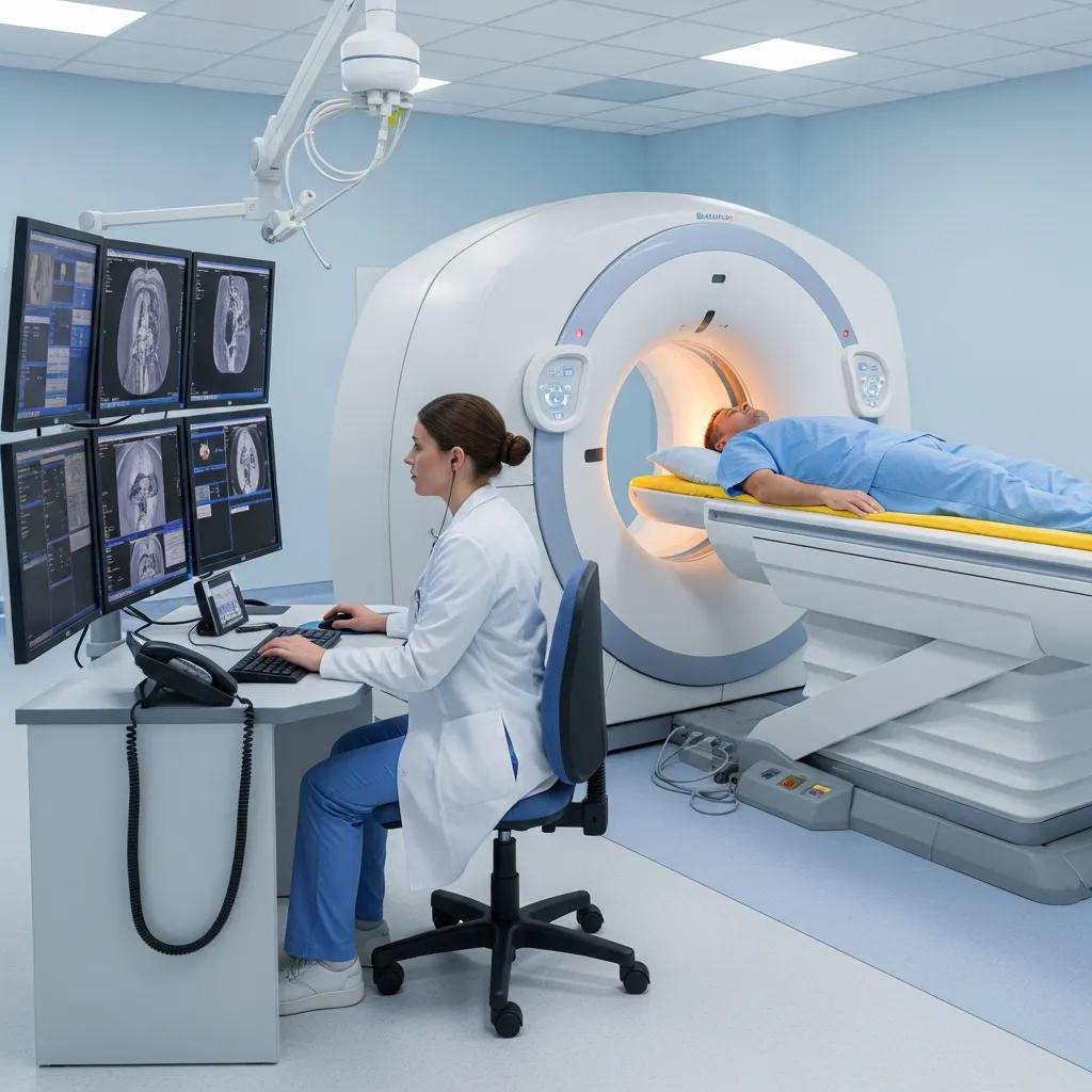
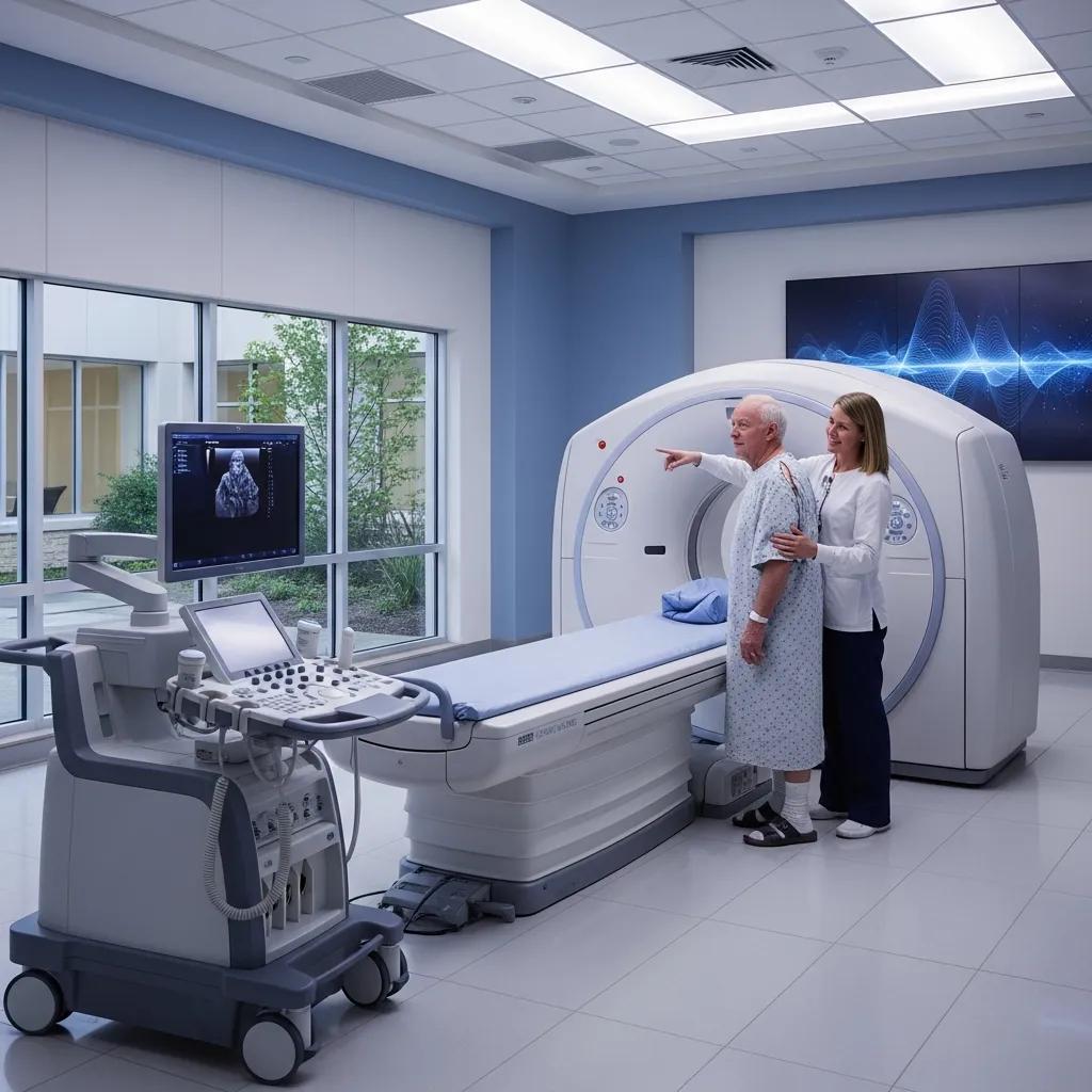
CT (computed tomography) uses a rotating X‑ray source and detector array plus computer reconstruction to produce detailed cross‑sectional images of bones, organs and blood vessels. Multiple projections are reconstructed into thin slices, giving CT its speed and high spatial resolution — advantages that are critical in emergencies and complex anatomy assessment. Your visit usually includes check‑in and consent, positioning, any required intravenous contrast, a short scan and brief post‑scan monitoring if contrast was used. Results proceed from image acquisition to radiologist reporting on a clinically appropriate timeline. Modern dose‑reduction methods such as iterative reconstruction and advanced detectors lower radiation while keeping diagnostic quality; many local providers use these low‑dose protocols routinely.
Different CT scans serve different purposes and have distinct preparation needs. The table below summarises common CT exams to help patients and referrers choose and prepare.
Introduction to CT type comparison table: the table that follows summarises modality names, typical indications, basic preparation requirements and radiation‑dose notes so you can see how scans differ.
This comparison helps referrers select the appropriate CT and helps patients prepare for a specific scan. Next we explain the typical patient pathway and what to expect at each stage.
What is a general CT scan and what are its diagnostic benefits?
A general CT scan acquires rapid, high‑resolution cross‑sectional images to help diagnose trauma, acute abdominal conditions, pulmonary embolus and to stage cancer by revealing detailed anatomy and pathology. Rapid slice acquisition gives better spatial resolution than plain X‑ray, allowing detection of subtle fractures, organ injury and lesion character. Patients benefit from quick scan times and clear information that supports timely clinical decisions; some studies use contrast to improve lesion conspicuity. Knowing the test’s strengths and typical reporting timeline helps set expectations and supports shared decision‑making between patient, referrer and imaging team.
How does low‑dose CT technology improve patient safety?
Low‑dose CT techniques reduce ionising radiation through advances in software and hardware — for example, iterative reconstruction, automated exposure control and more sensitive detectors. These improvements enhance signal processing and reconstruction so radiologists can see the necessary detail with less X‑ray flux. When clinicians and patients weigh risks and benefits, low‑dose protocols offer reassurance for routine follow‑up and screening without compromising diagnostic performance. Some local clinics highlight low‑dose/high‑definition CT systems as part of their commitment to patient safety and image quality.
Dose optimisation in digital radiography: ALARA principles and image quality
The ICRP optimisation principle aims to protect patients by using doses that are As Low As Reasonably Achievable (ALARA). Optimisation techniques include adjusting exposure factors (kV and mAs) and using exposure indicators as dose‑management tools. Research into computed radiography and digital systems has explored how these parameters influence image quality and dose, and methods such as visual grading and ROC analysis help assess clinical image performance. While studies sometimes show mixed results, the overall goal remains: balance lowest reasonable dose with diagnostic image quality.
Dose Optimization in Digital Radiography, 2019
- CT typically follows these steps: registration and consent, positioning, contrast if required, scanning and brief post‑scan checks.
- Tell staff about allergies, pregnancy or kidney problems before contrast studies.
- Hydration and following fasting instructions where required reduce risk and improve image quality.
Next we explain how ultrasound works and what preparation looks like for different types of scans.
What should patients know about ultrasound scans?
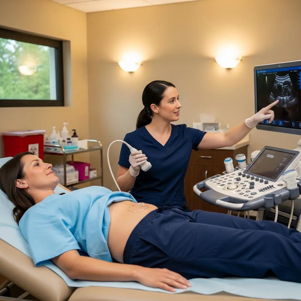
Ultrasound uses high‑frequency sound waves from a transducer to create real‑time images of soft tissues, blood flow and developing fetuses — all without ionising radiation. The physics — sound‑wave reflection and Doppler shifts — allow dynamic assessment of structure and function, and ultrasound is commonly used for abdominal, vascular, musculoskeletal and obstetric/gynaecological imaging. Preparation varies by subtype: fasting for some abdominal scans or a comfortably full bladder for pelvic exams improves acoustic windows and reduces the need for repeat studies. Knowing what to expect and following simple preparation steps helps your sonographer get the best images first time.
Introduction to ultrasound comparison table: the following table compares common ultrasound types, transducer choices, typical exam lengths and clinical uses to guide patients and referrers.
This table clarifies expectations by modality and supports the preparation guidance that follows.
How are different ultrasound types used in diagnostic imaging?
Ultrasound subtypes are tailored to clinical questions by transducer frequency and Doppler capabilities. Vascular Doppler studies measure flow velocity and direction to detect blockages or thrombosis; MSK ultrasound visualises tendons and ligaments and supports guided injections; obstetric ultrasound provides dating, anatomy and growth assessments central to prenatal care; and gynaecological scans evaluate uterine and ovarian pathology. Referrers choose the subtype to match the clinical problem, and the sonographer’s experience helps ensure high‑quality images and useful reports.
How should patients prepare for an ultrasound scan?
Preparation depends on the exam type. For abdominal scans, fasting for several hours reduces bowel gas and improves visibility. For pelvic or early pregnancy scans, a comfortably full bladder separates pelvic structures for clearer imaging. Wear loose, two‑piece clothing and bring your referral and any prior imaging to speed check‑in and give the sonographer clinical context. For children or anxious patients, let the clinic know in advance so staff can plan distraction techniques, parental presence or other supports.
- Follow fasting and full‑bladder instructions where specified to improve diagnostic accuracy.
- Bring referral paperwork and any previous imaging to your appointment.
- If the patient is a child or prone to anxiety, inform the clinic beforehand to arrange support.
Next we look at digital X‑ray: how it works, when it’s useful and what safety measures are in place.
Patient guide to X‑rays and digital radiography
Digital X‑ray uses electronic detectors to capture X‑ray transmission and produce high‑resolution images immediately for rapid assessment of bones, chest and dental structures. The technique creates 2‑D projections that are ideal for fracture detection, chest infection screening and routine bony follow‑up. Safety is governed by the ALARA principle — exposures are justified and minimised, especially for pregnant and paediatric patients. Knowing what X‑rays can show, their limits compared with cross‑sectional tests and the typical reporting pathway helps patients understand when X‑ray is the appropriate first‑line investigation and when further imaging may be needed.
Introduction to X‑ray comparison table: the table below describes common X‑ray types, body areas imaged, simple preparation steps and relative radiation exposure so you can compare risk and utility.
This comparison puts X‑ray benefits and exposure into context and prepares patients for what to expect during imaging and follow‑up.
How do digital X‑rays diagnose common conditions?
Digital X‑rays reveal structural changes such as fractures, lung consolidation, joint space narrowing and dental issues that correlate with clinical exams and symptoms. Fractures show alignment and displacement directly, while chest X‑rays demonstrate air‑space patterns consistent with infection or fluid. X‑rays are often the first‑line test because they’re fast and widely available; results inform whether further imaging — for example CT for complex fractures or MRI for soft‑tissue concerns — is needed. Clinicians always interpret X‑ray findings alongside clinical context before recommending next steps.
What safety measures are used in X‑ray imaging?
X‑ray safety follows ALARA: exposures are kept as low as reasonably achievable by optimising technique, using shielding and avoiding unnecessary repeats. Technologists use protective aprons, collimation to narrow the beam and automated exposure controls to reduce dose. Paediatric protocols further reduce exposure for children. Putting dose into perspective — most single X‑rays equal only a few days of background radiation — helps patients assess risk. Pregnant patients should always tell staff so alternatives or shielding options can be considered. These safeguards sit alongside clinic accreditation and quality assurance practices.
- ALARA, shielding and collimation protect patients and staff during X‑rays.
- Automated exposure controls and paediatric protocols minimise unnecessary dose.
- Always inform the imaging team if you are pregnant so protocols can be adjusted.
Choosing the right local clinic — one with accreditation and modern equipment — matters for both quality and convenience.
Why choose a diagnostic radiology clinic on the Central Coast?
Local diagnostic radiology clinics combine convenience with clear quality markers: accreditation, experienced staff and up‑to‑date equipment that together improve diagnostic certainty and patient experience. Accredited clinics follow documented quality‑assurance and equipment maintenance schedules that reduce variability in imaging and reporting. Modern capabilities — low‑dose CT, digital radiography and specialist ultrasound probes — enhance safety and diagnostic yield. Patient‑centred practices such as clear preparation guidance, child‑friendly approaches and efficient eReferral workflows reduce anxiety and administrative friction. Staying local also lowers travel for follow‑up imaging and helps clinical teams communicate more quickly about results.
How does Life Medical Imaging Central Coast ensure quality and accreditation?
Life Medical Imaging Central Coast is an independent, accredited radiology clinic serving the Central Coast. Accreditation confirms structured quality assurance, routine equipment maintenance and standardised protocols that support consistent imaging outcomes and patient safety. Our team of radiographers, sonographers and reporting radiologists apply these processes across a range of services so referrers and patients can rely on the results for clinical decision‑making. Accreditation is an important quality marker when choosing a provider that prioritises safety and reproducibility.
What advanced technologies and patient care practices are offered?
Some Central Coast clinics offer advanced systems such as low‑dose/high‑definition CT, digital X‑ray detectors and specialist ultrasound probes that improve image quality across many clinical applications. These hardware and software improvements reduce radiation where applicable, enhance soft‑tissue visualisation and support subspecialty work including cardiac CT, obstetric ultrasound and paediatric imaging. Patient‑centred care — clear preparation instructions, supportive approaches for children and streamlined eReferral processes — makes appointments easier and reduces delays. Accreditation plus modern technology provides technical performance and practical benefits that support diagnostic confidence and convenience.
- Look for accreditation, modern equipment and experienced staff as quality markers.
- Advanced modalities can improve diagnostic accuracy while lowering risk.
- Local clinics combine accessibility with clinical‑grade imaging capabilities.
Next, practical steps for booking and accessing imaging services, including eReferral and online image access.
How can patients book appointments and access imaging services?
Booking starts with a valid referral that specifies the exam and clinical question, plus clear communication about preparation and any pre‑authorisation. Clinics accept referrals in different ways; many referrers use eReferral systems to send requests electronically, which speeds the process and reduces paperwork. After your scan, secure online platforms let patients and authorised referrers view images and reports within usual clinical timeframes, aiding continuity of care. Below we outline a straightforward booking workflow and what to bring to appointments.
Introduction before booking steps: the numbered steps below describe the typical booking workflow and what to bring to ensure a smooth imaging visit.
- Make sure you have a valid referral that specifies the required modality and the clinical question.
- Contact the clinic by phone to schedule an appointment or confirm eReferral submission with your referrer.
- Follow exam‑specific preparation instructions such as fasting or arriving with a full bladder.
- Bring referral documentation, photo ID and any prior imaging to your appointment.
These steps clarify responsibilities for patients and referrers and reduce check‑in delays; below we explain what to expect when booking with local services.
What is the process for booking diagnostic imaging services?
Booking usually involves confirming referral details, choosing a suitable appointment time and receiving preparation instructions from the clinic. Clear, specific referrals speed scheduling and reduce repeat scans. When eReferral is available, referrers can submit electronically and the clinic will contact the patient to arrange a slot and pre‑test instructions. Mention pregnancy, allergies and current medications when booking; the clinic will advise if pre‑test blood tests are needed for contrast studies. For convenience, the clinic phone number for scheduling enquiries is 02 4326 7000 — calling ahead with preparation questions helps ensure an efficient visit.
How can patients access their medical images online?
Many providers use secure online systems — for example, Life Images — that let patients and authorised clinicians view studies and reports once the radiologist has finalised the report. Images and reports typically become available within a clinically appropriate timeframe after your exam, and the clinic will give instructions for access or offer assistance if you have difficulty. Robust security and privacy safeguards protect your information, and clinics provide contact points for troubleshooting or for sharing images with other health professionals. If you need help accessing Life Images or have questions about availability, contact the clinic by phone for guidance.
- Use eReferral where available to streamline bookings and reduce paperwork.
- Expect images and reports to be accessible online after radiologist sign‑off via secure systems.
- Contact the clinic by phone for help with booking or image access.
This completes our practical guide to radiologic sciences, modality expectations, safety considerations, local clinic quality markers and how to book and access imaging on the Central Coast.
Frequently asked questions
What should I expect during my first imaging appointment?
At your first appointment you’ll check in at reception where staff confirm your referral and ID. A technologist will explain the procedure, answer questions and guide you to the imaging room. Depending on the test you may change into a gown. The imaging process varies by modality, but you’ll be positioned comfortably while the scanner or sonographer captures the required images. After the exam you’ll receive any post‑scan instructions and information about how and when results will be available.
Are there any risks associated with medical imaging procedures?
Most imaging procedures are safe. The primary risk with X‑ray and CT is radiation exposure, which is minimised by following ALARA (As Low As Reasonably Achievable) principles. Ultrasound and MRI do not use ionising radiation; however, MRI may be contraindicated for some implants and ultrasound may have limitations for certain conditions. Always tell your healthcare team about allergies, pregnancy or relevant medical history before imaging so the team can manage risks appropriately.
How can I prepare for my imaging appointment?
Preparation depends on the exam. Typical instructions include fasting for abdominal ultrasound or having a full bladder for pelvic scans. Follow the specific guidance given by your referrer or the clinic. Bring any prior imaging, your referral and identification to speed check‑in. If you’re unsure about preparation steps, call the clinic ahead of your appointment.
What happens if I need a follow‑up imaging study?
If additional imaging is needed your healthcare provider will explain why and what type of study is required. They will issue a new referral for the follow‑up, which you can use to book the appointment. Make sure to tell the clinic about any changes in symptoms since your last study, as this can influence the follow‑up protocol. The clinic will explain any preparation required for the next scan.
How do I access my imaging results and reports?
Results are usually available through a secure online portal such as Life Images after the radiologist has finalised the report. The clinic will provide instructions for accessing your images and report, and can assist if you have trouble logging in. Your referrer or GP can also discuss the findings with you at a follow‑up appointment.
What should I do if I have concerns about the imaging procedure?
If you have concerns, tell the technologist or radiologist before the exam — they can explain the procedure and safety measures and address specific worries. If you have anxiety about scans, mention this when booking so the team can offer support strategies or schedule extra time to help you feel more comfortable.
Conclusion
Knowing how radiologic sciences work and what each imaging modality offers helps you make informed decisions about your care on the Central Coast. With clear preparation steps, an understanding of safety measures and a focus on accredited local clinics, you can navigate imaging with confidence. If you’d like more information or to book an appointment, please contact us today.