Medical Imaging on the Central Coast — Comprehensive Radiography Services and Patient Safety
Radiography covers the range of imaging techniques that create pictures of the body using controlled ionising radiation, sound waves or specialised detectors. These images help clinicians diagnose disease, plan treatment and monitor recovery with clear visual evidence. This guide describes how radiography works, the common modalities we use, and what you can expect at each stage — with a strong focus on safety, preparation and practical value. Many people worry about radiation or discomfort; by explaining dose‑reduction strategies, non‑radiation alternatives and straightforward preparation steps we aim to reduce uncertainty and improve outcomes. We map services to clinical needs, describe our low‑dose and high‑definition equipment, and offer practical booking and eReferral guidance for patients across the Central Coast. You’ll learn how different tests compare, how to prepare for CT and ultrasound, how radiologists interpret images, and how accredited clinics manage quality and safety — all in clear, evidence‑based language you can act on.
What Radiography Services Are Available at Life Medical Imaging Central Coast?
Radiography includes several different imaging methods that together address most diagnostic needs — from broken bones to pregnancy checks. Each modality works on different physical principles: X‑ray detectors record transmitted radiation, CT produces cross‑sectional X‑ray images, ultrasound reflects sound waves to build live images, and DEXA measures bone mineral density. Choosing the right test depends on the clinical question and the need to minimise risk. Understanding your options helps clinicians select the most accurate test first, reducing delays and unnecessary repeats. Below we list the main modalities, their typical uses, and a short guide to preparation and duration so you can plan your visit.
Which Diagnostic Scans and Imaging Modalities Do We Offer?
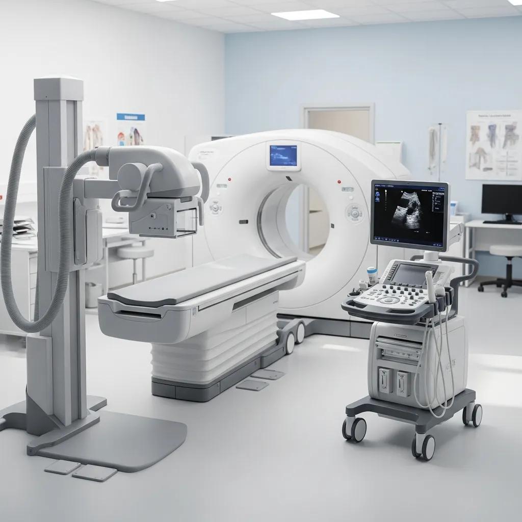
Here we define each modality and give practical examples of when it’s used. Digital X‑ray is fast and low dose — ideal for suspected fractures and chest checks and often used first in trauma. CT scanning (including low‑dose and cardiac CT angiography) provides detailed cross‑sectional images for acute abdominal pain, lung disease or coronary assessment. Ultrasound (general, vascular, musculoskeletal, obstetric & gynaecological) is the first‑line choice for pregnancy and many soft‑tissue problems because it does not use ionising radiation. DEXA measures bone density to assess fracture risk. Image‑guided interventional procedures (spinal or joint injections, biopsies, aspirations) combine diagnostic sampling with targeted treatment. Paediatric imaging uses child‑specific protocols for comfort and safety, and dental imaging supports oral and maxillofacial care.
The short summary table below is designed to help patients and referrers compare options quickly and prepare appropriately for appointments.
This quick reference helps patients and referrers pick the right test and plan logistics; the next section shows how these modalities map to common medical conditions.
How Do Our Radiography Services Support Specific Medical Conditions?
Different problems need different imaging because sensitivity, specificity and safety vary by test. For suspected kidney stones or appendicitis, CT gives rapid, sensitive detection and helps with surgical planning — its cross‑sectional views reveal complications and guide management. For suspected deep vein thrombosis or carotid disease, vascular ultrasound assesses blood flow non‑invasively and is usually the first choice, avoiding radiation. In pregnancy and gynaecology, pelvic ultrasound safely evaluates fetal development and ovarian or uterine conditions. Musculoskeletal complaints such as tendon tears are often best seen with targeted musculoskeletal ultrasound, which allows dynamic assessment. Bone health screening with DEXA quantifies fracture risk and informs osteoporosis treatment. Choosing the right modality reduces unnecessary tests and moves care forward faster.
After a test is selected, safety practices make sure it’s done with the lowest reasonable risk while still providing the diagnostic detail needed. If you want to book or ask about services, follow the clinic booking steps later in this article.
How Does Life Medical Imaging Ensure Patient Safety in Radiography?
We base patient safety on three key principles: justification, optimisation and quality assurance. Justification means each test should be clinically necessary and targeted to a clear question from your referrer. Optimisation focuses on keeping exposure as low as possible while preserving image quality — for example, choosing ultrasound when appropriate or using low‑dose CT settings. Quality assurance covers accreditation, regular equipment calibration and ongoing staff training so performance is consistent and documented. Together these measures create transparent safety governance and reassure patients that every scan is purposeful and carefully managed.
- Iterative reconstruction and advanced image processing reduce noise, letting us use lower exposure levels.
- Automatic exposure control adjusts X‑ray dose to the patient’s size and the body region being scanned.
- Tailored protocols include paediatric and low‑dose settings for children and patients needing repeat imaging.
These approaches lower cumulative exposure for vulnerable groups and are combined with clinical justification to meet ALARA (as low as reasonably achievable) principles. The following section details the specific CT and X‑ray measures we use to minimise dose.
What Radiation Dose Minimisation Techniques Are Used in CT and X-ray Scans?
We reduce CT and X‑ray dose by combining modern hardware with optimised protocols and operator controls. Contemporary CT scanners offer ultra‑low dose modes and iterative reconstruction algorithms that keep images clear while lowering mGy exposure. Automatic exposure control modifies tube current in real time based on patient attenuation, reducing dose for smaller patients. For X‑ray, sensitive digital detectors, careful beam collimation and adjusted kVp settings limit exposure to the area of interest. Paediatric protocols further reduce dose with smaller fields of view, adapted exposure parameters and shielding when appropriate. Dose monitoring systems record exposure metrics so clinicians can review cumulative exposure and avoid unnecessary repeats.
These technical measures work alongside clinical governance that reviews appropriateness and outcomes, ensuring dose reductions don’t compromise diagnostic performance and that our MRI and ultrasound safety practices continue this patient‑centred approach.
How Are Safety Protocols Applied in Ultrasound and MRI Procedures?
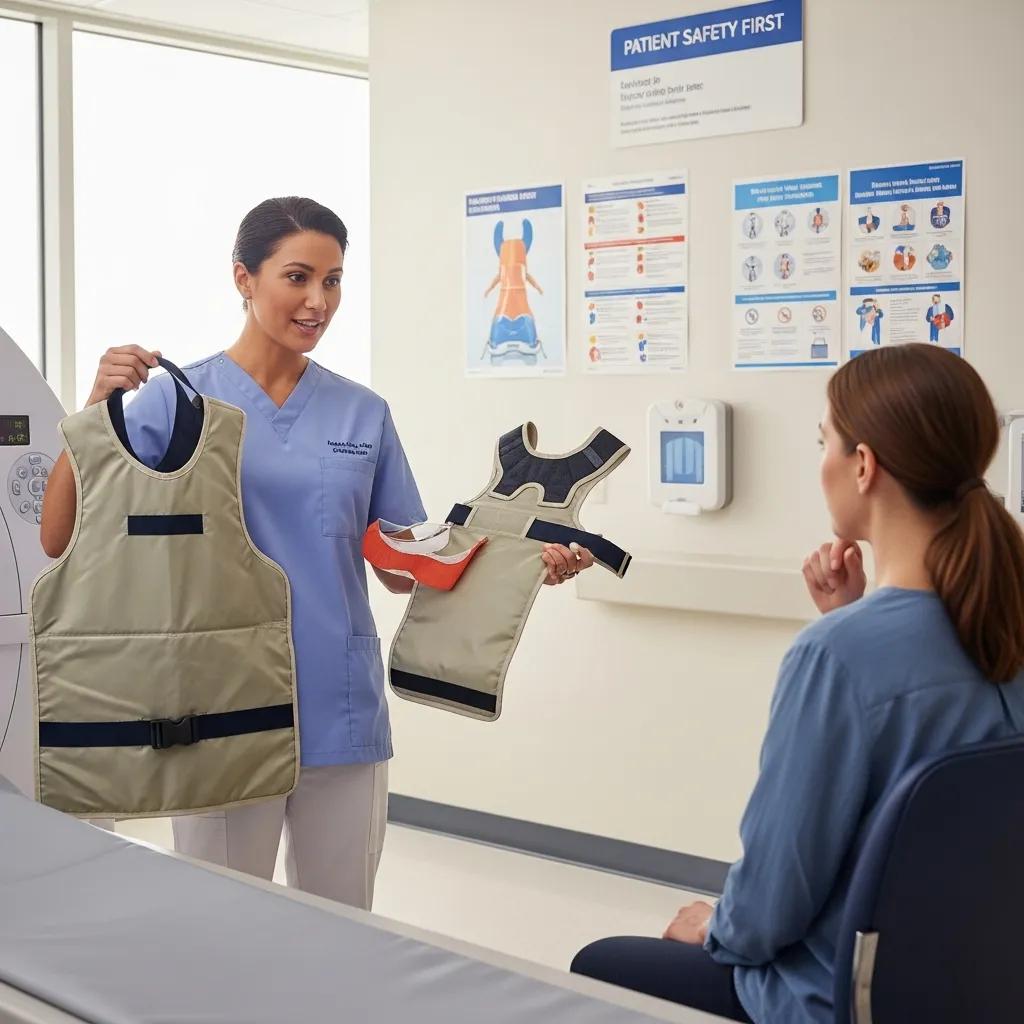
Although ultrasound and MRI don’t use ionising radiation, they still require specific safety checks. Ultrasound operators use ALARA‑oriented settings for Doppler (limiting thermal and mechanical indices) and rely on operator skill to obtain diagnostic images with minimal exposure. MRI safety emphasises screening for implants, devices or metal fragments; a structured MRI safety questionnaire and staff review prevent adverse interactions. When contrast agents are needed for MRI or CT, we screen kidney function and allergy history, obtain informed consent and have resuscitation‑ready protocols in place. Well‑trained staff and sub‑specialist oversight reduce repeat scans and improve diagnostic yield.
External accreditation and routine equipment maintenance provide additional reassurance and lead naturally into the practical patient preparation steps below.
How Can Patients Prepare for Their Radiography Appointments?
Good preparation improves image quality, shortens appointments and increases comfort. Requirements vary by test but common points include bringing your referral and ID, following fasting or hydration instructions when required, and wearing loose, metal‑free clothing for easy positioning. If you are pregnant, always tell the imaging team so we can consider non‑ionising alternatives. For children, distraction and parental presence often improve cooperation. Practical tips — arrive 10–15 minutes early, bring prior imaging if available, and follow pre‑scan guidance — keep appointments on time and reduce the chance of repeats.
- Gather Documents: Bring your referral, Medicare card if applicable, and a list of current medications and allergies.
- Follow Fasting Instructions: For some CT exams, fasting reduces the risk of contrast‑related nausea and improves image quality.
- Prepare Bladder Status: For pelvic ultrasounds, drink water and avoid urinating when instructed to ensure a full bladder.
- Wear Suitable Clothing: Choose loose clothing and avoid metal fasteners to simplify positioning and reduce artefacts.
- Notify Pregnancy or Implants: Tell staff if you are pregnant or have implanted devices so we can choose the safest option.
Following these steps helps the team capture diagnostic images quickly and comfortably. The table below summarises common preparation requirements by procedure.
This practical reference helps patients follow instructions that make imaging faster and more reliable. The next section gives specific preparation guidance for CT and ultrasound.
What Are the Preparation Guidelines for CT Scans and Ultrasound Appointments?
CT preparation commonly includes fasting for abdominal studies and screening for contrast allergies or reduced kidney function. You may be advised to arrive hydrated and to temporarily withhold certain medications if directed by your clinician. For contrast‑enhanced CT we obtain informed consent and review your medical history for allergies, thyroid disease or renal impairment to minimise adverse events. Ultrasound prep depends on the exam: abdominal scans often require fasting to reduce bowel gas, pelvic scans usually need a full bladder, and vascular or musculoskeletal exams require comfortable, metal‑free clothing to allow probe access. Parents should follow site‑specific guidance for children to improve cooperation and avoid repeat imaging.
Preparing correctly lowers the chance of an incomplete study and helps the team obtain diagnostic images on the first attempt, which also reduces cumulative exposure when radiation is used.
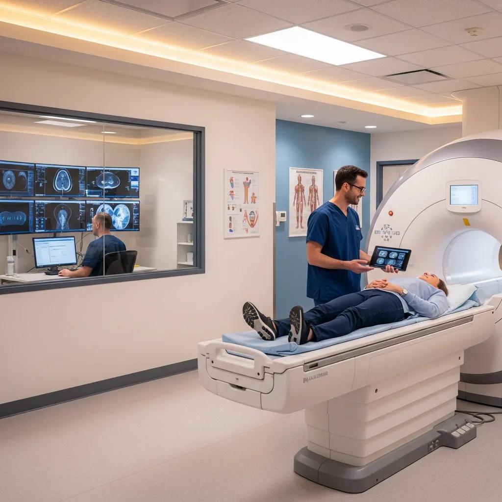
What Should Patients Expect During Their Radiography Procedure?
At check‑in we confirm the referral, medical history and consent. A technologist reviews any special instructions and prepares the equipment. Positioning and gentle immobilisation are used as needed while staff explain each step to reduce anxiety. Digital X‑rays take only seconds; CT scanning involves a few minutes of actual scanning within a slightly longer appointment slot; ultrasound exams typically last 20–40 minutes and involve real‑time interaction. For contrast studies, patients are observed briefly after injection and given post‑scan hydration advice. The technologist acquires the images and a radiologist later interprets them — results are sent to your referring clinician according to clinical urgency.
Knowing this workflow — from check‑in to report — helps you understand timing and what to expect, which usually reduces stress during the visit.
What Are the Benefits of Advanced Radiography Technologies at Life Medical Imaging?
Modern imaging technology improves diagnostic confidence while often lowering risk and appointment time. Low‑dose CT combines hardware and software advances to keep image detail while reducing radiation — useful for screening and repeat surveillance. 3D/4D obstetric ultrasound enhances fetal visualisation and supports more detailed assessment of anatomy and growth. Cardiac CT angiography provides high‑resolution, non‑invasive coronary imaging to assist planning without catheterisation for selected patients. These technologies speed diagnosis, reduce repeat imaging and improve pre‑treatment planning, all of which support better patient outcomes.
When matched to the right clinical problem, these advances deliver clear benefits. Below we explain how low‑dose CT and advanced ultrasound translate into real‑world safety and diagnostic improvements.
How Does Low Dose CT Improve Diagnostic Accuracy and Patient Safety?
Low‑dose CT uses iterative reconstruction, automatic exposure control and tailored scan ranges to lower radiation while keeping diagnostic detail. Noise reduction algorithms and tube‑current modulation let clinicians see subtle findings — such as small lung nodules or kidney stones — with confidence even at lower mGy levels. For screening or patients who need repeated imaging, the cumulative dose reduction is meaningful and lowers long‑term risk. Examples include lung nodule surveillance with low‑dose protocols and paediatric trauma imaging using age‑adjusted settings to balance safety and diagnostic performance. Dose records and governance ensure reductions are tracked and reviewed for quality.
These technical strategies balance image quality with patient‑centred safety and support clinical decision‑making without unnecessary radiation burden.
What Advantages Do 3D/4D Ultrasound and Cardiac CT Angiography Offer?
3D/4D ultrasound gives volumetric views that improve assessment of fetal anatomy and structural abnormalities, helping with perinatal planning and parental counselling by providing clearer morphological detail. It supports multiplanar review and dynamic assessment of moving structures, revealing findings that may be less obvious on 2D scans. Cardiac CT angiography delivers rapid, non‑invasive visualisation of coronary arteries and cardiac anatomy, supplying precise maps for cardiologists and surgeons when planning treatment. Both techniques reduce diagnostic uncertainty, can avoid more invasive tests, and often shorten the path to definitive care thanks to superior visualisation.
How Can Patients Book Ultrasound and CT Scan Services in Central Coast NSW?
Bookings can be made by patients online or via clinician‑submitted referrals; eReferral systems streamline information transfer and reduce admin delays. Generally, patients need a referral from their clinician that states the clinical question and requested modality, then follow the clinic’s booking instructions to supply the referral and any pre‑scan details. eReferral allows doctors to upload structured referrals electronically, include clinical notes and receive confirmations, which reduces transcription errors and speeds appointments. Some services may be eligible for bulk billing — ask your referrer about coverage or cost before booking.
- Obtain a valid referral from your clinician that states the clinical concern and requested modality.
- Submit the referral via the clinic’s online booking process or ask the referring clinician to send an eReferral.
- Receive appointment confirmation and any pre‑scan instructions by phone or the clinic’s communication channel.
- Attend the appointment with necessary documents, follow preparation instructions, and notify staff of any allergy or pregnancy concerns.
Following these steps helps secure timely imaging and ensures referrals contain the clinical detail needed for protocol selection and safety checks.
What Is the Process for Online Booking and eReferral Submissions?
Online booking usually asks for the referral, contact details and preferred times; the clinic confirms availability and issues preparation instructions. eReferral lets the referring clinician upload the referral and clinical notes electronically and receive an electronic booking confirmation, shortening turnaround and improving accuracy. For urgent or same‑day needs, clinicians should mark the referral as urgent so triage can prioritise it. Have your referral, Medicare number (if applicable) and a list of medications ready when booking to speed the process.
Which Locations Offer Radiography Services Across the Central Coast?
Life Medical Imaging Central Coast operates multiple sites to make imaging convenient across the region, including Bateau Bay, Killarney Vale, Umina Beach and Erina. Each location offers a selection of services — digital X‑ray, ultrasound, CT and specialist imaging — with some sites providing sub‑specialist women’s and cardiac imaging, paediatric services and interventional procedures. When booking, check which clinic provides the specific service you need so the right equipment and expertise are available. Bulk billing for eligible services is offered at participating locations; confirm details when you book.
What Are Common Questions About Radiography and Diagnostic Imaging?
Patients commonly ask which test is best, how radiologists read images, and what safety steps are in place. Clear answers reduce anxiety and support appropriate use. The short guide below outlines imaging types and one‑line use cases to help you spot which test might be relevant. Understanding the radiologist’s role — integrating images with clinical history, producing a structured report and recommending follow‑up — helps patients and clinicians act on results promptly. We also address radiation risk, preparation differences and typical reporting timelines so you can give informed consent and keep care moving.
What Are the Different Types of Medical Imaging and Their Uses?
Each imaging type has strengths and limits and is chosen to answer a specific clinical question while considering patient safety. X‑ray is quick and effective for fractures and chest checks but has limited soft‑tissue contrast. CT provides cross‑sectional detail for complex anatomy and acute conditions but uses more radiation than X‑ray. MRI offers superior soft‑tissue contrast without ionising radiation and is preferred for neurological and many musculoskeletal problems, though MRI safety screening is required. Ultrasound is radiation‑free and excellent for real‑time assessment of the fetus, abdominal organs and vascular flow. DEXA measures bone density for osteoporosis assessment. Choosing the right test balances diagnostic accuracy with safety and logistics.
How Do Radiologists Interpret Radiography Images for Accurate Diagnosis?
Radiologists read images in the context of the clinical history, prior studies and laboratory results to produce a structured report that answers the referrer’s question. Reporting includes image review, measurements, comparison with previous imaging if available, and an assessment of diagnostic certainty with recommended follow‑up if needed. Sub‑specialist reporting or double reading may be used for complex cases to improve accuracy, and urgent findings are communicated directly to the referring clinician to speed care. Turnaround times depend on urgency, but established reporting pathways help clinicians make timely decisions and maintain continuity of care.
Life Medical Imaging Central Coast is a NATA‑accredited independent radiology clinic offering CT (general CT, Cardiac CT, CT Angiography), Dental Imaging, Digital X‑ray, General / Vascular / Musculoskeletal / Obstetric & Gynaecological Ultrasound, Interventional procedures (spinal/joint injections, biopsies, aspirations), Paediatric Imaging, Body Composition DEXA Scan, Bone Mineral Densitometry, and Platelet‑Rich Plasma (PRP) therapy injections. We highlight sub‑specialist women’s and cardiac imaging expertise, ultra‑low dose and high‑definition equipment, experienced clinical and support staff, a modern patient environment, online booking and eReferral capability, and bulk billing for eligible services. To arrange an appointment or ask a question, call 02 4326 7000 or use our online booking and eReferral options; confirm at booking which location (Bateau Bay, Killarney Vale, Umina Beach or Erina) best suits your requested service.
Frequently Asked Questions
What should I do if I have a fear of medical imaging procedures?
Feeling anxious about imaging is common. Tell the imaging staff about your concerns before the appointment — they’ll explain the steps, answer questions and offer reassurance. Bringing a friend or family member for support can help, and many clinics provide distraction options such as music or guided breathing techniques. If needed, staff can suggest strategies to make the experience more comfortable.
Are there any alternatives to ionising radiation imaging?
Yes. Ultrasound is a safe, non‑ionising option for many conditions, especially in pregnancy and soft‑tissue assessments. MRI is another non‑ionising alternative that gives excellent soft‑tissue contrast and is often chosen for neurological and musculoskeletal problems. Your clinician will discuss the most appropriate option based on your condition and safety needs.
How can I access my imaging results after the procedure?
Results are usually sent to the referring clinician, who will discuss them with you during a follow‑up appointment. Some centres also offer online patient portals where you can view reports directly. If you need copies of images or reports, request them from the imaging facility — there may be a small fee. Ask your referrer how and when you should expect your results.
What should I do if I experience discomfort during the imaging procedure?
If you feel any discomfort during the scan, tell the technologist straight away. They’re trained to ensure your comfort and can adjust positioning, equipment or timing, and provide breaks if needed. Your safety and comfort are priorities, so please speak up at any time.
Can I eat or drink before my imaging appointment?
Whether you can eat or drink depends on the test. Some CT scans requiring contrast need fasting for a few hours beforehand, while many X‑rays and ultrasounds have no specific dietary restrictions. Always follow the preparation instructions provided by the clinic or your referrer to ensure the best possible images.
What are the qualifications of the staff performing the imaging procedures?
Imaging is performed by qualified radiologic technologists and interpreted by radiologists. Technologists operate the equipment and manage patient safety during scans and hold recognised certifications. Radiologists are medical doctors specialising in image interpretation and reporting. Both groups undertake ongoing training to stay current with advances in imaging and safety.
How does Life Medical Imaging ensure the quality of its services?
We maintain quality through accreditation, scheduled equipment maintenance and continuous staff training. Our protocols include routine calibration, internal audits and ongoing professional development. We also welcome patient feedback to improve services. These measures help ensure accurate diagnoses and safe imaging experiences.
Conclusion
Knowing the range of radiography services at Life Medical Imaging Central Coast helps you make informed choices about diagnostic care. With a focus on safety, modern technology and clear preparation advice, you can expect reliable imaging that supports accurate diagnosis and reduces anxiety. Follow the booking and preparation steps outlined here for a smooth visit that puts your health first. To learn more or to book an appointment, visit our website or contact us today.