Diagnostic imaging and radiation technology on the Central Coast — a practical patient and service guide
Diagnostic imaging that uses radiation creates internal pictures of the body to help diagnose illness, guide treatment and track recovery. This guide is written for people on the Central Coast who want clear, usable information about those tests and local services.
In the pages that follow you’ll find straightforward explanations of how X‑rays and CT scanners make images, when ionising tests are chosen instead of non‑radiation options, what to do before common exams, and the safety systems that keep doses as low as possible while still delivering diagnostic detail. We also answer common questions about dose, accreditation and how to book services nearby. Sections cover: what radiation technology is and how it’s used, clinic safety and ultra‑low dose approaches, the radiation‑based services available locally, patient preparation and expectations, modality comparisons, and how referring doctors can submit eReferrals and access reports. Keywords such as CT scans Central Coast, low dose CT scan, digital X‑ray benefits and radiation safety Australia are used to connect clinical concepts with local service options.
What is radiation technology and how is it used in diagnostic imaging?
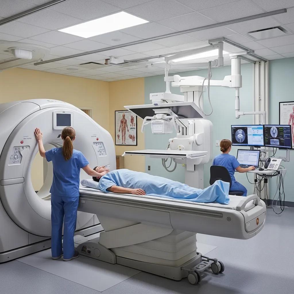
Radiation technology in medical imaging uses carefully controlled ionising radiation to produce images of the body’s internal structures. These images help clinicians spot fractures, internal bleeding, tumours and vascular problems quickly and accurately. Different machines work in different ways: projection X‑rays send a beam through the body to make a single flat image, while CT scanners rotate X‑ray sources and detectors to build cross‑sectional slices that can be viewed in three dimensions. These methods give high‑resolution views of bone, lungs, blood vessels and complex soft tissues — information that’s often vital in emergency and surgical care. Any clinical benefit is balanced against exposure risk through dose optimisation, which we explain in the next section.
How does radiation technology work in CT scans and X‑rays?
An X‑ray image is created when a controlled beam of photons passes through the body and is absorbed differently by various tissues; dense structures such as bone absorb more photons and appear lighter on the detector.
CT scanners use the same ionising photons but collect many angled projections while the X‑ray tube and detectors rotate around the patient. Reconstruction algorithms then assemble those projections into thin cross‑sectional images that reveal internal detail.
Detectors convert incoming X‑ray energy into electrical signals, and modern digital processing — including iterative reconstruction and AI‑assisted denoising — improves image clarity while allowing lower radiation doses.
Knowing how detectors and reconstruction work helps patients understand why a CT can show far more anatomical detail than a plain X‑ray. That technical foundation leads into the practical clinical benefits outlined next.
What are the benefits of using radiation technology for medical diagnosis?
Radiation‑based imaging is fast and delivers excellent spatial resolution, which supports quick diagnosis of fractures, acute bleeding, lung disease and vascular conditions such as pulmonary embolus or arterial narrowing. CT and X‑ray produce objective, reproducible images that can be shared electronically with referring clinicians and specialists for team‑based care and follow‑up. These modalities are especially valuable in emergency settings — where speed and whole‑body assessment can be lifesaving — and in cardiology, where cardiac CT angiography non‑invasively visualises coronary anatomy. Although ionising radiation is a consideration, modern dose‑reduction tools and careful clinical justification make these tests safe and effective when they’re the appropriate choice.
How does Life Medical Imaging Central Coast ensure medical radiation safety?
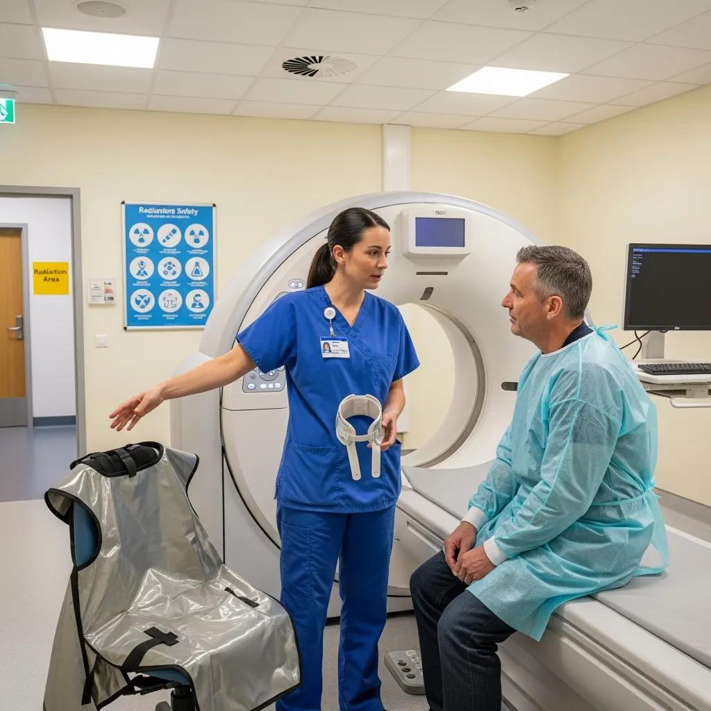
At Life Medical Imaging Central Coast patient safety is central to everything we do. We combine accredited quality systems, dose‑optimised technology and ongoing staff training to meet national regulations and international best practice. Our approach follows ALARA — As Low As Reasonably Achievable — using modern scanners that can perform ultra‑low dose studies without losing diagnostic value, plus routine quality assurance and dose monitoring. Our team includes radiographers and radiologists with sub‑specialist training in areas such as women’s and cardiac imaging, so protocols are chosen to match the clinical question, patient size and age. We also have paediatric dose‑reduction pathways where needed. The table below outlines key safety measures, how they’re applied and the benefit for patients and referrers.
This summary shows how systematic safety measures minimise risk while enabling high‑definition diagnostic imaging. The next section explains what “ultra‑low dose” means in practice for patients.
What is ultra‑low dose radiation technology and why is it important?
“Ultra‑low dose” describes scanning protocols and post‑processing techniques that substantially reduce the radiation needed to make diagnostic images while keeping the detail clinicians require. Enablers include advanced iterative reconstruction, more sensitive detectors and AI‑assisted denoising — technologies that preserve contrast and spatial resolution at lower photon counts. For patients, ultra‑low dose protocols lower lifetime stochastic risk, which is especially important for younger people and anyone needing repeated scans, without forfeiting clinical information. Understanding this balance explains why a referrer may choose a low‑dose CT for follow‑up but a standard protocol for an initial, more detailed assessment.
Iterative reconstruction for ultra‑low dose CT pulmonary angiography
Objective: Evaluation of a new iterative reconstruction algorithm (IMR) for the detection or exclusion of pulmonary embolism (PE) using ultra‑low dose computed tomography pulmonary angiography (CTPA). Methods: Lower dose CT datasets were simulated from CTPA exams of 16 patients with PE, using dose levels (DL) at 50%, 25%, 12.5%, 6.3% and 3.1% of the original tube current.
Ultra low dose CT pulmonary angiography with iterative reconstruction, A Sauter, 2016
How are radiation safety standards maintained at our clinics?
We maintain standards through formal accreditation, scheduled equipment maintenance, regular dose audits and structured staff competency programs that benchmark practice. NATA accreditation provides external assurance of our laboratory and imaging quality systems and confirms compliance with national testing and safety requirements. On a day‑to‑day level we follow procedures such as patient identity checks, pregnancy screening when relevant, appropriate shielding where indicated, and radiologist review of protocol choice for complex cases. These operational controls work together to deliver safe, consistent imaging services for the Central Coast community. The next section lists the radiation‑based services available locally.
What diagnostic imaging services using radiation technology are available on the Central Coast?
Patients on the Central Coast can access a full range of radiation‑based imaging suited to routine and specialised needs: general CT for body imaging, cardiac CT and CT angiography for vascular assessment, digital X‑ray for musculoskeletal and chest imaging, dental OPG for dental planning, and bone densitometry (DEXA) for bone health. Each test is matched to a clinical question — for example, fracture detection, stroke and trauma assessment, coronary artery evaluation, dental diagnostics and osteoporosis screening — and preparation varies by exam. Life Medical Imaging Central Coast offers these modalities across local clinics and helps patients and referrers with appointment bookings; please phone the clinic to book or enquire. The table below compares these procedures and gives practical patient information.
This comparison helps patients prepare and choose the right exam. The next two subsections look in more detail at CT services and the advantages of digital X‑ray.
What CT scan services does Life Medical Imaging offer?
Our CT services include general CT for broad anatomical surveys, cardiac CT for non‑invasive coronary assessment and CT angiography for vessel visualisation. The choice of study follows the clinical referral. General CT is used for trauma, chest and abdominal problems and oncological staging and may require breath‑hold cooperation and, in some cases, intravenous contrast. Cardiac CT often involves heart‑rate control and ECG gating to reduce motion artefact. CT angiography needs precise timing of contrast to opacify the vessels and may include hydration instructions. We apply low‑dose cardiac and vascular protocols when clinically appropriate and can assist with bookings via our patient enquiries phone line.
How does digital X‑ray technology improve patient care?
Digital X‑ray replaced film with direct digital detectors, giving immediate images, fast post‑processing and easy electronic sharing with referrers — all of which speeds clinical decisions. Benefits include fewer repeat scans because images can be checked instantly, dose efficiency from modern detectors and automatic exposure control, and improved image enhancement for subtle fractures or lung changes. In practice this means quicker diagnoses in emergency care and simpler follow‑up for outpatients, while dental OPGs deliver panoramic dental views with minimal radiation. The digital workflow also supports faster report turnaround and better access to images for referrers.
How should patients prepare for radiation‑based imaging procedures?
Preparation depends on the test but follows common principles: follow fasting or hydration instructions for contrast studies, remove metal that could affect images, and tell staff if you’re pregnant or have had recent imaging. Let the team know about medications, allergies — particularly to iodinated contrast — and prior surgeries so protocols can be tailored and risks minimised. For children or people who feel anxious, clinics provide clear preparation guidance and comfort measures to help cooperation and get the best images. The next sections give step‑by‑step preparation tips and explain the patient experience before, during and after imaging.
Follow these preparation steps for most radiation‑based exams:
- Confirm appointment details and preparation instructions: Check whether fasting, extra fluids or medication changes are required for your specific exam.
- Disclose medical history: Tell staff about pregnancy, allergies, kidney problems or recent contrast studies.
- Remove metal and wear comfortable clothing: Loose clothes and no jewellery improve comfort and image quality.
- Arrive early with referral and ID: Early arrival lets staff complete administrative checks and answer any last‑minute questions.
Following these steps reduces delays and supports high‑quality diagnostic images. The following subsection lists exam‑specific preparation details.
What are the preparation steps for CT scans and X‑rays?
When CT uses intravenous contrast, patients are often asked to fast for a few hours and to drink extra fluids before and after the scan to help clear contrast from the kidneys; specific medication instructions depend on the clinical situation. Plain X‑rays usually need little preparation beyond removing metal and wearing comfortable clothing. For dental OPGs you may be asked to remove dental prostheses. If you are pregnant or might be pregnant, tell the imaging team before the exam so staff can consider alternative tests or adapt protocols to reduce dose. Following these directions helps ensure safe, diagnostic imaging and lets the team choose the best protocol for your needs.
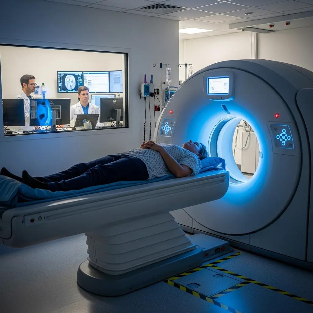
What can patients expect during and after radiation imaging?
On arrival you’ll complete identity and clinical checks and staff will explain the procedure, including any contrast use; consent is obtained when required. X‑rays are usually quick and painless. CT scans need careful positioning and short breath‑holds; contrast injections can cause a brief warm or taste sensation and patients are monitored for rare reactions. After most scans you can resume normal activities unless sedation or a reaction occurred. Radiologists review images and produce a report that is sent to the referring clinician within standard turnaround times; urgent findings are escalated directly to the referrer so appropriate care can proceed without delay.
How does radiation technology compare across different diagnostic imaging modalities?
Comparing modalities helps decide when CT or X‑ray is appropriate versus non‑ionising options such as ultrasound or MRI. Each modality has its own mechanism, presence or absence of radiation and a typical dose profile that guides clinical choice. CT gives high‑detail cross‑sectional images at a higher dose, X‑ray provides quick projection images at a low dose suitable for bones and chest, and ultrasound avoids ionising radiation and excels at soft‑tissue, obstetric and vascular flow assessment. The table below summarises these attributes to help patients and referrers weigh trade‑offs when selecting an imaging pathway.
This comparison clarifies clinical pathways and leads into examples where radiation‑based imaging is preferred over ultrasound.
What are the differences between CT scans, X‑rays, and ultrasound imaging?
CT produces volumetric slices reconstructed into cross‑sectional views, giving high contrast resolution for complex anatomy. X‑ray projects anatomy into a single plane and is ideal for rapid fracture or chest assessment. Ultrasound uses reflected sound, has no ionising radiation and is preferred for obstetrics, superficial soft tissue and targeted organ assessment, though it is operator‑dependent and limited by air or bone. Choosing a modality balances resolution, speed, safety and the clinical question: CT for detailed cross‑sectional anatomy, X‑ray for quick bone and chest checks, and ultrasound when radiation avoidance and real‑time assessment are priorities.
When is radiation‑based imaging preferred over ultrasound?
Radiation‑based imaging is chosen when bone detail, air‑filled structures or complex three‑dimensional anatomy need to be resolved — for example suspected skull or pelvic fractures, lung disease and vascular mapping with CT angiography. In emergency or polytrauma situations, whole‑body CT rapidly finds life‑threatening injuries that ultrasound can miss. Imaging of the coronary arteries or detailed abdominal and pelvic staging also often requires CT for accurate characterisation. These clinical use‑cases explain why referrers sometimes select CT or X‑ray despite the availability of non‑ionising alternatives.
How can referring doctors access and utilise radiation technology services on the Central Coast?
Referring clinicians can use eReferral systems or standard referral letters to book radiation‑based imaging. Include the clinical indication, prior imaging, relevant labs and the urgency level to help with scheduling and protocol choice. Clear referral details — specifying the diagnostic question, whether contrast is required and any comorbidities — let imaging teams prepare the right protocol and consent process. Reports and images are delivered securely via PACS and encrypted portals, with routine turnaround times and expedited pathways for urgent cases. The steps below offer practical guidance for efficient referrals and smooth communication.
Referrers should follow these steps for eReferral and booking:
- Provide a clear clinical question: State the suspected diagnosis and what the imaging should answer.
- Include relevant history and prior imaging: Attach previous reports or note recent studies to avoid unnecessary duplication.
- Specify urgency and contrast needs: Indicate if the study is urgent and whether intravenous contrast is required.
- Ensure patient contact details are accurate: Correct contact information reduces scheduling delays.
These steps reduce administrative delays and increase the chance of the right investigation being done quickly. The next subsection outlines the operational eReferral steps and what to expect.
What is the eReferral process for radiation imaging appointments?
eReferral typically involves submitting a structured electronic referral or a standard referral letter with patient identifiers, clinical history, indication and recent imaging so the service can triage and book the most suitable slot. Once received, imaging teams choose the protocol and schedule an appointment; routine wait times depend on modality and clinical urgency, while urgent cases are prioritised. Referrers should provide contact details and preferred communication channels for any clarifications, and patients receive tailored preparation instructions once booked. Using eReferral improves documentation, reduces duplicated studies and speeds up access to diagnostic imaging within multidisciplinary care pathways.
How are imaging reports and patient images shared securely?
Reports and images are shared with referrers via secure imaging systems (PACS) and encrypted portals to protect patient privacy while enabling timely clinical review and integration with electronic medical records. Referrers receive a formal radiology report that includes key findings, an impression and any recommendations; images are available through secure links or direct PACS access depending on local arrangements. Turnaround times follow agreed service levels, with expedited reporting for urgent or inpatient findings. A named contact is available for clinical questions or case discussion so referrers can act quickly on important results.
For referral or booking enquiries, clinicians and patients on the Central Coast can contact Life Medical Imaging Central Coast by phone to discuss appointments, eReferral arrangements and exam preparation.
- Clinical triage and booking via referral submission: Ensures the correct protocol is chosen and appointments are allocated in a timely way.
- Secure report and image delivery: Protects patient data and supports prompt clinical decisions.
- Designated contact pathways for urgent queries: Speeds communication for critical findings.
These operational steps close the loop between referrers, imaging teams and patients, helping deliver safe, efficient radiation‑based diagnostic care on the Central Coast.
Frequently Asked Questions
What should I expect during a CT scan or X‑ray procedure?
When you arrive you’ll complete identity and clinical checks and staff will explain the test and any contrast use. X‑rays are usually very quick and painless. CT scans require careful positioning and short breath‑holds; if contrast is used you may feel a brief warm sensation. Most people leave able to continue normal activities unless sedation was given. Radiologists review the images and send a report to the referring clinician within standard turnaround times; urgent results are communicated immediately to the referrer.
How can I ensure my safety during radiation‑based imaging?
Follow the clinic’s preparation instructions (for example fasting or hydration when contrast is planned) and tell staff about medical history, allergies or pregnancy so protocols can be adapted. Life Medical Imaging Central Coast also uses strict safety measures — such as ultra‑low dose protocols and regular equipment checks — to minimise exposure while preserving image quality. If you’re unsure about any step, ask the team — they’ll explain what’s being done to keep you safe.
Are there any risks associated with radiation‑based imaging?
Radiation‑based tests like CT and X‑ray are generally safe, but there is a very small long‑term increase in cancer risk associated with ionising radiation, especially for younger people or those needing multiple scans. Clinics use dose‑optimisation strategies to keep exposure as low as possible while ensuring diagnostic quality. Discuss any concerns with your healthcare provider so you can weigh the benefits and risks for your situation.
What are the differences between various imaging modalities?
Each imaging modality has a specific role. CT gives detailed cross‑sectional images for complex anatomy, X‑ray provides quick, low‑dose views for bones and chest, and ultrasound uses sound waves (no radiation) for obstetrics and soft‑tissue assessment. Knowing the differences helps clinicians and patients choose the most appropriate test for the clinical question.
How can I prepare for a radiation‑based imaging procedure?
Preparation varies by exam but typically includes confirming instructions, disclosing medical history, and removing metal objects that could interfere with the image. For CT with contrast you may need to fast for a few hours and drink extra fluids before and after the scan. Always check the clinic’s specific instructions and raise any questions before your appointment.
What should I do if I have concerns about radiation exposure?
If you’re worried about radiation exposure, speak with your referring clinician or the imaging team. They can explain why the test is recommended, the expected benefits and the safety measures in place. Depending on the clinical question, alternatives such as ultrasound or MRI may be considered. Open discussion will help you make an informed choice.
Conclusion
Knowing how diagnostic imaging and radiation technology work makes it easier to make informed healthcare decisions on the Central Coast. These tests offer timely diagnosis, high‑quality images and robust safety controls — and Life Medical Imaging Central Coast is here to help patients and referrers navigate the options. If you have questions or need to arrange a scan, contact the clinic to discuss the right test for your needs.