Life Medical Imaging Central Coast: Benefits, Scans, Safety & Booking
Ultrasound uses high‑frequency sound waves to produce real‑time images of internal organs and soft tissues. It’s a safe, non‑ionising tool used for diagnosis and monitoring across many clinical situations. This guide explains the main benefits of ultrasound, the common scan types we provide at Life Medical Imaging Central Coast, practical preparation steps, and the safety and accreditation standards that underpin reliable results. If you’re wondering which scan you need, how to prepare, or how results are reported, this page answers those questions and walks you through booking and what to expect on the day.
What Are the Key Benefits of Medical Ultrasound Imaging?
Ultrasound creates diagnostic images by sending and receiving sound waves and converting returning echoes into live images. Because it does not use ionising radiation it is suitable for repeated exams and pregnancy care. Ultrasound also gives immediate, dynamic views of moving structures — for example, blood flow with Doppler or fetal activity in obstetric scans. It’s a versatile, cost‑effective first‑line tool used in abdominal, vascular, musculoskeletal and women’s health imaging. Knowing these strengths helps patients and referrers choose the right test and understand when further imaging (CT or MRI) might be needed.
Core benefits for patients and clinicians include:
- Safe and radiation‑free: uses sound rather than ionising radiation, making it appropriate for pregnancy monitoring.
- Real‑time imaging: allows assessment of movement, blood flow and guidance for procedures using Doppler and dynamic views.
- Versatile and accessible: effective across many organ systems and readily available in outpatient clinics.
- Cost‑effective and efficient: often quicker and less expensive than cross‑sectional imaging for initial assessment.
These strengths make ultrasound a practical choice for diagnosis and for tracking progress over time. The table below summarises typical scan focuses and their benefits.
This comparison shows how different scan types address distinct diagnostic questions and helps referrers select the most appropriate investigation.
Life Medical Imaging Central Coast is an independent, NATA‑accredited radiology service with experienced sonographers and specialist radiologist reporting. We focus on patient‑centred care and modern equipment. To arrange a scan or discuss referrals and preparation, please call 02 4326 7000.
How Does Ultrasound Provide Safe, Non-Invasive Diagnostic Imaging?
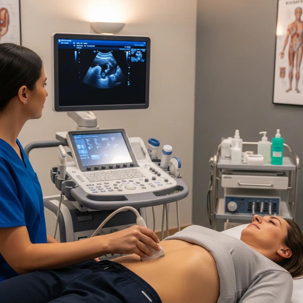
Ultrasound works by emitting high‑frequency sound from a transducer; echoes from tissue interfaces are turned into live images. There is no ionising radiation involved, so ultrasound is inherently safer than X‑ray based tests for repeated imaging and for use during pregnancy. The technique is non‑invasive — the transducer is moved over the skin with gel to improve contact. Because image quality depends on operator skill and equipment settings, accredited centres and trained sonographers play an important role in ensuring accurate results.
What Conditions Can Ultrasound Effectively Diagnose?
Ultrasound is used as a first‑line or supportive test across many areas. It detects problems in soft tissues, fluid collections and blood flow, and visualises moving anatomy. Common abdominal uses include gallstones, liver disease and kidney conditions. Gynaecological scans assess ovarian and uterine problems. Vascular studies look for deep vein thrombosis and carotid disease. Musculoskeletal scans evaluate tendon tears, bursitis and joint pathology. Breast ultrasound helps characterise lumps and complements mammography. When ultrasound is insufficient, it often indicates whether further CT or MRI is required.
- Abdominal: liver, gallbladder, pancreas — identifies stones, masses and organ enlargement.
- Gynaecological / Obstetric: uterus, ovaries, fetus — monitors pregnancy and investigates pelvic symptoms.
- Vascular: veins and arteries with Doppler — detects DVT and assesses blood flow.
- Musculoskeletal: tendons, ligaments and joints — evaluates tears, inflammation and guides injections.
Which Types of Ultrasound Scans Are Available at Life Medical Imaging Central Coast?
We offer a broad range of diagnostic ultrasound services across abdominal, women’s health, vascular, musculoskeletal and breast imaging. Availability varies by site, and many scans can be booked locally using our online system or by phone. The sections below outline the common scan types, what they assess and typical indications to help patients and referrers choose the right study.
Our ultrasound services include general abdominal, pelvic and thyroid scans, obstetric and gynaecological studies, vascular Doppler investigations, musculoskeletal ultrasound, breast imaging and advanced 3D/4D obstetric scans. We use grey‑scale and Doppler techniques as required to answer specific clinical questions. Knowing which scan matches a patient’s symptoms helps ensure accurate referrals and timely investigation.
- General abdominal ultrasound: assesses liver, gallbladder, pancreas and kidneys for stones, masses or inflammation.
- Obstetric & gynaecological ultrasound: dating, anatomy and growth scans, plus transvaginal imaging for pelvic assessment.
- Vascular ultrasound (Doppler): evaluates for venous thrombosis, arterial disease and carotid pathology.
- Musculoskeletal ultrasound: visualises tendons, ligaments and joints to diagnose tears, strains and inflammatory conditions.
This outline helps referrers match clinical questions to the correct scan and prepares patients for the focus of their appointment.
This service table links common clinical problems with the appropriate ultrasound type so patients and clinicians can make informed choices before booking.
What General Ultrasound Services Are Offered for Internal Organs?
General ultrasound examines abdominal organs — liver, gallbladder, pancreas, spleen and kidneys — and superficial structures such as the thyroid and testes. Typical referrals include abdominal pain, abnormal liver function tests, suspected gallstones, renal colic or palpable masses. Scans are non‑invasive and usually take 20–45 minutes depending on complexity. A sonographer will move the transducer across the skin while you lie in positions that optimise views; Doppler may be added to assess blood flow. Images are reviewed by a radiologist who issues the final report; abnormal results often lead to targeted follow‑up or specialist input.
What Women’s Health Ultrasound Scans Are Provided?
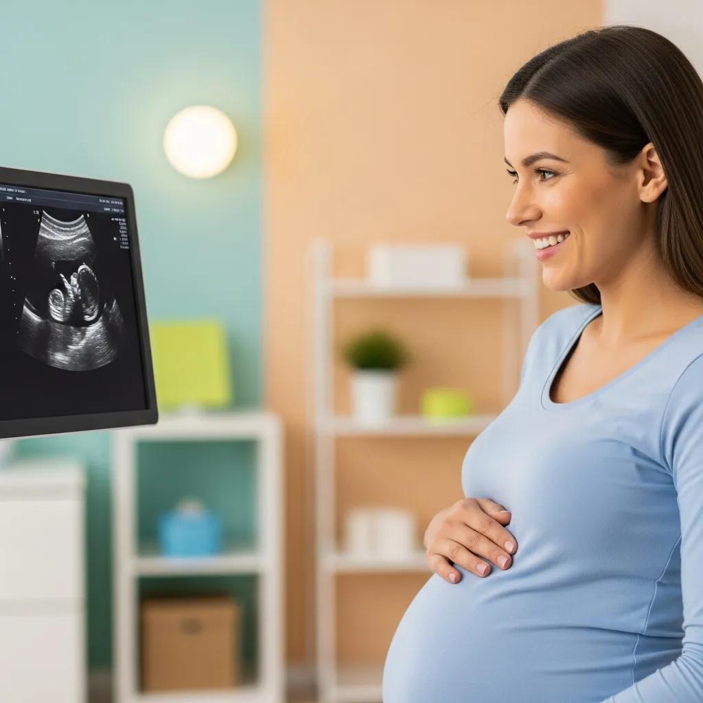
Our women’s imaging services include obstetric scans (dating, anatomy and growth) and gynaecological studies such as transvaginal pelvic imaging. Dating scans determine gestational age, anatomy scans assess fetal development in mid‑pregnancy, and growth scans monitor fetal size and wellbeing later in pregnancy. We also offer 3D/4D imaging for enhanced anatomical views and parental reassurance when appropriate. Gynaecological scans investigate pelvic pain, abnormal bleeding, ovarian cysts or fibroids and can support fertility assessments. Preparation and timing differ between early and later pregnancy scans, so accurate referral details help ensure the correct study is scheduled.
How Do Musculoskeletal and Vascular Ultrasound Scans Support Diagnosis?
Musculoskeletal ultrasound provides dynamic imaging of tendons, ligaments, joints and soft tissues and can be performed during movement. It’s useful for diagnosing rotator cuff tears, Achilles pathology and inflammatory bursitis, and it supports ultrasound‑guided injections or aspirations. Vascular ultrasound uses Doppler to measure blood flow and detect venous thrombosis, arterial stenosis or peripheral arterial disease; it combines functional information with anatomy in a non‑invasive exam. If results are equivocal, further cross‑sectional imaging or specialist referral may be recommended as part of the diagnostic pathway.
How Should Patients Prepare for Different Ultrasound Scans?
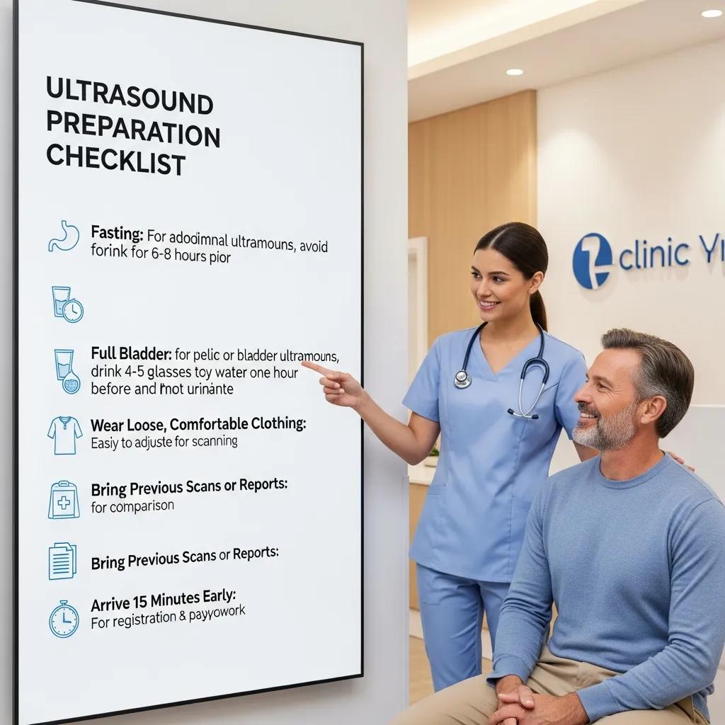
Good preparation improves image quality and reduces the chance of needing a repeat appointment. Requirements vary by scan type — fasting for abdominal studies, a full bladder for some pelvic or early pregnancy scans, and appropriate clothing for musculoskeletal or breast scans. Bring your referral, any relevant prior imaging and identification (including Medicare details where relevant). If you have diabetes, mobility issues or other health concerns, contact the clinic before your appointment so we can advise and make suitable arrangements.
Short, practical preparation checklists help prevent delays and improve diagnostic success.
- Abdominal scans: fast for 6–8 hours where instructed to reduce bowel gas and improve organ visualisation.
- Pelvic/early pregnancy scans: arrive with a comfortably full bladder to improve acoustic windows for pelvic structures.
- Musculoskeletal/breast scans: wear loose clothing and expose the area needing scanning to expedite the procedure.
This preparation matrix clarifies expectations and practical tips to improve imaging success and reduce the need for repeat scans.
What Are the Preparation Guidelines for Obstetric and Pelvic Ultrasounds?
Early pregnancy and some pelvic scans often require a full bladder because it elevates the uterus and improves the acoustic window. Later pregnancy scans typically do not need a full bladder — instructions vary for dating versus anatomy or growth studies, so follow the appointment confirmation carefully. Wear comfortable clothing; you may be asked to change into a gown depending on the site. Partners are usually welcome within space and privacy limits. If you require special medication timing (for example, diabetes management), contact your referrer or our clinic in advance for tailored advice.
How to Prepare for General, Musculoskeletal, and Vascular Ultrasound Procedures?
For abdominal scans, fasting reduces bowel gas and gives clearer views of organs; the usual guidance is 6–8 hours depending on the referral. For musculoskeletal scans, ensure the area is accessible — bring shorts or a top that allows direct scanning of shoulders, hips or knees. For vascular studies, avoid lotions or creams on the limbs and wear clothing that allows easy access. Continue regular medications unless advised otherwise and let us know about mobility needs when booking so we can schedule appropriately.
What Safety Measures and Expertise Ensure Accurate Ultrasound Results?
Accurate ultrasound depends on well‑maintained equipment, skilled operators and robust quality systems. Accredited centres follow regular audits, equipment servicing and standardised reporting to maintain diagnostic reliability. NATA accreditation provides an external framework that checks procedures, equipment calibration and staff competency — a reassuring sign for patients. Sonographers perform the scans and acquire images; radiologists review those images and provide the formal report. This team approach supports accurate diagnosis and clear clinical follow‑up.
This page aims to inform patients about ultrasound and to help you access our services with confidence about quality standards. NATA accreditation demonstrates our commitment to tested procedures and equipment performance. For questions about staff, reporting times or scan indications, call 02 4326 7000 to speak with our team.
How Does NATA Accreditation Guarantee Quality and Safety?
NATA accreditation involves regular external review of technical procedures, equipment maintenance and staff competency, ensuring diagnostic services meet national standards. It promotes consistent image quality through scheduled calibration, proficiency testing and documented quality assurance processes, reducing technical variability. For patients, accreditation is a clear trust signal that the centre participates in ongoing audits and corrective procedures, which supports confidence in both imaging and reporting timelines.
What Qualifications Do Sonographers and Radiologists Have at Life Medical Imaging?
Sonographers are allied health professionals trained in ultrasound physics, anatomy and scanning techniques and maintain their skills through continuing professional development. Radiologists are medical specialists who interpret imaging studies and produce diagnostic reports, with access to subspecialty opinions when complex findings require further expertise. Our workflow — sonographer acquisition followed by radiologist reporting — ensures both image quality and interpretation meet clinical expectations, and staff will explain their roles so you know who performs the scan and who reviews the results.
How Can Patients Book Ultrasound Appointments and Access Services Locally?
Booking an ultrasound generally requires a valid clinical referral, patient ID and scheduling via our online portal or by phone. Life Medical Imaging Central Coast supports online bookings and operates multiple Central Coast locations to improve local access. Knowing the booking steps and what to bring reduces delays on the day and ensures referrals and reports are processed correctly.
Follow these steps to book an ultrasound efficiently:
- Obtain a valid referral from your GP or specialist that specifies the clinical question and the requested scan type.
- Use the clinic’s online booking system or call 02 4326 7000 to request an appointment, providing referral details and preferred dates.
- Confirm preparation instructions and bring the referral, identification, and any prior imaging to the appointment.
- Arrive early for registration and ask reception about reporting timelines and how results will be sent to your referrer.
What Are the Steps to Book an Ultrasound Scan Online or by Phone?
Online bookings usually prompt you to upload a referral and choose the scan type. If you book by phone, have your referral, Medicare details and prior imaging information ready so staff can advise on preparation and site availability. For urgent or complex referrals, ask about priority appointments or specialist reporting pathways. Clear information at booking helps us allocate the correct appointment length, the right sonographer skillset and the expected reporting turnaround.
Where Are Life Medical Imaging’s Central Coast Locations and What Services Are Offered?
Life Medical Imaging operates across several Central Coast sites, offering general ultrasound, women’s imaging, vascular studies, musculoskeletal scans and breast ultrasound; some clinics provide specialised women’s and cardiac imaging. Not all services are available at every location, so choose the site that lists the required scan or call our bookings team to confirm the nearest availability. Our multiple‑site model reduces travel time for patients while maintaining consistent quality across locations. For location‑specific services and appointment availability, call 02 4326 7000.
Frequently Asked Questions
What should I expect during an ultrasound appointment?
You’ll be asked to lie on a couch while a sonographer applies a water‑based gel to the area being examined. The transducer is gently moved across your skin to capture images. The test is usually painless and takes 20–45 minutes depending on complexity. You may be asked to hold your breath or change position to improve views.
Can I eat or drink before my ultrasound?
Preparation depends on the scan. For abdominal ultrasounds, fasting for 6–8 hours is commonly required to reduce bowel gas and improve image quality. For pelvic or early pregnancy scans, you may need a full bladder. Always follow the specific instructions given when you book.
How long does it take to receive ultrasound results?
Preliminary observations are available soon after the scan, but the formal report is reviewed by a radiologist and can take a few hours to a few days. Urgent findings are prioritised and communicated immediately to the referring clinician. Your referrer will discuss results and next steps with you.
Are there any risks associated with ultrasound scans?
Ultrasound is widely regarded as a safe imaging method because it uses sound waves rather than ionising radiation. The main variables affecting accuracy are operator skill and equipment quality. Accredited centres and trained staff minimise these risks. Discuss any concerns with your referrer or our team before the procedure.
What should I bring to my ultrasound appointment?
Bring your referral, identification and any prior imaging or reports that may be relevant. Follow any preparation instructions provided (for example, fasting or a full bladder). Comfortable clothing that allows easy access to the scan area will help speed the appointment.
Can I bring someone with me to the ultrasound appointment?
Yes — companions are generally welcome, and many patients find support helpful, particularly for obstetric scans. Check with the imaging centre beforehand, as space and privacy constraints may limit the number of people in the exam room.
Is Ultrasound Safe During Pregnancy and Other Conditions?
Yes. Ultrasound is considered safe in pregnancy and for most clinical situations because it uses sound rather than ionising radiation. International guidelines and current research support its use for routine pregnancy surveillance and diagnostic evaluation when performed by accredited providers. Operators apply the ALARA principle (as low as reasonably achievable) to acoustic output and exam duration to ensure safety while obtaining diagnostic information. When additional imaging is needed, clinicians balance diagnostic benefit and modality‑specific risks to select the safest option.
How Are Ultrasound Results Delivered and Interpreted?
After the scan, the sonographer compiles images and preliminary observations which are reviewed by a radiologist who issues the formal report. Turnaround times vary; urgent findings are prioritised and communicated promptly to the referrer. Reports are usually sent to the referring clinician, who will discuss implications and recommended next steps with you. If further investigation is needed, the report will advise on targeted imaging, specialist referral or clinical correlation with blood tests.
Conclusion
Ultrasound is a safe, non‑invasive and cost‑effective imaging option for diagnosing and monitoring a wide range of conditions. Its real‑time imaging and absence of ionising radiation make it particularly valuable in pregnancy and for assessing many organ systems. Understanding the available scan types, preparation requirements and how results are reported helps you and your referrer make informed decisions. To learn more about our ultrasound services or to book an appointment, contact Life Medical Imaging Central Coast on 02 4326 7000.