Diagnostic Imaging — Central Coast: Advanced Radiology and Patient‑centred Care
Diagnostic imaging creates clear visual information about the body to help diagnose, monitor and guide treatment. This guide explains the core radiologic technologies used locally on the Central Coast, practical preparation steps for patients, the specialist services we offer, and how image‑guided interventions support diagnosis and pain relief. You’ll find straightforward, actionable advice for common scans, an overview of interventional options such as spinal injections and biopsies, and a plain explanation of safety standards and recent advances — including ultra‑low dose imaging. We organise content around modalities, indications and equipment to make complex topics easier to follow and to help referring clinicians optimise imaging requests.
Which medical imaging technologies are most commonly used?
Medical imaging uses several core technologies that differ by how they create images, their typical clinical uses and their safety profiles. The main modalities are CT, MRI, ultrasound, digital X‑ray and DEXA. Each converts a physical signal — X‑rays, sound waves or magnetic fields — into diagnostic images using specialised detectors and reconstruction software. Knowing the strengths and limits of each modality helps clinicians choose the best test for a clinical question and balance diagnostic yield with safety issues such as radiation exposure. The table below summarises the primary modalities, how they work and their common applications to support quick decision‑making for referrers and patients.
This comparison makes it easier to select the right modality for common clinical scenarios.
The table highlights when one modality is preferred and how mechanism and clinical use guide decision‑making. Next we look more closely at CT and ultrasound.
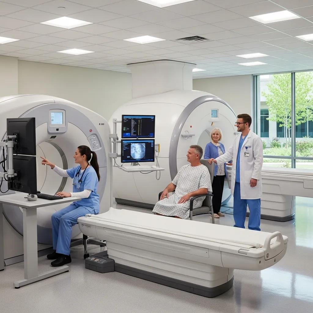
How do CT scans work and what do they detect?
CT acquires multiple X‑ray projections as the scanner rotates, then reconstructs those datasets into high‑resolution cross‑sectional images. CT is excellent for detecting acute haemorrhage, complex fractures, lung disease (including pulmonary emboli) and detailed abdominal pathology — which makes it a core tool in emergency and outpatient care. Modern iterative reconstruction and ultra‑low dose protocols lower radiation exposure while keeping diagnostic quality high. For example, low‑dose chest CT can speed diagnosis of pulmonary conditions with a limited increase in radiation, and many local clinics now offer high‑definition, low‑dose CT for timely assessment of urgent problems.
CT’s speed and detail complement ultrasound’s strengths in safe, real‑time assessment.
What are the benefits of ultrasound imaging on the Central Coast?
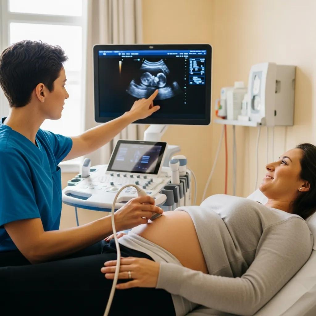
Ultrasound uses sound waves to produce live images without ionising radiation, making it ideal for pregnancy scans and repeat follow‑up studies. It’s especially useful for obstetrics and gynaecology (including 3D/4D fetal imaging), vascular studies that assess blood flow, and musculoskeletal exams that show tendons and joints in motion. Because ultrasound is portable and immediate, it can be performed at the bedside or in clinic and often complements CT and X‑ray by providing functional, dynamic information that guides further imaging or intervention.
With this overview of CT and ultrasound, we’ll move on to practical preparation guidance for common procedures.
How should patients prepare for common radiology procedures?
Proper preparation improves image quality, diagnostic accuracy and patient comfort. Preparation varies by modality and clinical question. Typical steps include following fasting instructions for certain abdominal CTs, checking contrast allergy history before contrast‑enhanced scans, and screening for metal implants that affect MRI safety. Bring referral paperwork, photo ID and any prior imaging to help the radiology team compare studies and speed reporting. Patients who follow instructions reduce the risk of rescheduling, shorten appointment times and help clinicians reach a reliable diagnosis sooner.
Below is a concise, scannable table covering pre‑ and post‑procedure advice for common radiology tests.
This quick reference helps patients and referrers find critical preparation points. Below are specific checklists for CT/MRI and ultrasound/X‑ray.
Before a CT or MRI appointment, make sure to:
- Confirm the Referral: Ensure the referral clearly states the clinical question so the correct imaging protocol is selected.
- Review Allergies and Kidney Function: Advise of contrast allergy history and provide recent kidney function tests if contrast is planned.
- Follow Fasting Requirements: Fast as instructed for abdominal contrast studies, typically 2–6 hours.
- Prepare for Comfort and Safety: Remove jewellery and metal objects and bring any implant documentation for MRI screening.
Completing these steps reduces delays and helps the imaging team focus on accurate diagnosis and care.
What should you know before a CT scan or MRI?
Before CT, tell the team about allergies, current medications and any kidney impairment — iodinated contrast can affect renal function. Fasting may be required for some abdominal studies to improve image quality. MRI requires careful screening for implanted metal devices, pacemakers or metallic fragments because magnetic fields can interact with metal; if you are claustrophobic, discuss anxiolytic options with the clinic in advance. Clear referral information allows technologists to choose the right protocol; consent is obtained for contrast and image‑guided procedures. Always inform staff if you are pregnant or have recent surgical implants so they can select the safest pathway.
Having this information ready helps tailor the scan and improves your overall experience.
Understanding contraindications for contrast‑enhanced imaging is important for safe care.
Patient preparation for contrast‑enhanced imaging
Contrast media is commonly used for CT and some MRI studies. Poor kidney function may preclude intravenous contrast and an alternative imaging option may be recommended.
How should patients prepare for ultrasound and digital X‑ray?
Ultrasound preparation depends on the exam: pelvic and early pregnancy scans usually need a full bladder to improve pelvic organ visualisation; abdominal scans often require fasting to reduce bowel gas. For musculoskeletal ultrasound, wear loose clothing and bring prior imaging or a list of symptoms to guide a dynamic assessment. Digital X‑ray needs minimal preparation but tell staff if you are pregnant and remove jewellery or clothing with metal that could obscure images. These simple steps help produce clearer images and speed diagnosis.
Aftercare for these non‑invasive tests is minimal — most patients can resume normal activities immediately and await results as advised by their referring clinician.
Life Medical Imaging Central Coast also supports streamlined logistics — online appointment booking and eReferral options simplify pre‑scan administration. Using these tools to upload referral details and relevant history before arrival reduces wait times and ensures paperwork is complete on the day. Efficient administrative workflows help deliver faster imaging and quicker results to referrers.
Which specialised radiology services are available at Life Medical Imaging Central Coast?
We provide a comprehensive range of specialist imaging services to meet local clinical needs, combining sub‑specialist reporting and modern equipment to support accurate diagnosis and treatment planning. Our offerings include General CT, Cardiac CT and CT angiography for vascular assessment; Digital X‑ray and dental imaging; a full ultrasound program (General, Vascular, Musculoskeletal, Obstetric & Gynaecological) with 3D/4D obstetric capability; and DEXA bone density scans. Interventional services include spinal and joint injections, biopsies and aspirations, and selected therapeutic options such as Platelet‑Rich Plasma (PRP) injections. We highlight NATA accreditation, sub‑specialist expertise in women’s and cardiac imaging, and ultra‑low dose techniques as differentiators that build diagnostic confidence.
This service summary helps referrers and patients match clinical needs to the tests and procedures available locally.
- Women’s Imaging: Breast ultrasound, obstetric 3D/4D scans, gynaecological ultrasound and image‑guided breast biopsies.
- Cardiac Imaging: Cardiac CT and CT angiography for non‑invasive assessment of coronary and structural heart disease.
- Interventional & Therapeutic: Image‑guided spinal and joint injections, biopsies/aspirations and PRP therapy.
- Diagnostic Imaging Suite: General CT, digital X‑ray, dental imaging, paediatric imaging and DEXA bone density assessments.
These descriptions clarify what referrers can request and what patients can expect when specialist imaging or intervention is indicated.
What does women’s imaging include and how does it help care?
Women’s imaging covers obstetric assessment, breast and gynaecological diagnostics. It supports screening, pregnancy monitoring and targeted diagnostic follow‑up using ultrasound and image‑guided procedures. Breast ultrasound and ultrasound‑guided biopsy are essential for characterising lesions found on exam or mammography, while 3D/4D obstetric imaging improves anatomical visualisation when clinically appropriate. Sub‑specialist reporting enhances diagnostic accuracy and helps coordinate timely follow‑up between patients, referrers and multidisciplinary teams. These services combine safe imaging modalities and procedural expertise to support early detection, accurate diagnosis and efficient patient pathways for women’s health.
High‑quality women’s imaging links with local referral networks to ensure continuity from screening through to diagnosis and treatment.
How is cardiac imaging performed and what are the benefits?
Cardiac CT and CT angiography use rapid, ECG‑synchronised X‑ray acquisition to produce detailed images of coronary arteries, cardiac chambers and surrounding structures without the need for invasive angiography in many cases. These studies help assess coronary artery disease, congenital abnormalities, cardiac masses and pericardial disease with speed and precision. Advantages include fast acquisition, excellent spatial resolution and reliable evaluation of coronary calcification and stenosis — information that supports risk stratification and treatment planning. Local access to cardiac CT within a sub‑specialist reporting framework gives clinicians a valuable non‑invasive option that complements functional testing in patient management.
Cardiac CT’s detailed, non‑invasive assessment can help guide decisions between medical therapy, further testing or invasive procedures.
How do interventional radiology procedures support diagnosis and pain management?
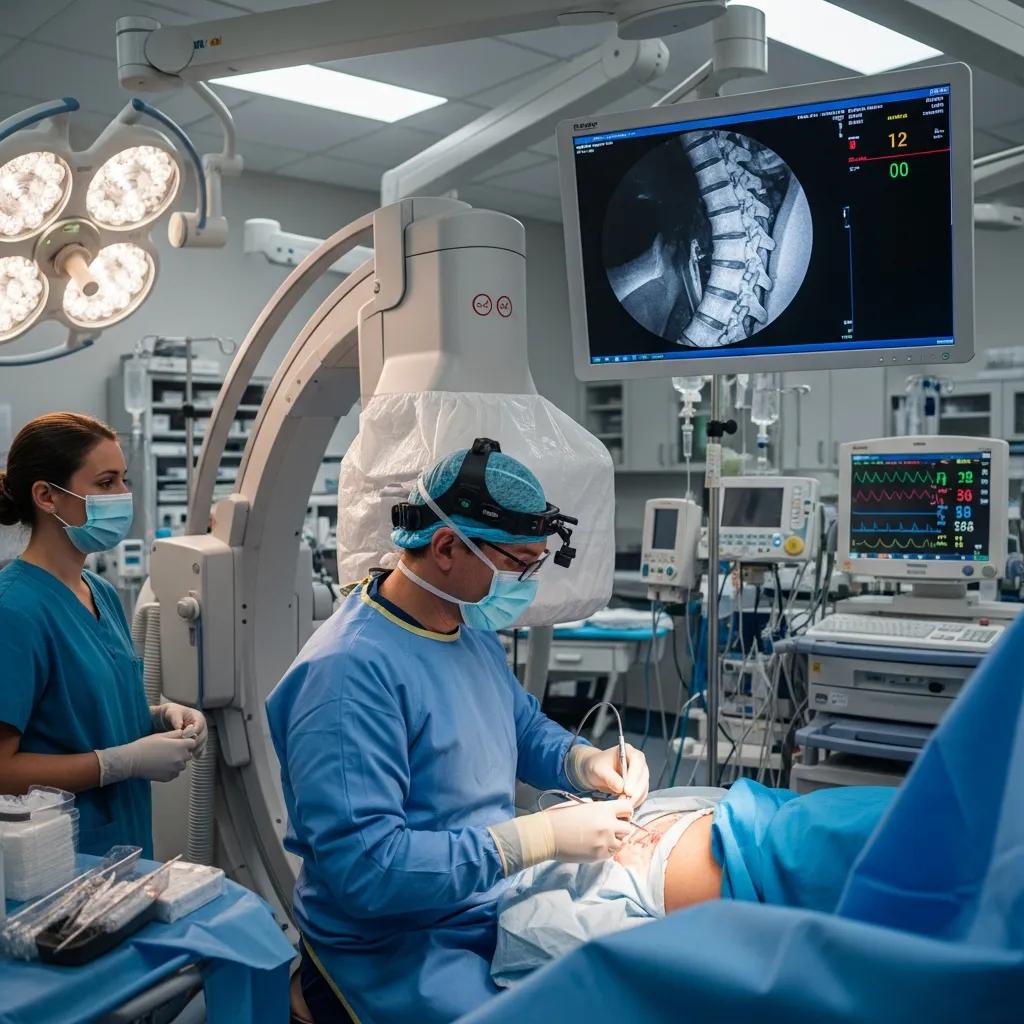
Interventional radiology uses imaging guidance to deliver diagnostic and therapeutic procedures precisely, reducing invasiveness and targeting the source of pain or diagnostic sampling sites. Common image‑guided procedures include spinal injections for radicular pain, joint injections for osteoarthritis, tissue biopsies and fluid aspirations, and regenerative treatments such as PRP for selected musculoskeletal injuries. CT or ultrasound guidance improves needle placement accuracy, raises procedure safety and usually shortens recovery compared with blind techniques — enhancing both diagnostic yield and therapeutic outcomes. Knowing indications, likely benefits and expected recovery timelines helps referrers and patients decide when interventional options are appropriate.
The table below summarises common procedures, indications and typical recovery times to aid rapid clinical decisions.
That table clarifies typical indications and recovery expectations. Below are procedural specifics for injections and PRP.
What are spinal and joint injections and when are they recommended?
Spinal and joint injections place anti‑inflammatory medication or analgesics directly into inflamed structures under image guidance to reduce pain and restore function. They are commonly used for radicular pain, facet arthropathy and inflammatory or degenerative joint conditions. CT or ultrasound guidance confirms accurate needle placement, maximising therapeutic effect and reducing risks. Choice between steroid injections and regenerative options such as PRP depends on diagnosis, past response to treatment and patient preference — steroids often give faster relief, while PRP aims to support longer‑term tissue healing in suitable cases. Most patients need brief activity modification and may notice improvement over days to weeks; follow‑up is arranged to assess response and plan further care.
Careful patient selection and precise imaging guidance make these interventions an effective part of multimodal pain management.
CT guidance is often preferred for certain spinal injection procedures.
CT‑guided spinal injections for pain management
CT guidance is commonly used for lumbar and thoracic facet joint injections with the patient prone. Contrast may be used to confirm needle position when indicated.
How do biopsies, aspirations and PRP therapy work in radiology?
Imaging‑guided biopsies and aspirations use real‑time imaging to target lesions or fluid collections for diagnostic sampling, providing pathologists with representative tissue while limiting procedural risk. Radiologists plan a safe needle path, use local anaesthesia and obtain samples via core or fine‑needle techniques; specimens are handled per laboratory instructions to preserve diagnostic integrity. PRP therapy involves drawing a patient’s blood, concentrating platelets and injecting the platelet‑rich fraction into tendons or joints under ultrasound guidance to promote healing through growth factors. Both diagnostic and regenerative procedures rely on sterile technique, precise image guidance and coordinated reporting to ensure clinical value and safe recovery.
Clear discussion of indications, risks and expected outcomes helps patients and referrers set realistic expectations and plan aftercare.
How does Life Medical Imaging Central Coast improve the experience for patients and referrers?
We focus on efficient administration, specialist reporting and comfortable facilities to deliver timely, high‑quality imaging. Our eReferral and online booking options streamline scheduling so referrers can submit investigations electronically and patients can book with minimal delay. Secure image access and timely reporting ensure clinicians receive results quickly and patients can obtain images or follow‑up instructions via established portals. Our emphasis on sub‑specialist reporting for women’s and cardiac imaging, NATA accreditation and up‑to‑date equipment underpins our commitment to reliable, patient‑centred service.
These administrative and clinical features reduce friction across the care pathway and support continuity from referral to definitive management.
How can patients book appointments and access imaging results online?
Booking and result access follow a simple workflow: referrers submit a clear electronic referral with the clinical question, patients receive appointment confirmation and attend with necessary documentation, and reports are sent to the referrer and made available to patients via secure portals or on request. Confirm preparation steps before arrival to avoid delays. If issues arise, contact the clinic’s bookings team or referral support for urgent arrangements. Using eReferral and online booking tools consistently improves scheduling accuracy and report turnaround.
Efficient electronic workflows help clinicians act on imaging findings sooner and reduce patient waiting time.
What resources help referring doctors optimise imaging requests?
Referrers benefit from clear guidance on modality selection, standardised referral forms and eReferral pathways that describe the clinical question, relevant history and prior imaging. Including details such as symptom onset, focal examination findings and prior interventions helps radiologists choose the most appropriate protocol and provide actionable recommendations. Educational protocols that outline indications, contrast requirements and expected turnaround times support efficient referral decisions and patient counselling. Sharing examples of well‑constructed referrals and keeping communication channels open improves collaboration between referrers and imaging specialists.
Providing complete clinical information via eReferral helps imaging services deliver targeted, high‑value studies that directly inform patient care.
What safety standards and technological advances shape modern radiology?
Quality and safety in imaging rest on formal accreditation and ongoing technological progress that lower risks while improving diagnostic confidence. Accreditation such as NATA verifies equipment performance, calibration, staff competency and quality control procedures, ensuring consistent imaging standards and documented safety practices. Advances like ultra‑low dose CT, iterative reconstruction and high‑sensitivity detectors improve image quality at lower radiation doses and widen diagnostic options for sensitive populations, including children and pregnant patients. Together, these safeguards and innovations support accurate diagnoses with reduced procedural risk and more efficient patient pathways.
A brief look at accreditation and dose‑reduction technology shows how standards and innovation work together to improve care.
How does NATA accreditation support quality and safety?
NATA accreditation is an independent quality process that assesses imaging services across equipment calibration, performance checks, staff training and documented quality control. For patients, accreditation means reproducible imaging standards, traceable equipment checks and formal procedures that reduce variation and error, increasing confidence in results. Accreditation also requires ongoing staff education and audit, which drives continuous improvement and alignment with best practice. The presence of NATA accreditation signals a commitment to formal quality systems that protect patient safety and support reliable diagnostic outcomes.
Knowing about accreditation helps clinicians and patients appreciate the systems behind trustworthy imaging services.
What are the benefits of ultra‑low dose imaging and modern equipment?
Ultra‑low dose techniques and contemporary hardware reduce radiation exposure while maintaining or improving diagnostic image quality through software advances like iterative reconstruction and more sensitive detectors. Benefits include lower lifetime radiation risk, broader use of CT where radiation concerns once limited it, and better visualisation of small or low‑contrast lesions that aids early detection. Faster scan times and improved patient comfort also reduce motion artefact and repeat scans. These improvements increase diagnostic confidence and support safer, more patient‑centred imaging services.
Ongoing equipment development and dose‑reduction strategies show how quality imaging and safety evolve together to enhance clinical care.
Frequently Asked Questions
What role does interventional radiology play in pain management?
Interventional radiology uses imaging to perform minimally invasive procedures that target the source of pain. Procedures such as spinal and joint injections deliver medication directly where it is needed, often reducing symptoms with lower risk and quicker recovery than surgery. By accurately treating pain generators, image‑guided interventions can significantly improve function and quality of life for people with chronic pain.
How can patients get imaging results quickly?
To help ensure timely results, follow pre‑procedure instructions, bring all required documents (referral, ID, prior imaging) and arrive on time. Results are typically sent to the referring clinician and made available to patients through secure online portals or on request. Ask about the expected reporting timeframe when you attend so you know when to expect results and the next steps for your care.
What should patients expect during an ultrasound?
An ultrasound is a non‑invasive, well‑tolerated test that uses sound waves to create real‑time images. A water‑based gel is applied to the skin and a transducer is moved over the area of interest. You may be asked to change position or hold your breath briefly for clearer images. The exam is usually quick and requires no recovery time — you can resume normal activities immediately afterwards.
What are the advantages of digital X‑ray technology?
Digital X‑ray offers faster image acquisition and processing, enabling immediate review and quicker clinical decisions. Modern detectors reduce radiation dose while delivering high‑quality images that are easy to store and share electronically. Digital image manipulation also aids diagnostic accuracy, making digital X‑ray the standard choice in contemporary practice.
How does Life Medical Imaging ensure patient safety during procedures?
Patient safety is central to our practice. We follow rigorous safety protocols and hold NATA accreditation to demonstrate compliance with quality assurance standards. Staff receive regular training, and we use advanced technologies such as ultra‑low dose protocols to minimise radiation exposure while maintaining diagnostic quality. Ongoing monitoring and quality improvement ensure procedures are performed safely in a supportive environment.
What follow‑up care is available after imaging procedures?
After imaging, your referring clinician will review the report and discuss results and next steps. For procedures that cause discomfort, we provide aftercare instructions and arrange follow‑up as needed. If additional imaging or specialist review is required, your referrer will coordinate those appointments. Clear communication about follow‑up helps you understand your care pathway and feel supported throughout recovery.
Conclusion
Understanding diagnostic imaging helps patients and referrers make informed choices about care. At Life Medical Imaging Central Coast we combine modern technology, specialist reporting and friendly, efficient processes to improve diagnostic accuracy and patient experience. Explore our services and online resources, or contact us to discuss how we can support your diagnostic needs.