Medical Imaging Technology: Advances, Applications and Patient Benefits
Medical imaging covers the tools clinicians use to see anatomy and function — from diagnosis and treatment planning to monitoring response. Recent innovations are making scans faster, safer and easier to access for patients. This guide explains how CT, MRI, ultrasound and digital X‑ray work, highlights practical innovations such as AI, ultra‑low‑dose CT and 3D/4D ultrasound, and outlines benefits for cardiac, women’s and musculoskeletal imaging. We also include clear guidance for referrers and patients on choosing the right test, preparing for scans and what to expect on the day. Where relevant we note local access: Life Medical Imaging Central Coast provides NATA‑accredited diagnostic services across multiple Central Coast sites and offers many of the modalities described, with appointments and referrals available by phone. The sections that follow cover recent technology advances, CT versus MRI comparisons, ultrasound uses, portable X‑ray, patient safety and preparation, and future trends shaping practice.
What Are the Latest Advancements in Medical Imaging Technology?
Progress in imaging combines better hardware, smarter software and more efficient workflows to improve diagnostic accuracy, speed reporting and reduce patient risk. Notable developments include AI for detection and triage, ultra‑low‑dose and high‑definition CT for cardiac and lung detail, 3D/4D ultrasound for obstetrics, and portable devices linked to cloud PACS for faster remote review. These advances help detect smaller lesions, shorten time to diagnosis and expand access in community and aged‑care settings. The table below maps key technologies to their main attributes and the direct patient benefits they deliver.
Each entry above links a technical improvement to a practical patient outcome and supports clinical decision making. The sections that follow describe how AI fits into radiology workflows and why ultra‑low‑dose, high‑definition CT is important for certain clinical questions.
How Is AI Enhancing Radiology Diagnostics?
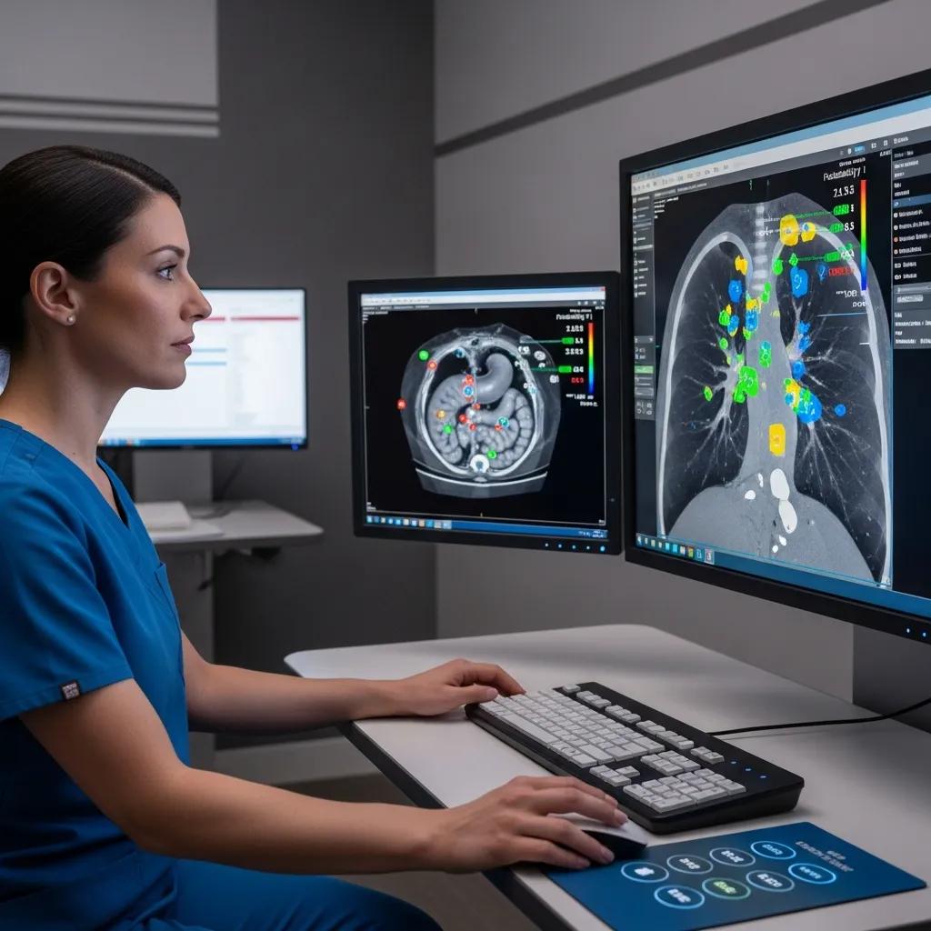
AI assists radiology with image reconstruction, anomaly detection and workflow prioritisation by learning patterns from large datasets and flagging findings for radiologist review. Typical uses include automated nodule detection on chest CT, intracranial haemorrhage triage in emergency CT and denoising to support lower‑dose protocols while keeping diagnostic clarity. AI is an augmentation tool — it speeds turnaround and raises sensitivity but does not replace specialist interpretation. Real‑world deployments show faster report times and improved detection in targeted tasks; importantly, human‑in‑the‑loop workflows keep clinical oversight and governance front of mind. Knowing how AI is used helps referrers and patients understand why reads can be quicker and, in many cases, more consistent.
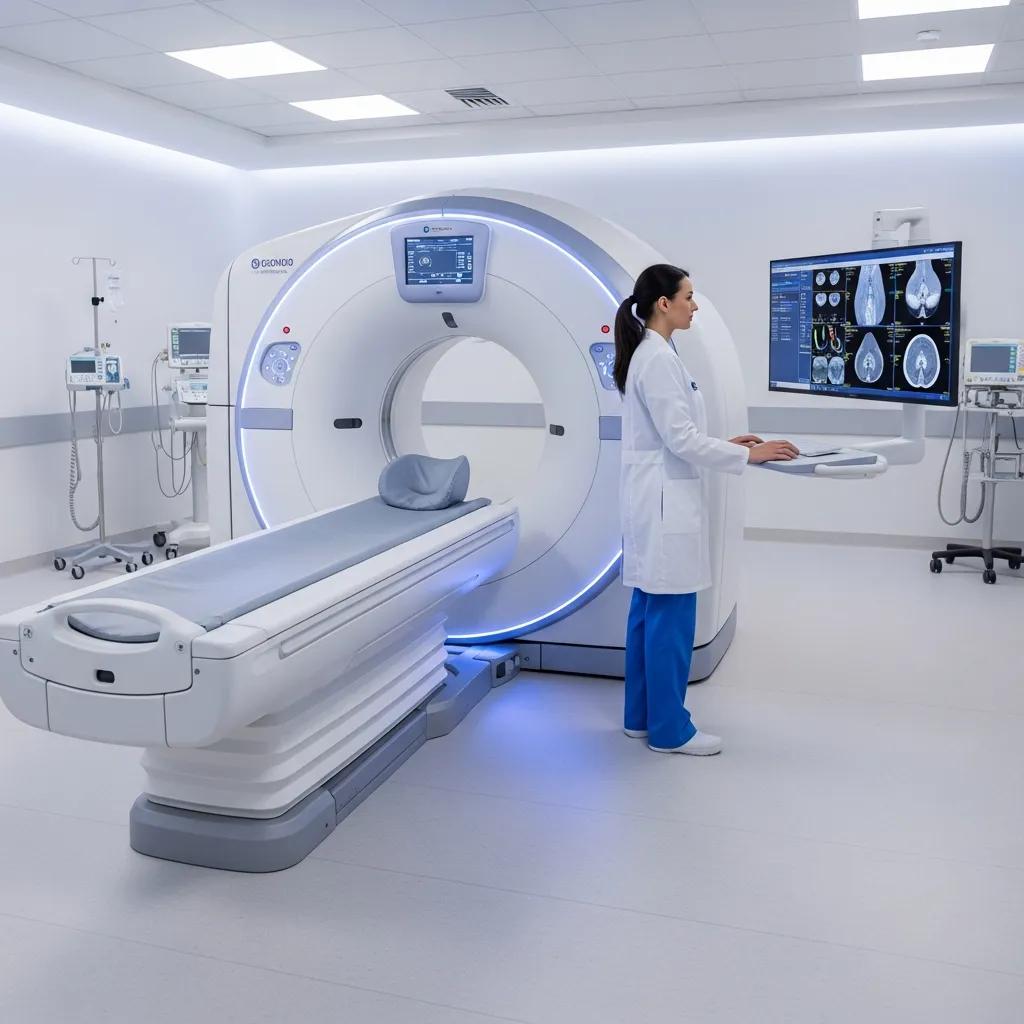
What Are the Benefits of Ultra-Low Dose and High Definition CT Scans?
Ultra‑low‑dose CT combines sensitive detectors, iterative reconstruction and optimised protocols to reduce ionising radiation while preserving fine spatial detail — valuable for serial imaging and screening. High‑definition CT improves visualisation of small structures such as coronary plaques or subtle lung nodules, which supports confident cardiac and thoracic assessment. Clinical advantages include safer repeat imaging, clearer depiction of tiny lesions and the ability to perform detailed angiographic studies with a lower cumulative dose. These technical gains are particularly useful for patients needing cardiac CT angiography or lung follow‑up, delivering accurate answers with a smaller radiation footprint. Key clinical benefits are summarised below.
Evidence shows ultra‑low‑dose CT protocols can substantially lower radiation exposure without sacrificing diagnostic quality, which is especially relevant in cardiac imaging.
Ultra-Low Voltage CT for Reduced Radiation and Contrast Agent Doses
Achieving low radiation dose and reduced contrast agent use in coronary CT angiography at 60‑kVp ultra‑low tube voltage — findings that support dose‑optimised cardiac protocols (2025).
- Lower Radiation Risk: Dose‑reduction techniques make repeat scans safer for chronic conditions.
- Improved Lesion Detection: High‑definition imaging reveals small nodules and fine vascular detail.
- Enhanced Cardiac Assessment: Clear coronary visualisation supports non‑invasive cardiac planning.
These benefits explain why many modern imaging pathways prioritise dose optimisation and high‑resolution acquisition for vulnerable patient groups. Next we compare CT and MRI to help choose the right modality.
How Do CT Scans and MRI Differ in Medical Imaging?
CT and MRI are complementary tools built on different physics. CT uses ionising X‑rays to produce cross‑sectional density images; it’s fast and excellent for bone injury, chest imaging and detecting haemorrhage. MRI uses magnetic fields and radiofrequency pulses to generate rich soft‑tissue contrast without radiation, making it ideal for neurological and musculoskeletal detail. Practical differences — accessibility, scan time, contraindications and contrast needs — influence which test is chosen in clinic or emergency settings. The table below gives an at‑a‑glance comparison of common modalities, their best uses and strengths or limitations.
What Are the Key Differences Between CT and MRI Scans?
CT measures X‑ray attenuation to create fast, high‑contrast images well suited to bone, air and acute bleeding. MRI measures proton behaviour to deliver superior soft‑tissue contrast and a range of functional sequences (T1, T2, diffusion). CT scans are typically shorter — often minutes — making them ideal for trauma and urgent vascular imaging; they do involve ionising radiation, which modern dose‑reduction methods minimise. MRI avoids radiation but needs longer scan times and careful screening for ferromagnetic implants. Understanding these differences helps referrers choose the most appropriate test for speed, tissue type and safety, and guides patient preparation covered next.
When Should You Choose CT Scan Over MRI?
Choose CT for acute trauma, rapid assessment of fractures or intracranial haemorrhage, and detailed pulmonary or bony evaluation where speed and bone contrast matter. CT angiography is preferred for urgent vascular questions — for example suspected pulmonary embolism or coronary artery assessment — because it delivers high‑resolution vascular maps quickly. Where local services provide specialised CT (such as high‑definition cardiac CT), referrals can be expedited to guide treatment planning. For non‑urgent soft‑tissue characterisation or when avoiding ionising radiation is a priority, MRI is usually the better option; knowing these scenarios helps clinicians make timely local referrals and schedule appropriately.
What Are the Diagnostic Uses of Ultrasound Imaging?
Ultrasound uses high‑frequency sound waves to produce real‑time images without ionising radiation, making it versatile for obstetric, gynaecological, vascular and musculoskeletal work. Its portability and Doppler capability enable dynamic blood‑flow assessment and image‑guided procedures such as injections or biopsies, and it supports bedside diagnostics in many settings. Ultrasound’s strengths are safety, immediate feedback and excellent assessment of fluid, soft tissue and fetal anatomy; limitations include operator dependence and restricted acoustic windows. The short lists below outline common applications and how 3D/4D technology extends diagnostic and patient‑experience benefits.
Advances in 3D and 4D ultrasound have increased prenatal diagnostic capability, offering new perspectives on fetal anatomy and improving communication with parents.
Advanced 3D/4D Ultrasound for Prenatal Diagnosis and Fetal Imaging
3D/4D ultrasound techniques provide volumetric views that complement traditional 2D imaging, enhancing detection of fetal anomalies, enabling dynamic assessment of cardiac motion and supporting patient counselling. Digital volume storage also aids review and teaching. (Current 3D/4D ultrasound technology in prenatal diagnosis, 2005.)
Common ultrasound applications include:
- Obstetric Imaging: Tracking fetal growth, anatomy checks and pregnancy surveillance.
- Vascular Studies: Detecting deep vein thrombosis and assessing carotid blood flow.
- Musculoskeletal Imaging: Evaluating tendon tears, joint effusions and guiding injections.
How Do 3D and 4D Ultrasound Improve Obstetric and Gynaecological Imaging?
3D and 4D ultrasound reconstruct volumetric datasets that give clinicians better spatial context for fetal and pelvic anatomy, helping detect anomalies and plan surgery when needed. 4D adds time, so clinicians and families can watch fetal movement in real time — which can aid assessment and increase parental reassurance. Volumetric imaging improves measurement reproducibility and can clarify complex anatomy for preoperative planning. For patients, these techniques offer clearer images and a more informative experience within women’s imaging pathways. This helps referrers pick the ultrasound modality that best matches the clinical question.
What Are the Uses of Vascular and Musculoskeletal Ultrasound?
Vascular ultrasound uses Doppler to assess blood flow and vessel structure — detecting stenosis, occlusion or thrombosis and monitoring grafts or carotid disease. Musculoskeletal ultrasound visualises tendons, ligaments and joint spaces to diagnose tears, inflammation and effusions and to guide injections or aspirations. Ultrasound is often the first‑line test because it’s quick, repeatable and effective for image‑guided procedures. In musculoskeletal care, dynamic scanning during movement can reveal problems missed on static imaging; in vascular work, it provides non‑invasive flow data to shape management decisions. These capabilities keep ultrasound central to outpatient, emergency and interventional practice.
How Is Portable X-Ray Equipment Transforming Medical Imaging?
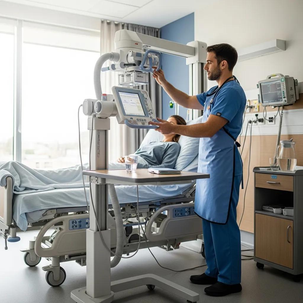
Portable X‑ray brings image acquisition to the bedside or community setting, reducing risky transfers for frail or infectious patients and enabling timely imaging where fixed suites aren’t practical. Modern portable units with digital detectors produce images quickly and connect to cloud PACS for rapid radiologist review, helping clinicians make faster treatment decisions and support infection control. Typical use cases include aged care facilities, emergency departments and rural clinics. The list below summarises direct patient advantages of portable digital X‑ray.
- Reduced Patient Movement: Bedbound or critically unwell patients avoid risky transfers to imaging departments.
- Faster Diagnosis: On‑site imaging speeds triage and treatment decisions in urgent care.
- Improved Infection Control: Bedside imaging cuts cross‑traffic through clinical areas during outbreaks.
What Are the Advantages of Portable X-Ray Devices for Patients?
Portable X‑ray devices improve convenience and safety by allowing imaging where the patient already is, lowering logistical burdens and improving comfort. In emergency triage, bedside radiography can quickly confirm pneumothorax, displaced fractures or line placement problems so staff can act immediately. While fixed high‑end units can deliver marginally higher image quality, modern digital detectors and post‑processing close the gap and support fast teleradiology reads. Overall, portable X‑ray expands access to diagnostics and shortens time to treatment, particularly in community and aged‑care settings. The next section outlines how digital X‑ray technology further boosts diagnostic performance.
How Does Digital X-Ray Technology Improve Diagnostic Accuracy?
Digital X‑ray replaces film with electronic detectors that capture a wider dynamic range, allow post‑processing (contrast, magnification) and enable instant transfer to PACS for remote specialist review. These features improve visualisation of subtle fractures, streamline workflows and speed clinical decisions; dose‑optimisation tools also minimise unnecessary exposure. Digital systems make teleradiology efficient by supporting secure, rapid sharing and archiving — crucial for multi‑site clinics and urgent reporting. Together, these improvements deliver faster, more accurate diagnoses and closer integration with multidisciplinary care pathways.
What Should Patients Know About Safety and Preparation for Medical Imaging?
Patient safety in imaging focuses on minimising radiation, managing contrast risks and giving clear pre‑scan instructions so appointments run smoothly. Modern scanners and protocols reduce dose through iterative reconstruction and tailored settings, while staff screen for allergies, pregnancy and implant safety to protect vulnerable patients. Simple preparation steps for CT, MRI, ultrasound and X‑ray reduce delays; the table below summarises common preparation needs, typical appointment lengths and aftercare advice.
This quick reference helps you prepare and sets expectations for typical scan experiences. The checklist below summarises essential safety measures and arrival tips.
- Disclose Medical History: Tell staff about allergies, pregnancy, implants or previous contrast reactions.
- Follow Fasting/Fluid Instructions: Adhere to CT fasting or ultrasound bladder guidance to ensure diagnostic quality.
- Arrive Early with Referral: Bring your referral and any prior imaging if available for comparison.
Following these steps reduces delays and improves image quality. For local bookings or questions, contact the NATA‑accredited Life Medical Imaging Central Coast by phone to arrange appointments or discuss referral pathways. Clear preparation helps keep scans safe and efficient.
What Are the Future Trends and Innovations in Medical Imaging Technology?
Looking ahead, we expect tighter integration of AI with multimodal clinical data, more personalised imaging protocols, growth in molecular imaging and wider deployment of portable and cloud‑based systems to deepen diagnostic reach and improve access. AI will increasingly combine imaging with electronic health records and lab or genomic data to produce richer diagnostic insights and prioritise urgent cases, while regulation and explainability will guide safe adoption. Portable devices with fast cloud review and 5G connectivity will decentralise imaging, bringing specialist input to rural and aged‑care settings and reducing time to treatment. The next sections explore how multimodal AI and cloud solutions will shape practice and access.
The interaction between AI and low‑dose CT is an active research area, aiming to boost image quality and diagnostic power while further cutting radiation exposure — especially for cardiac studies.
AI and Low-Dose CT for Cardiac Imaging
Combined low‑dose simulation and deep learning for CT denoising: application to ultra‑low‑dose cardiac CTA — an example of how AI can enhance image quality in low‑dose cardiac protocols (CK Ahn, 2019).
How Will AI and Multimodal Diagnostics Shape Medical Imaging?
AI linked with multimodal diagnostics will connect imaging features to clinical notes, lab results and genomics to generate personalised risk profiles and aid triage. In practice, an AI‑driven workflow could prioritise scans with suspected critical findings, recommend protocol adjustments to avoid unnecessary sequences and produce integrated reports that help multidisciplinary teams. This promises faster, more precise decisions and tailored follow‑up but requires strong validation, clinician oversight and governance to ensure safety. As these systems mature, adoption will be driven by demonstrable improvements in outcomes and efficiency.
What Role Do Portable and Cloud-Based Imaging Solutions Play in Healthcare?
Portable and cloud‑based imaging expand access by enabling image acquisition outside hospitals and allowing rapid specialist review via cloud PACS. Mobile ultrasound and portable X‑ray linked to secure cloud platforms let radiologists provide timely teleradiology reads, shortening time to diagnosis and supporting local treatment. These solutions also help multi‑site practices by centralising reporting and archives, which supports quality control and specialist access across distributed clinics. Together, portability and cloud connectivity are reshaping where and how diagnostic imaging is delivered, improving equity of access and operational resilience for health services.
Frequently Asked Questions
What should patients expect during a medical imaging appointment?
On arrival you’ll check in and provide relevant medical history, including allergies and prior imaging. Depending on the test you may change into a gown. Procedures vary: CT scans are generally quick, while MRI appointments take longer. Our staff will explain the process, show you how to lie or position for the best images and answer any questions to keep you comfortable and informed.
How can patients prepare for a CT scan?
Preparation depends on the type of CT. If contrast will be used you may be asked to fast for a few hours and to disclose allergies or kidney issues. Wear comfortable clothing without metal fasteners. Tell staff about any prior contrast reactions. Following these instructions helps get the best images and reduces the chance of complications.
Are there any risks associated with medical imaging procedures?
Most imaging tests are safe, but there are some risks to consider. CT involves ionising radiation; repeated scans increase lifetime exposure, though modern protocols minimise dose. Contrast agents can cause allergic reactions in a small number of people, and some tests have implant‑related contraindications. Your referrer and imaging team will balance risks and benefits and answer questions before proceeding.
What advancements are being made in patient safety for medical imaging?
Safety improvements include ultra‑low‑dose imaging protocols, better contrast management and integration of AI to support detection and workflow. Staff training and strict safety checks (for pregnancy, implants and allergies) are standard. These measures aim to keep care effective while reducing risk and improving outcomes.
How do patients access their imaging results?
Results are usually reported by a radiologist to the referring clinician. Many facilities also offer patient portals where you can view reports and images online. You can request copies of your imaging directly from the centre. We recommend discussing results with your referrer to understand next steps.
What role does telemedicine play in medical imaging?
Telemedicine allows radiologists to review images remotely and enables virtual consultations, which is especially helpful in rural or underserved areas. Cloud‑based imaging lets specialists share images securely, speeding diagnosis and treatment decisions. Telemedicine also supports follow‑up discussions so patients can review results without extra travel.
Conclusion
Modern medical imaging is delivering clearer answers faster, with improved safety and broader access. Understanding the strengths of CT, MRI and ultrasound helps patients and referrers choose the right test and prepare effectively. These innovations improve the patient experience and support timely treatment. For more information on preparing for your scan or to learn about our services, contact Life Medical Imaging Central Coast today.