Medical Imaging Education: Degrees, Training & Careers
Medical imaging education covers the formal degrees, hands‑on clinical training, professional registration and ongoing CPD that prepare radiographers, sonographers and imaging technologists to deliver safe, accurate diagnostic services. This guide explains why structured education matters for patient outcomes and workforce readiness, and maps the common routes from undergraduate study to specialist practice — including part‑time or online study and targeted CPD. If you’re a trainee, referrer or employer wondering which qualifications lead to HCPC eligibility, how placements are organised, or which CPD best keeps skills current, you’ll find clear comparisons of degree types, plain‑English summaries of core modalities (MRI, CT, ultrasound, X‑ray), a sample “day in the life”, and a look at technology trends — such as AI and photon‑counting CT — that are changing training needs. Later sections cover accredited CPD, how placements and apprenticeships boost employability, and resources to help patients and referrers prepare for imaging.
What qualifications are needed for medical imaging technologists in the UK?
To practise as a medical imaging technologist you generally need an accredited undergraduate or postgraduate qualification plus professional registration, supported by supervised clinical placements and ongoing CPD. Undergraduate BSc programmes in Diagnostic Radiography usually lead to HCPC‑eligible registration; MSc courses and postgraduate diplomas provide routes into advanced practice and modality specialisation. Apprenticeships are an employer‑sponsored alternative in some regions. Registration with the Health and Care Professions Council (HCPC) and membership of professional bodies such as the Society and College of Radiographers (SCoR) set professional and ethical standards, and sustained CPD keeps skills current. The table below summarises the common academic pathways, typical duration and expected outcomes.
HCPC standards and learner expectations for diagnostic radiography (M. Hardy, 2023)
This UK Delphi study explored how learners’ expectations align with the 2023 HCPC Standards of Proficiency, highlighting areas where training and regulatory expectations still need closer alignment.
Core qualifications compared for radiography entry and registration:
This comparison shows how each pathway leads to practice and why placement hours are critical for readiness. The next section outlines the most common degree types and typical entry requirements.
Which diagnostic radiography degrees are available in the UK?
Most routes start with an undergraduate BSc in Diagnostic Radiography; conversion and specialist MSc programmes are available for those with related degrees or clinical experience. Courses combine theory and supervised clinical placements so students gain practical competence alongside academic learning. Typical entry requirements include A‑levels (or equivalent) in science subjects such as physics or biology, plus evidence of commitment to healthcare; postgraduate options usually expect a relevant first degree or professional experience. Core modules cover anatomy, imaging physics, radiation protection, patient care and image interpretation, with placements used to demonstrate workplace proficiencies assessed by universities and clinical mentors. Understanding the balance of classroom learning and hands‑on exposure helps applicants choose the best route for their career goals.
How do online radiology training courses support career development?
Online training complements formal degrees by offering flexible CPD modules, image‑interpretation case banks and remote learning that fit around shift work and placements. Accredited e‑learning can contribute CPD points recognised by professional bodies and typically blends recorded lectures, interactive cases and formative assessments to sharpen diagnostic reasoning and safety practice. For working radiographers, part‑time or distance MSc programmes let you advance without pausing employment; short online modules enable targeted upskilling in areas such as MRI safety or ultrasound optimisation. When paired with supervised clinical practice, blended learning efficiently reinforces both theory and hands‑on competence.
How can you build a successful radiography career pathway in the UK?
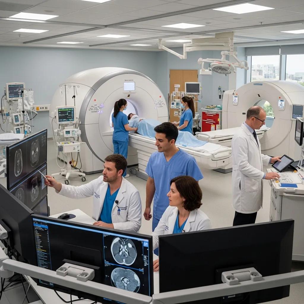
A radiography career typically starts with an accredited qualification, progresses through HCPC registration and early clinical posts, and develops via specialisation, CPD and leadership training. Early steps include securing high‑quality placements during study, applying for rotational posts to gain multimodality exposure, and networking with mentors and professional groups. Common specialisms include CT, MRI, ultrasound, paediatrics and women’s imaging; each requires focused CPD and supervised practice to move into advanced reporting or consultant roles. Below is a practical step‑by‑step pathway trainees can follow to plan progression and promotions.
A practical progression plan for radiography careers:
- Complete an accredited degree: Obtain an HCPC‑eligible qualification with embedded placements.
- Register and secure your first post: Take an entry‑level role to consolidate hands‑on skills.
- Pursue modality training: Complete CPD and supervised practice in chosen specialisms.
- Apply for advanced roles: Move into specialist, reporting or leadership positions.
These steps form a clear ladder from graduate to specialist practitioner. The next section describes a typical working day to show how these elements come together in practice.
What does a day in the life of a radiographer or sonographer look like?
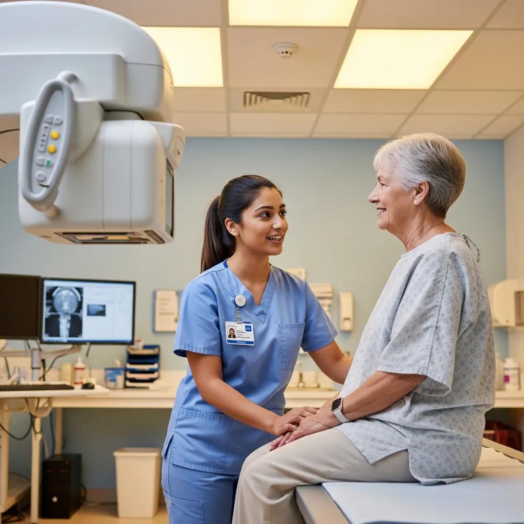
A clinical day mixes patient care, equipment operation and image‑quality checks with reporting and MDT coordination. Mornings often start with equipment checks, caseload planning and urgent inpatient work. Radiographers execute patient identification, safety screening, positioning and acquisitions across modalities such as X‑ray, CT and ultrasound; sonographers perform real‑time scanning and explain findings to patients. Afternoons commonly include image review, case discussions with radiologists, MDT meetings and documentation of doses or incidents, followed by equipment cleaning and handover. That blend of technical skill and patient‑centred communication is why substantial clinical placements are essential for developing both technique and professional judgement.
Support for students on temporary HCPC registration (R.M. Strudwick, 2021)
This study explored the experiences of academics and practice educators who supported final‑year radiography students working on the temporary HCPC register, highlighting lessons for supervision and readiness during practice placements.
What are local career opportunities in medical imaging on the Central Coast?
Regional diagnostic centres and independent clinics on the Central Coast offer both entry‑level and specialised roles, often giving early access to CT, MRI, ultrasound and digital X‑ray—valuable for rapid skills development. Local employers frequently host student placements and rotational posts that broaden multimodality experience; attending CPD events and networking with clinical educators raises your visibility for vacancies. Life Medical Imaging Central Coast is an example of an independent diagnostic clinic in the region providing CT, MRI, ultrasound, digital X‑ray and specialised women’s and cardiac imaging, and it lists education and careers resources for applicants and referrers. Reaching out to local provider pages and careers contacts helps candidates understand vacancy cycles and available training pathways.
What are the key medical imaging procedures and technologies to understand?
Knowing the strengths and limits of core modalities — MRI, CT, ultrasound and digital X‑ray — helps clinicians, referrers and patients choose the right test, prepare correctly and set realistic expectations. Each modality relies on different physics and suits different clinical problems: MRI offers excellent soft‑tissue contrast without ionising radiation; CT delivers fast cross‑sectional detail, useful in trauma; ultrasound provides real‑time, radiation‑free imaging ideal for obstetrics and vascular work; and digital X‑ray is efficient for bone and chest imaging. The table below summarises how each modality works, common uses and simple patient preparation steps to aid clear communication.
Comparative summary of imaging modalities:
This snapshot helps referrers select the right test and prepares patients for what to expect. The following subsections explain MRI mechanics and contrast ultrasound and CT in practical terms.
How does an MRI scan work and what should patients expect?
MRI uses strong magnetic fields and radiofrequency pulses to align hydrogen atoms and capture signals that are reconstructed into detailed soft‑tissue images — ideal for brain, spine and joint assessment. Patients lie on a motorised table that moves into a gantry; scan times vary from around 15 minutes to over an hour depending on the protocol. Safety steps include removing metal items and disclosing implants. Claustrophobia is common, so clear staff communication, ear protection and, if needed, pre‑arranged mild anxiolytics help improve tolerance. Good pre‑scan information reduces anxiety and improves cooperation, which in turn raises image quality and diagnostic value.
What are the benefits of ultrasound and CT scans in diagnostic imaging?
Ultrasound and CT play complementary roles. Ultrasound is portable, low‑cost and radiation‑free, offering real‑time assessment for obstetrics, abdominal and vascular problems; it excels with Doppler flow but is operator‑dependent and affected by body habitus. CT is fast and reproducible, giving high‑resolution cross‑sectional detail useful in trauma, lung and vascular studies, but involves ionising radiation and sometimes intravenous contrast. Preparation differs: ultrasound may require a full or empty bladder for specific exams, while CT often needs fasting and checks for contrast allergy or renal function. Knowing these trade‑offs helps clinicians balance diagnostic accuracy, safety and patient comfort.
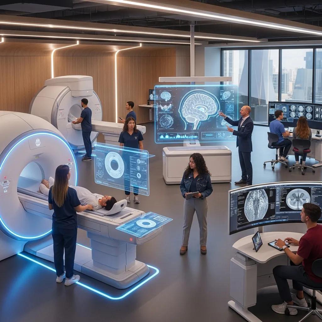
How is advanced technology shaping medical imaging education and practice?
New technologies — artificial intelligence, photon‑counting CT and enhanced 3D/4D obstetric imaging — are changing diagnostic practice and the skills imaging professionals need. AI assists with triage, lesion detection and workflow automation but requires training in validation, recognising bias and integrating algorithm outputs with clinical judgement. Hardware advances such as ultra‑low dose CT and higher‑fidelity 3D imaging affect protocol design and patient counselling, so hands‑on training is vital to use these safely. Modern education must therefore combine informatics, physics and practical modality training; the next sections review AI use‑cases and recent modality innovations.
How is artificial intelligence enhancing radiology diagnostics?
AI supports radiology by automating repetitive tasks, flagging urgent findings and assisting image interpretation through pattern recognition. When validated and clinically integrated, these tools can improve throughput and sensitivity for targeted pathologies. Current applications include triage (prioritising urgent studies), detection (for example intracranial haemorrhage or lung nodules) and workflow optimisation (scheduling, dose tracking). Limitations remain — generalisability, regulation and the need for clinician oversight — so CPD should include AI literacy: understanding performance metrics, failure modes and how to explain AI‑assisted findings. Training that pairs clinicians with curated clinical datasets, supervised interpretation and governance safeguards ensures AI augments rather than replaces professional judgment.
What are the latest innovations in CT and 3D/4D obstetric imaging?
CT advances include ultra‑low dose protocols and photon‑counting detectors that enhance contrast resolution and tissue characterisation while reducing radiation, prompting updates to protocol design and dose monitoring. Obstetric imaging has benefited from higher‑quality 3D/4D ultrasound that improves structural visualisation and parental counselling without ionising radiation. These developments require updated scanning skills and reporting standards; CPD and simulation training should cover new protocols, dose optimisation and patient counselling. Hardware and software improvements change everyday workflows and underline the continued value of hands‑on placements for consolidating modern practice.
What continuing professional development options are available for imaging professionals?
CPD for imaging professionals includes accredited online modules, workplace workshops, postgraduate degrees and employer‑led in‑house training — all aimed at maintaining competence and enabling specialisation. Accredited providers offer modular learning that carries measurable CPD points recognised by professional bodies; universities run masters and postgraduate diplomas for advanced practice. Apprenticeships and supervised placements remain key for hands‑on consolidation. The table below summarises common CPD formats, typical providers and what to consider when planning a learning pathway.
Overview of CPD formats and providers:
This overview helps clinicians choose a blend of learning formats for sustained development. The following subsections detail accredited modules and the role of placements.
Which accredited CPD modules support radiographer skill advancement?
High‑value CPD modules include image interpretation, modality‑specific safety (MRI safety, radiation protection), contrast administration and specialist scanning techniques such as cardiac or paediatric imaging. When selecting modules, check provider accreditation, CPD point allocation and whether practical assessment or supervised practice is included — hands‑on skills need workplace confirmation. Reputable programmes combine case‑based learning with assessment and reflective practice logs that align with professional revalidation. A progressive pathway — starting with core updates and advancing to specialist reporting or leadership modules — helps build both competence and career credentials.
How do clinical placements and apprenticeships enhance practical training?
Placements and apprenticeships convert classroom learning into procedural competence by giving supervised real‑world exposure to positioning, patient communication and safety workflows under experienced mentors. Placements let students see diverse clinical scenarios, learn multidisciplinary processes and accumulate required practice hours; apprenticeships often blend paid employment with structured learning and assessment and can lead directly to job offers. To secure placements, contact university placement coordinators, be flexible with shift patterns and demonstrate professionalism; during placements, set clear learning objectives, reflect on practice and meet regularly with mentors to maximise learning. These workplace experiences are essential for moving from learner to confident practitioner.
Student experiences on the temporary HCPC register during COVID‑19 (R.M. Strudwick, 2021)
This evaluation explored the experiences of final‑year radiography students who worked on the temporary HCPC register during the pandemic, offering insights into supervision, readiness and workforce support under exceptional circumstances.
How can patients and referrers benefit from medical imaging education resources?
Clear educational resources reduce patient anxiety, explain preparation steps and set expectations, and they help referrers choose the right imaging pathway and supply higher‑quality referrals that speed diagnosis. Patient materials that describe procedure length, safety checks and what images reveal support informed consent and cooperation. Referrer resources that outline indications, contraindications and eReferral best practice reduce inappropriate tests. The subsections below provide concise patient guidance for common scans and practical tips for referrers seeking education and workflow improvements.
What should patients know about common imaging procedures?
Patients should understand a procedure’s purpose, any preparation required and typical duration: MRI commonly needs removal of metal and can take 15–60+ minutes; CT is fast but may require fasting or contrast checks; ultrasound is real‑time and sometimes needs a full or empty bladder; X‑rays are quick with minimal preparation. Safety considerations include optimised radiation doses for CT and X‑ray, MRI safety screening for implants and the need to disclose pregnancy or contrast allergies. Report turnaround times vary; referrers usually receive formal reports electronically and will discuss results and next steps with patients. Clear pre‑scan instructions and staff communication help reduce anxiety and improve diagnostic quality.
How can referring doctors access educational support and best practice?
Referrers benefit from concise guidance that sets out imaging indications, preferred modalities and the key clinical information to include on referral forms — history, examination findings and the diagnostic question that will guide protocols. Best practice includes using pre‑test probability to choose imaging wisely, indicating urgency, and following local eReferral workflows so images and reports are accessible to the care team. Many diagnostic centres publish referrer education pages and decision‑support tools; engaging with these local resources helps align referrals with current protocols and streamlines patient pathways.
For further help & bookings
For local clinical experience, placements or questions about services on the Central Coast, Life Medical Imaging Central Coast operates an independent diagnostic clinic providing CT, MRI, ultrasound, digital X‑ray, dental imaging and specialised women’s and cardiac imaging. The clinic maintains referrer, education and careers resources that can assist with enquiries and bookings. NATA accreditation and other service details are listed on the provider pages; clinicians seeking placements or referral guidance should consult the clinic’s referrer and careers sections for next steps and contacts.
Frequently Asked Questions
What role does technology play in medical imaging education?
Technology underpins modern imaging education: virtual simulations, online learning platforms and AI tools create interactive, flexible training that complements workplace experience. These resources provide timely feedback, controlled practice scenarios and updatable content so students and clinicians can stay current with evolving techniques and safety standards.
How can students finance their medical imaging education?
Students typically fund study through government loans, university scholarships or targeted grants for healthcare courses. Some employers offer sponsorship or apprenticeship funding for trainees. Early research into available funding, scholarships and employer schemes will help manage costs and identify sponsored pathways.
What are the career prospects for medical imaging professionals?
Career prospects are strong. Demand for radiographers, sonographers and imaging technologists is growing as imaging technology advances and the population ages. Specialisation in MRI, CT or ultrasound can open higher‑grade roles, reporting opportunities and leadership or teaching positions, making the field both varied and rewarding.
What common challenges do students face in medical imaging programmes?
Students often need to balance academic work with clinical placements, which can be time‑intensive and stressful. Mastering complex equipment and adapting to busy clinical environments are common pressures. Robust support — mentorship, peer networks and wellbeing resources — is important to manage workload and sustain progress.
How important is continuing professional development (CPD) in medical imaging?
CPD is essential. It keeps practitioners up to date with new techniques, safety requirements and regulatory standards, supports career progression and underpins high‑quality patient care. Regular engagement with workshops, online modules and university courses helps professionals maintain registration and expand scope of practice.
What skills are essential for success in medical imaging careers?
Key skills include technical proficiency with imaging equipment, meticulous attention to detail, clear communication and strong patient care. Problem‑solving, teamwork and a commitment to lifelong learning are also vital as technologies and clinical practices evolve.
Conclusion
Medical imaging education gives professionals the practical skills and clinical knowledge needed to work confidently in diagnostic radiography and related roles. Understanding the available pathways, qualification requirements and CPD options helps aspiring technologists plan their careers. Using local resources and targeted learning improves patient care and career prospects. Explore our resources and local contacts to take the next step in your medical imaging journey.