Imaging Technology Course — A practical guide to medical imaging careers and technologies
Medical imaging technology describes the diagnostic tools that create visual views of the body to support clinical decisions, diagnosis and treatment planning. This guide walks through how the main modalities work, why clinicians choose one over another, and what patients and prospective students can expect from training and clinical practice. Many readers want clear patient-facing explanations plus a step‑by‑step roadmap into careers like radiography and sonography; we aim to balance practical, plain-language descriptions with pathway guidance and checklists for preparation. You’ll find side‑by‑side comparisons of modalities, career steps and accreditation notes, updates on innovations such as AI and low‑dose imaging, and straightforward advice for common scans. We also point to clinic-level resources to help you move from learning to booking or enquiry, while keeping clinical accuracy front and centre. Keywords such as medical imaging training, imaging technology course, how CT scans work, and become a sonographer UK are used thoughtfully to support both patient and student needs.
What are the main types of medical imaging technology?
Medical imaging includes several complementary modalities, each based on different physical principles to produce diagnostic images and support care. Knowing the core options helps referrers and patients understand strengths, limits and what to expect during an exam. The principal types covered here are CT, MRI, ultrasound, digital X‑ray and DEXA — each differs by components, imaging mechanism and typical clinical uses. The table below gives a quick comparison to help patients and referrers make informed choices.
The table below compares major imaging modalities by core technology and common uses.
This comparison shows how choice of modality balances image contrast, spatial resolution and patient factors such as radiation exposure. Those trade‑offs lead into the modality‑specific mechanics and patient considerations described in the sections that follow.
How do CT scans work and what are their applications?
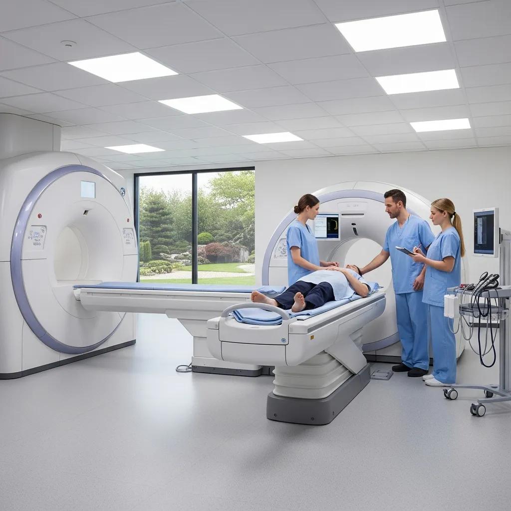
Computed Tomography (CT) acquires cross‑sectional images by rotating an X‑ray source and detectors around the patient while reconstruction software builds thin slices of anatomy. CT delivers high spatial resolution quickly, which is why it’s the go‑to for emergency trauma, lung and abdominal assessment, and vascular studies like CT angiography. Protocols are tailored to the clinical question and may use iodinated contrast to highlight vessels and soft tissues; modern scanners also use iterative reconstruction and other software tools to reduce dose. During a CT you lie on a motorised table that passes through the gantry, and you may be asked to hold your breath for chest or abdominal scans. If contrast is needed, staff complete safety checks beforehand.
The speed of CT makes it essential in emergency care, and that influences patient preparation and workflow. Low‑dose protocols and targeted scanning help keep radiation exposure as low as reasonably achievable without losing diagnostic value — we cover dose‑reduction strategies and evolving scanner technology later in this guide.
What is MRI technology and how is it used in diagnosis?
Magnetic Resonance Imaging (MRI) uses strong magnetic fields and radiofrequency pulses to elicit tissue‑specific signals that coils detect and software reconstructs into high‑contrast images. MRI gives excellent soft‑tissue contrast without ionising radiation, making it especially useful for the brain, spinal cord, joints and many abdominal applications where tissue characterisation matters. Examinations use different pulse sequences to highlight fluid, fat or tissue structure, and protocols are tailored to the clinical question to maximise diagnostic value. Patients are screened for implants, metallic fragments and claustrophobia; ear protection, open‑bore systems and sedation pathways are available to improve comfort and safety.
Because MRI relies on magnetism rather than X‑rays, it complements CT when radiation exposure or bony detail are less helpful. The modality’s safety profile and sequence versatility make it central to many diagnostic pathways and a key area of learning during training and placements.
Life Medical Imaging Central Coast uses CT, MRI, ultrasound and digital X‑ray across our sites on the Central Coast to support diagnostic care for patients and referrers. Our service pages and education resources offer practical preparation tips and appointment details so patients and referrers know what to expect before a scan.
How can you pursue a career in medical imaging?

Careers in medical imaging typically follow a structured path of academic study, supervised clinical placements and professional registration — details vary by country. Radiographers and sonographers usually complete degree‑level training that combines university teaching with hands‑on placements to build technical skills and patient care competency. After qualifying, registration with the relevant professional body is normally required to practise; continuing professional development and postgraduate options help you specialise or advance into senior roles.
This table links common educational routes to everyday clinical duties and highlights why placements and registration are essential steps toward independent practice.
Typical steps to become a registered imaging professional are practical milestones you can plan for:
- Meet academic entry requirements: Achieve prerequisite subjects and grades for your chosen undergraduate or postgraduate programme.
- Complete accredited training: Enrol in a recognised imaging degree or conversion course with supervised clinical placements.
- Undertake supervised clinical placements: Build hands‑on experience in hospital or clinic settings under qualified mentors.
- Apply for professional registration: Register with your national regulator or professional body and maintain CPD to practise.
These steps show the usual progression from applicant to practising professional and where choices — such as elective modules or placement types — shape future specialisations. Clinics play an important role in supporting placements and offering insight into day‑to‑day practice.
Life Medical Imaging Central Coast provides educational information, FAQs and referrer resources to help prospective students understand daily practice and local placement opportunities. Our materials are designed to complement accredited university programmes and guide students and referrers toward suitable clinical experiences.
What are the latest advancements in diagnostic imaging technology?
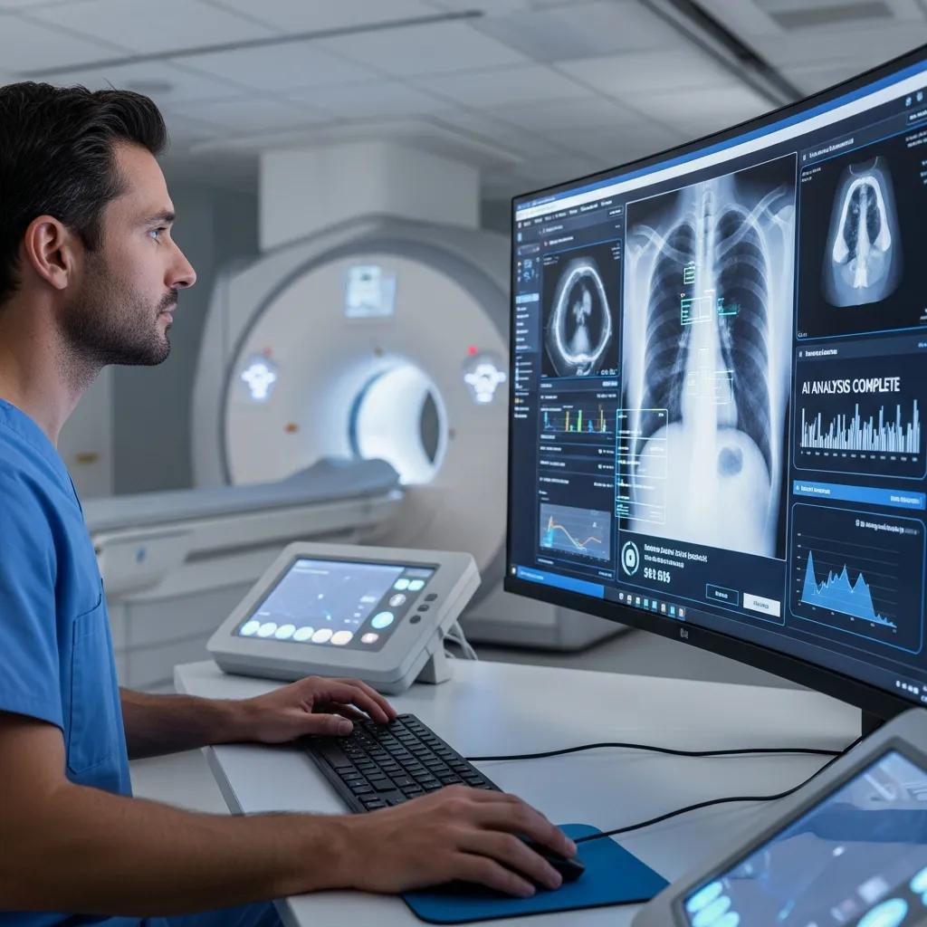
Recent advances in diagnostic imaging focus on artificial intelligence, low‑dose techniques, higher‑definition detectors and workflow automation — all aimed at improving accuracy, speed and patient safety. AI tools now help with image triage, automated measurements and quality checks, while hardware and software improvements reduce radiation dose in CT and X‑ray without compromising image quality. These changes affect how clinicians work and how students learn, increasing the need for basic computational literacy and familiarity with dose‑optimised protocols.
This summary shows how technical improvements translate into measurable clinical gains and why modern curricula increasingly cover these topics.
AI supports imaging workflows by automating repetitive tasks, aiding detection and quantification, and prioritising urgent studies for reporting. Typical AI uses include automated organ or bone segmentation, pulmonary nodule detection and vessel lumen measurements; these tools speed workflows and improve consistency but need validation and human oversight to manage bias and clinical relevance. Ethical and regulatory frameworks shape deployment, so students should learn both capabilities and limitations to use AI safely.
- AI can flag urgent cases early, reducing time to critical intervention.
- AI standardises measurements, which helps track disease progression reliably.
- AI can reduce routine reporting load, freeing clinicians to focus on complex interpretation.
These points highlight practical AI benefits while stressing the need for clinical governance and ongoing validation.
AI in Radiology: Workflow automation, accuracy and efficiency gains
Artificial intelligence is rapidly transforming diagnostic and interventional radiology, with growing regulatory approvals and clinical adoption. Integration varies by modality and procedure. This structured narrative review compares four representative workflows — MRI and CT screening, coronary stenting and liver cryoablation — to quantify automation readiness, accuracy improvements and efficiency gains, and to highlight future opportunities for AI‑driven workflow optimisation.
AI in radiology and interventions: a structured narrative review of workflow automation, accuracy, and efficiency gains of today and what’s coming, M Friebe, 2025
AI integration in radiology workflows is a major development area, with differing maturity across research and production environments.
Integrating AI into radiology workflow: maturity levels
A framework for integrating AI into radiology workflows, describing research, production and feedback maturity levels and how they affect clinical deployment and monitoring.
Integrating AI into radiology workflow: levels of research, production, and feedback maturity, E Dikici, 2020
Low‑dose imaging reduces patient radiation through hardware design, optimised acquisition protocols and advanced reconstruction. In CT and digital X‑ray, strategies such as tighter collimation, automated exposure control and iterative reconstruction keep image quality high at lower doses. These benefits are especially important in screening programmes, paediatric imaging and patients who need repeated follow‑up, where cumulative exposure matters. Knowing how dose optimisation works helps with patient consent, education and equipment selection during placements.
Life Medical Imaging Central Coast notes the clinical adoption of low‑dose and high‑definition equipment across our centres, showing how local clinics implement these innovations to improve safety and diagnostic detail. This is an example of clinical uptake rather than an endorsement of specific vendors, and it reinforces the value of hands‑on experience with modern scanners during training.
What does a medical imaging technologist do?
A medical imaging technologist — commonly called a radiographer or sonographer — acquires diagnostic images, keeps patients safe and comfortable, and performs quality checks so images are fit for interpretation. The role blends technical operation of advanced equipment with direct patient care: taking histories, positioning patients and monitoring them during procedures. Technologists run equipment QA, manage radiation safety where relevant, and liaise with radiologists and referrers to ensure imaging answers the clinical question. The list below summarises typical day‑to‑day skills expected on shift.
Typical daily responsibilities for imaging technologists include:
- Patient preparation and screening: Confirm identity, clinical indication, and check for implants or allergies.
- Image acquisition and protocol management: Choose the right protocol, position patients, operate imaging systems and manage contrast when indicated.
- Quality control and documentation: Review image quality, complete examination records and perform routine equipment checks.
- Multidisciplinary communication: Discuss urgent findings with radiologists, coordinate with referring clinicians and join MDTs as required.
What are the daily responsibilities of a radiographer?
Radiographers carry out a sequence of tasks to ensure diagnostic images are captured safely and to a high standard. They start with patient identification, history taking and safety screening, then select the appropriate protocol and position the patient precisely. They set equipment parameters to balance image quality with dose or scan time. After acquisition, radiographers check image quality, document the study in the imaging information system and — where local practice allows — communicate urgent findings to radiologists or the referrer. The role also includes routine equipment checks and participation in quality assurance to maintain diagnostic standards. Daily work ranges from routine chest X‑rays to complex CT protocols, which is why supervised placements are essential for building competence and decision‑making.
The global evolution of diagnostic radiography shows varied career pathways and the importance of accreditation for professional development.
Global evolution of diagnostic radiography and career pathways
Diagnostic radiography continues to evolve across the UK, NHS and internationally. Accreditation pathways support radiographers as they develop advanced practice and specialisations.
A global evaluation of advanced practice recognition pathways for diagnostic radiography, J Hewis, 2025
How do radiologists and radiographers collaborate?
Radiographers and radiologists work in a coordinated workflow where acquisition and interpretation are separate but interdependent. Radiographers make sure images meet diagnostic standards and provide clear clinical context to the reporting radiologist through accurate referrals and focused imaging protocols. Radiologists interpret the images, issue reports and advise on further imaging or interventions. Urgent or unexpected findings prompt direct communication and faster reporting. Collaboration also extends to interventional procedures and multidisciplinary meetings, where technologists support image‑guided work and contribute to procedural safety.
How can patients prepare for different medical imaging procedures?
Preparation varies by modality, clinical question and whether contrast is required. Clear pre‑appointment instructions help improve comfort, image quality and safety. Good preparation reduces the chance of cancellations or repeat scans and increases the chance of getting diagnostically useful images on the first visit. Below are practical, modality‑specific checklists and tips for patients and referrers.
Patients can use the following preparation checklist for common imaging types:
- For CT scans: Tell us about allergies or kidney problems, follow fasting instructions if contrast is planned, and wear loose clothing with no metal.
- For MRI scans: Remove all metal items, disclose implants or devices, let staff know about claustrophobia and follow fasting instructions for certain abdominal studies.
- For ultrasound exams: Follow bladder or fasting instructions for pelvic and abdominal scans, and wear comfortable two‑piece clothing for easy access.
What should you know before a CT or MRI scan?
Before a CT or MRI, confirm the clinical reason for the scan, disclose allergies and medical history, and follow any fasting or medication advice from your referrer or clinic. For CT with contrast, staff will check kidney function and contrast allergy history; for MRI, screening for implants, pacemakers or metal fragments is mandatory because of the strong magnetic field. During the scan you’ll lie on a motorised table and may be asked to hold your breath or receive contrast injections; staff monitor patients throughout and provide ear protection for MRI noise. Most people return to normal activities afterwards, though those given sedation or contrast may have a short observation period for safety.
Clear communication between referrer, clinic and patient reduces the likelihood of rescheduling and keeps diagnostic pathways efficient.
How is ultrasound imaging performed and what are its safety considerations?
Ultrasound uses high‑frequency sound waves from a transducer to produce real‑time images of organs and tissues. A water‑based gel improves contact between the probe and skin, and exams usually take 15–45 minutes depending on complexity. Patients may be asked to change position or briefly hold their breath to get better views. Ultrasound is safe for most people because it uses no ionising radiation, making it the preferred choice in obstetrics, many paediatric cases and repeat follow‑ups. Specific preparation such as a full bladder for pelvic scans or fasting for some abdominal studies helps improve image quality.
Because ultrasound is operator dependent, the sonographer’s skill and experience significantly influence diagnostic quality — another reason why accredited training and supervised placements matter.
Where can you find reliable educational resources about medical imaging technology?
Reliable resources include accredited university programmes, professional bodies and focused short courses that combine theoretical learning with supervised clinical placements. Prospective students should prioritise programmes recognised by national regulators and professional organisations, and check placement arrangements and assessment methods when comparing courses. Online resources such as professional society guidance, university course pages and curated MOOCs can supplement formal training, but hands‑on clinical experience remains essential for competence.
The table below lists resource types, what they offer and how they help students choose the right pathway.
This mapping helps you compare different educational formats and shows why accredited placements are central to professional readiness.
Which external institutions offer accredited imaging technology courses?
Many universities and higher‑education providers in the UK and Australia offer accredited diagnostic radiography and imaging programmes, typically validated by regional regulators and professional bodies. Prospective applicants should check course accreditation with relevant registries and confirm that clinical placement arrangements meet training requirements. When comparing institutions, look for clear curriculum outlines, placement durations and assessment methods, and read student or placement testimonials about clinical experience. Short courses and CPD modules can complement degree training by focusing on advanced modalities, AI literacy and dose‑optimised protocols.
Choosing a course means balancing academic content, placement quality and professional recognition to make the transition into practice as smooth as possible.
How does Life Medical Imaging support career and educational insights?
Life Medical Imaging Central Coast offers an information hub, education links and FAQs to help students, referrers and patients understand imaging practice and appointment logistics. While we don’t run accredited courses directly, our resources explain procedural expectations, modality capabilities and referral pathways, and can help prospective trainees find suitable clinical placement opportunities and local referrer contacts. These materials are intended to complement accredited university programmes by giving practical insight into everyday imaging workflows and patient preparation.
Students and referrers can use our educational information alongside university course pages and professional body guidance to build a rounded view of training and clinical expectations. For bookings or enquiries about procedural preparation and referrer resources, Life Medical Imaging Central Coast provides patient and referrer information to guide next steps.
For appointments or enquiries about services, patients and referrers can contact Life Medical Imaging Central Coast through the clinic’s published patient and referrer resources and online booking pathways. This guide focuses on clinical education and preparation, with our local resources offered as a practical complement to accredited courses and national professional advice.
Frequently Asked Questions
What qualifications are needed to become a medical imaging technologist?
Most imaging technologists complete an accredited degree in diagnostic radiography or sonography that combines theory with clinical placements. After graduation, registration with the appropriate professional body — for example the HCPC in the UK — is usually required to practise. Ongoing continuing professional development (CPD) is important to keep skills current and to progress into specialised or advanced roles.
What are the common challenges faced by students in medical imaging courses?
Students commonly face the challenge of mastering technical skills and detailed anatomy while also developing confident patient communication. Clinical placements can be demanding since students apply classroom learning in live settings while maintaining patient safety. Rapid technological change, including AI and low‑dose techniques, means continuous learning is necessary, which some students find overwhelming at times.
How does AI impact the future of medical imaging?
AI is changing medical imaging by improving diagnostic consistency and speeding workflows. It automates routine tasks such as image triage and measurements, helping reporting teams focus on complex cases. AI can also improve image quality and reduce repeat scans through smarter protocols. As AI matures, it will play an increasing role in personalised imaging care, so future imaging professionals should understand how to use these tools safely and critically.
What safety measures are in place for patients undergoing imaging procedures?
Safety measures include thorough pre‑scan screening for risks such as contrast allergies or metal implants, strict radiation protection protocols for CT and X‑ray, and continuous monitoring during procedures. Technologists follow established guidelines to minimise exposure and ensure patient comfort. Clear pre‑scan instructions and good communication also reduce anxiety and improve safety.
What role do medical imaging technologists play in patient care?
Imaging technologists ensure diagnostic images are acquired safely and accurately. They prepare and reassure patients, operate complex equipment, choose appropriate protocols and verify image quality for reporting. Their technical skills and patient care make a direct contribution to diagnosis and to the overall patient experience.
How can prospective students find clinical placement opportunities in medical imaging?
Many accredited programmes arrange placements through established hospital and clinic partnerships. Students can also network with professionals, attend career events and join relevant societies for leads. Contacting local imaging centres directly — including Life Medical Imaging Central Coast — can provide information about placement availability and the application process.
Conclusion
Understanding medical imaging technology opens doors to rewarding healthcare careers and equips students with practical skills for patient care and diagnostic accuracy. This guide outlines the main modalities, career pathways and emerging developments, while emphasising the value of accredited training and hands‑on experience. If you’re ready to take the next step, explore our resources, speak with local clinics and connect with university programmes to plan your path into medical imaging.