Comprehensive Imaging Requirements — How we ensure quality, safety and compliance
Medical imaging requirements set the technical, clinical and regulatory standards that make diagnostic imaging reliable and safe. This guide outlines what those requirements cover — from accreditation and radiation protection to equipment specs, staff training and patient preparation — so clinicians, referrers and patients can see how consistent, high-quality imaging is delivered. Poorly defined practice increases repeat scans, diagnostic uncertainty and unnecessary exposure; meeting recognised standards reduces those risks by aligning everyday clinic processes with national codes and professional guidance. Below we compare key Australian frameworks with UK equivalents, summarise core patient-safety procedures and dose-optimisation approaches, detail equipment and maintenance expectations, explain ionising radiation regulation and referral justification, and offer clear preparation steps for patients and referrers. Throughout, we show how clinic-level systems — policies, audits, qualified staff and current technology — turn high-level requirements into safer, better imaging.
What are the key medical imaging quality standards in the UK and Australia?
Quality standards for medical imaging are formal documents and inspection frameworks that set minimum expectations for clinical governance, equipment performance, dose management and staff competence. Their purpose is simple: ensure imaging is justified, optimised and reproducible so diagnostic confidence is high and risks are minimised. In Australia, RANZCR guidance and ARPANSA radiation safety codes are central; in the UK, the Royal College of Radiologists and national inspection frameworks provide comparable direction. Both systems stress accreditation, audit and continuous improvement. Knowing the practical changes these standards require helps patients and referrers judge service quality and helps clinics prepare for inspections and ongoing audits.
Improving quality and safety in medical imaging through systematic evaluation
In high‑income health systems, average exposure from medical imaging can equal about 80% of natural background radiation, yet doses for the same procedure can vary significantly between facilities. That variability — and the risks it implies — supports the need for systematic evaluation of radiology services to raise quality and protect patients.
Clinical audit and practice accreditation, 2013
Typical clinic-level actions that map to these standards include:
- Documented policies for justification, optimisation and informed consent.
- Regular equipment calibration and QA logs supported by scheduled preventive maintenance.
- Clear evidence of staff qualifications, CPD and competency assessments.
Those practical steps translate regulatory aims into day‑to‑day practice. Below we examine how UK and Australian guidance compare in more detail.
How do UK diagnostic imaging guidelines compare to Australian standards?
UK guidelines emphasise standardised protocols, inspection frameworks and patient‑centred governance. Australian regulation focuses strongly on radiation protection via ARPANSA, supported by clinical guidance from RANZCR; both require documented justification and optimisation. The UK approach often uses national inspection regimes to enforce standards, while Australia combines radiation safety codes, professional college guidance and accreditation processes to reach similar outcomes. For clinics, this means implementing local policies that cover both technical checks (equipment performance, dose reference levels) and governance expectations (audit schedules, patient information).
For patients and referrers the practical message is consistent: clinics should maintain traceable QA records, accessible pathways for clinical justification and clear documentation of staff competency. Those measures reduce variation between sites and show how accreditation is interpreted in everyday clinical care.
What role does NATA accreditation play in imaging quality and compliance?
NATA accreditation evaluates technical and laboratory services against recognised standards and provides independent assurance that a clinic’s quality systems, equipment calibration and staff competence have been reviewed. Accreditation inspects protocols, maintenance records and competency evidence, giving patients and referrers confidence that procedures, measurements and reporting meet accepted benchmarks.
Clinics retain accreditation by planning recurrent audits, recording corrective actions and keeping thorough QA logs. That ongoing cycle supports continuous improvement and makes accreditation a meaningful, maintained assurance — not a one‑off certificate.
How is patient safety ensured through radiology patient‑safety protocols?
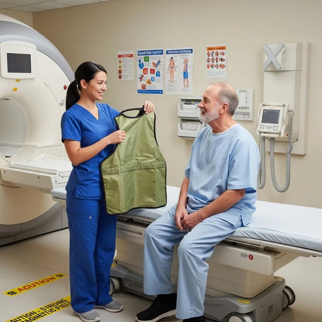
Patient safety in radiology combines technical radiation protection, standardised procedural checks and clinical governance to prevent avoidable harm while maximising diagnostic value. Core principles such as ALARA (as low as reasonably achievable), formal justification of each exam and routine dose monitoring create a system where scans are performed only when clinically needed and at the lowest reasonable exposure. Operational measures — multi‑point patient identification, documented consent and pregnancy screening — reduce procedural errors and unexpected risk. Together these systems ensure each imaging episode is traceable, appropriate and recorded for quality review.
Common safety protocols in contemporary imaging services include:
- ALARA‑based optimisation with documented dose‑reference values and optimisation plans.
- Multi‑point patient identification, written informed consent and pregnancy checks when relevant.
- Routine dose audits, incident reporting systems and periodic staff radiation monitoring.
These protocols protect patients and staff and inform equipment and workflow choices that lower exposure while preserving diagnostic performance.
Life Medical Imaging Central Coast follows these patient‑safety principles, using accredited quality systems, trained staff and evidence‑based optimisation to minimise dose and improve image quality. To book or ask about our safety measures call Life Medical Imaging Central Coast on 02 4326 7000. Referrers can discuss pathways and eReferral options directly with the clinic.
What are radiation dose optimisation practices in medical imaging?

Dose optimisation means choosing scanner settings, protocols and shielding so the study answers the clinical question with the lowest reasonable ionising dose. Practical measures include:
- Tailoring protocols to body size and the specific clinical question.
- Using paediatric‑specific dose settings for children.
- Applying iterative reconstruction algorithms on CT to reduce noise at lower doses.
- Collimation and appropriate shielding for X‑rays.
- Optimising contrast and slice parameters for the indication.
Automated dose‑monitoring systems log dose metrics for each exam, enabling audit against local and national reference levels and informing protocol adjustments.
Effective optimisation relies on a data‑driven feedback loop: measure dose by protocol, benchmark results, tweak parameters and retrain staff. That cycle keeps exposures low without sacrificing diagnostic value and supports transparent communication with referrers about expected dose for specific studies.
How are patient identification and consent managed in imaging procedures?
Identification and consent use standardised, multi‑point verification and documented consent workflows that ensure the planned procedure matches the clinical question. Best practice confirms identity at booking, on arrival and immediately before imaging, alongside a review of clinical justification and pregnancy screening where applicable. Informed consent explains the purpose, benefits, likely risks (including radiation exposure) and reasonable alternatives; this is documented in the patient record and linked to the imaging request.
These steps create a clear audit trail connecting referral, clinical justification and consent to the performed study, reducing the risk of wrong‑patient or wrong‑site imaging and meeting legal and ethical obligations. Next, we consider how equipment supports these safety practices.
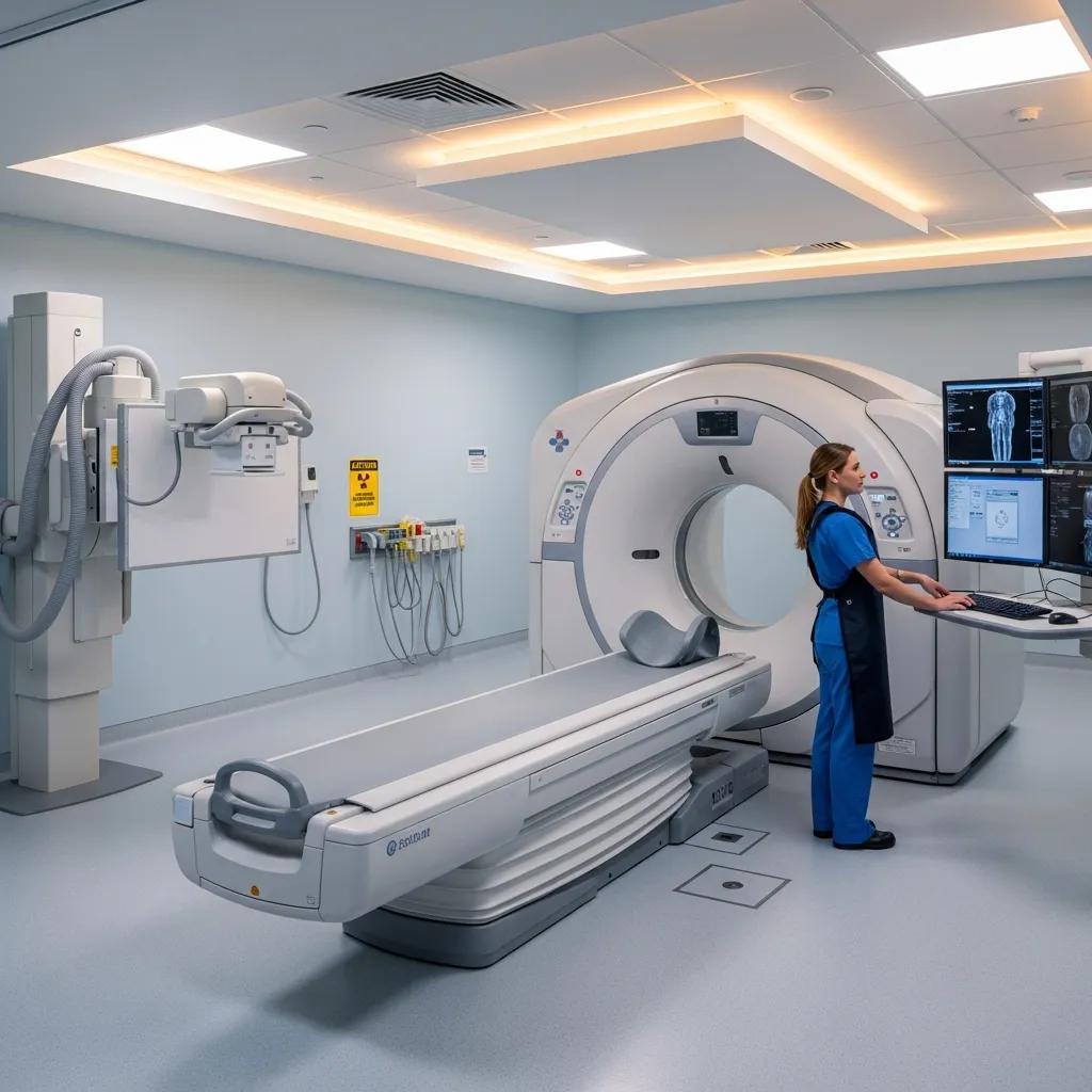
What are the diagnostic imaging equipment specifications and technology requirements?
Equipment specifications describe the technical performance needed to produce diagnostic images while supporting radiation safety and reproducibility. Important attributes include detector resolution, expected dose ranges for common CT protocols, ultrasound transducer frequency ranges, and dose‑reduction features such as iterative reconstruction or ultra‑low‑dose modes. Modern systems with high‑definition detectors and optimisation tools reduce repeat imaging and support better clinical decisions; older hardware can increase variability and maintenance needs. Regular maintenance and calibration ensure equipment stays within specification and that dose measurements remain accurate over time.
Below is a comparative table to help clinicians and informed patients compare common modalities by typical dose range, resolution and maintenance rhythm.
This comparison shows how modality choice balances diagnostic need, exposure and image detail — and how specifications guide protocol selection and procurement decisions.
Life Medical Imaging Central Coast uses “ultra‑low dose, high definition” CT and modern digital detectors to meet these specifications, reduce exposure and preserve diagnostic information. To enquire about equipment or to book an appointment, call Life Medical Imaging Central Coast on 02 4326 7000.
How does ultra‑low dose CT technology improve imaging safety?
Ultra‑low dose CT lowers ionising exposure by reducing tube current/voltage and using advanced reconstruction algorithms to preserve image quality. In practice this means acquiring less raw signal and using computation to suppress noise and enhance detail, producing images that are sufficient for many clinical questions such as surveillance, chest imaging and selected abdominal studies. This approach is especially valuable for repeat imaging, paediatric care and screening pathways where cumulative dose matters.
Successful implementation requires validated protocols and trained radiographers to maintain diagnostic confidence. When used correctly, ultra‑low dose CT significantly reduces population exposure while keeping images clinically useful.
What are the maintenance and calibration standards for imaging equipment?
Regular maintenance and calibration keep imaging equipment within manufacturer and regulatory tolerances. Typical practices include daily QC checks, scheduled medical physics calibrations, detector calibration and planned preventive maintenance visits. Recording these activities in QA logs is essential for accreditation and audit; calibration intervals follow manufacturer guidance and regulatory codes. Inadequate maintenance can increase artefacts, introduce dose measurement variability and raise the risk of misdiagnosis, while a rigorous schedule reduces downtime and preserves image fidelity.
Clearly assigned responsibilities — manufacturer service for major checks, in‑house technologists for daily QC and medical physicists for periodic calibration — create redundancy and accountability that protect patients and maintain diagnostic reliability.
What are the ionising radiation regulations affecting medical imaging?
Ionising radiation in medical imaging is regulated to protect patients and staff by requiring optimisation, justification, dose limits and monitoring. In Australia, ARPANSA radiation safety codes set out obligations for equipment performance, dose management and worker protection; RANZCR guidance complements these with clinical governance and justification principles. These frameworks require clinics to adopt local radiation‑protection policies, keep dose records and ensure staff competence so legal duties become everyday clinical practice.
Regulation drives practical clinic tasks such as setting diagnostic reference levels, using dose‑monitoring software, investigating incidents and keeping written justification for higher‑dose studies. These measures control risk and maintain transparency with referrers and patients.
How do ARPANSA radiation safety codes protect patients and staff?
ARPANSA codes require optimisation, monitoring and reporting to limit unnecessary exposure and ensure protective measures for staff and patients. Facilities must adopt ALARA practices, maintain equipment standards, run worker monitoring programs and implement safety management systems. At clinic level this translates into written radiation safety policies, routine dose audits and designated personnel who oversee compliance and training.
Those obligations — dose recording, incident reporting and regular review — make radiation governance auditable and support referral and justification processes that reduce inappropriate imaging.
This mapping makes regulatory expectations tangible and helps patients and referrers see how compliance protects safety and quality.
What are the referral guidelines and clinical justification requirements for imaging?
Clinical justification means every imaging request should state a clear clinical question and that the expected diagnostic benefit outweighs risk. Guidelines provide criteria for common presentations and suggest alternatives when appropriate.
Referrers should include relevant history, prior imaging and the diagnostic query so radiologists can choose the most suitable modality and protocol. eReferral systems and structured requests support justification by ensuring essential information is present and flagging high‑dose exams for extra review.
Appropriate referrals reduce unnecessary exposure and improve diagnostic yield. Practical examples include selecting ultrasound for a straightforward soft‑tissue issue instead of immediate CT, or noting prior studies to avoid repeats.
How are radiology teams qualified and trained to meet imaging requirements?
Radiology teams meet standards through accredited education, professional registration and ongoing competency assessment. A multidisciplinary team — radiologists, radiographers and sonographers — provides the skills for safe, high‑quality imaging. Roles are clear: radiologists interpret images and advise on protocols, radiographers operate equipment and manage dose, and sonographers perform specialised ultrasound examinations. Each role requires demonstrated competence, documented CPD and participation in local audit and peer review to keep skills current.
What training and certification do radiographers and sonographers require?
Radiographers and sonographers typically hold accredited tertiary qualifications and maintain registration or membership with relevant professional bodies, supported by documented competency assessments for specialised procedures. Certification pathways include formal education, supervised clinical training and periodic reassessment for advanced or interventional tasks. Verifiable credentials and CPD records let referrers and patients confirm staff competence.
Transparent verification processes ensure personnel performing or assisting with imaging are appropriately trained, which reduces procedural risk and improves diagnostic consistency.
How does ongoing professional development support imaging standards?
Ongoing professional development (CPD) keeps staff up to date with new protocols, technologies and safety expectations. CPD activities include case reviews, peer audits, manufacturer training and attendance at professional courses. Clinics that document CPD and link it to competency assessments provide evidence of continual improvement and a clear commitment to quality. Regular audit cycles and multidisciplinary review feed CPD, creating a feedback loop that refines protocols and improves outcomes.
This culture of continuous learning directly improves image quality and safety, strengthening the trust referrers and patients place in the imaging service.
How can patients prepare for imaging procedures to meet safety and quality standards?
Good preparation helps scans deliver the information needed while reducing risk and discomfort. Preparation varies by modality but common elements include arriving with ID and referral documentation, telling staff about medications and allergies, and following fasting or hydration instructions when required. Clear pre‑scan instructions and checklists reduce the chance of rescan, contrast reactions or incomplete studies, and allow staff to complete pregnancy screening and other safety checks before scanning.
- Bring ID and referral: Have your referral form and photo ID ready at reception.
- Disclose medical history: Tell staff about allergies, current medications and recent surgeries.
- Follow modality instructions: Observe fasting for some CTs, remove metal for MRI and wear comfortable clothing for ultrasound.
Following these steps reduces delays and supports important safety checks. Below are expectations for common modalities.
What should patients expect during different imaging scans?
Modalities offer different experiences: CT is a short, table‑based scan with brief breath‑holds and sometimes contrast; X‑ray is quick with minimal prep; ultrasound involves a sonographer using gel and a probe, often with real‑time assessment; MRI can be longer, requires stillness inside a magnet and needs metal screening. Staff will explain expected duration, sensations and any possible side effects such as contrast reactions.
Clear expectations help patients arrive prepared and keep the clinical pathway moving smoothly.
How are pregnancy screening and special safety protocols implemented?
Pregnancy screening uses standard questions at booking and on arrival to identify potential fetal exposure and to offer alternatives to ionising imaging when appropriate. If pregnancy is confirmed or suspected, clinicians favour non‑ionising options such as ultrasound or MRI where clinically suitable, or adjust protocols and shielding to minimise exposure. Urgent imaging during pregnancy balances maternal benefit and fetal risk and follows documented multidisciplinary decision‑making and consent procedures.
Documented screening and alternative‑pathway procedures protect both mother and fetus while ensuring necessary diagnostic information can still be obtained when clinically required.
Life Medical Imaging Central Coast supports patient‑centred safety through NATA‑style quality systems, qualified staff and referrer support including eReferral and secure image access. To book or enquire about scans, safety procedures or referrer workflows, contact Life Medical Imaging Central Coast on 02 4326 7000.
Frequently Asked Questions
What are the benefits of using NATA‑accredited imaging services?
NATA accreditation shows a facility has been independently assessed against strict quality standards. It confirms that quality systems, equipment calibration and staff competence have been reviewed. For patients this means greater confidence in diagnostic accuracy and safety. For clinics, accreditation promotes consistent practice and operational improvements that align care with national safety expectations.
How can patients ensure they are adequately prepared for imaging procedures?
Follow the clinic’s pre‑scan instructions, bring photo ID and your referral, and tell staff about allergies, medications and recent procedures. If fasting or hydration are required, follow those directions closely. If you’re unsure, call ahead — clear preparation reduces the chance of repeat scans and helps the appointment run smoothly.
What should patients do if they have concerns about radiation exposure during imaging?
Talk with your referring clinician or the imaging team before the scan. Ask why the test is recommended, what measures are in place to minimise dose (for example ALARA practices) and whether a non‑ionising alternative such as ultrasound or MRI is appropriate. Understanding the expected benefit and risk will help you make an informed decision.
What role do radiologists play in ensuring imaging quality?
Radiologists interpret images and advise on the most appropriate protocols for each clinical question. They review justification for requests, contribute to quality assurance through audits and peer review, and help keep reporting standards high. Their clinical input is central to both patient safety and diagnostic accuracy.
How do imaging facilities maintain compliance with safety regulations?
Facilities comply through documented policies, regular audits, equipment maintenance and staff training. They implement safety management systems that include routine QC, calibration and quality assurance checks, and ensure staff undertake CPD. By following ARPANSA and RANZCR guidance, clinics keep services safe and compliant.
What are the implications of not following imaging safety protocols?
Failing to follow safety protocols raises the risk of misdiagnosis, unnecessary radiation exposure and patient harm. It can lead to repeat imaging, delayed care and possible regulatory consequences such as loss of accreditation. Maintaining strict protocols is essential to protect patients and clinic reputation.
How can patients access their imaging results and reports?
Patients can access results through the clinic’s patient portal where available, or by contacting the clinic directly. Many services offer secure online access to reports and images, and patients can request copies to share with other clinicians. Discussing results with the referring clinician helps interpret findings and plan next steps in care.
Conclusion
Clear, enforced imaging requirements are the foundation of safe, accurate diagnostic care. When clinics follow recognised standards and maintain robust systems — from accreditation and equipment maintenance to staff training and patient preparation — they reduce risk and build confidence for referrers and patients alike. If you’d like to learn more about how these practices support your imaging experience, explore our resources or contact us for further information. Together we ensure every imaging study is performed with care, precision and patient safety at the centre.