Comprehensive Hospital Imaging on the Central Coast: Diagnostic Tests, Preparation and Patient-Focused Care
Hospital imaging brings together several diagnostic technologies that let clinicians look inside the body to diagnose conditions, plan treatment and monitor recovery. This guide explains how the common imaging tests work, what you should do to prepare, and how imaging supports care across the Central Coast. You’ll find clear, plain‑language descriptions of MRI, CT, ultrasound, X‑ray and DEXA scans, practical preparation checklists for each test, and an overview of how imaging supports children’s and women’s health. We also cover how private imaging costs are structured and the usual booking and referral pathways for private scans. Expect concise lists, comparison tables and actionable CT/MRI preparation steps designed to answer common questions about hospital imaging and private MRI bookings.
What Are the Main Types of Hospital Imaging Services Available on the Central Coast?
Hospital imaging includes several distinct modalities, each using different physical principles to answer clinical questions efficiently and accurately. MRI uses magnetic fields and radiofrequency to produce detailed soft‑tissue images without ionising radiation. CT uses X‑rays to create rapid cross‑sectional images, ideal for bone and acute conditions. Ultrasound uses sound waves for real‑time imaging of organs and pregnancy. Digital X‑ray captures quick 2D images for bone and chest assessment, and DEXA measures bone density to assess osteoporosis risk. Knowing these differences helps you understand which test your doctor may request and why. The next section looks more closely at MRI and CT so you can see how each suits specific clinical needs.
Each modality has strengths that guide clinical decisions:
- MRI — superior soft tissue contrast for brain, spine and joint assessment.
- CT — fast cross‑sectional imaging for trauma, lungs and emergency assessment.
- Ultrasound — real‑time, portable imaging for pregnancy, abdominal and vascular studies.
- Digital X‑ray — quick, low‑dose 2D images for bones and chest screening.
- DEXA — precise bone mineral density measurement for osteoporosis management.
These differences determine which test a referring doctor will choose and what preparation is needed before your scan.
The table below summarises how each modality works and where it’s most useful.
This comparison clarifies why clinicians select particular tests and what patients can expect in terms of safety and diagnostic focus.
How Do MRI Scans Work and What Conditions Do They Diagnose?
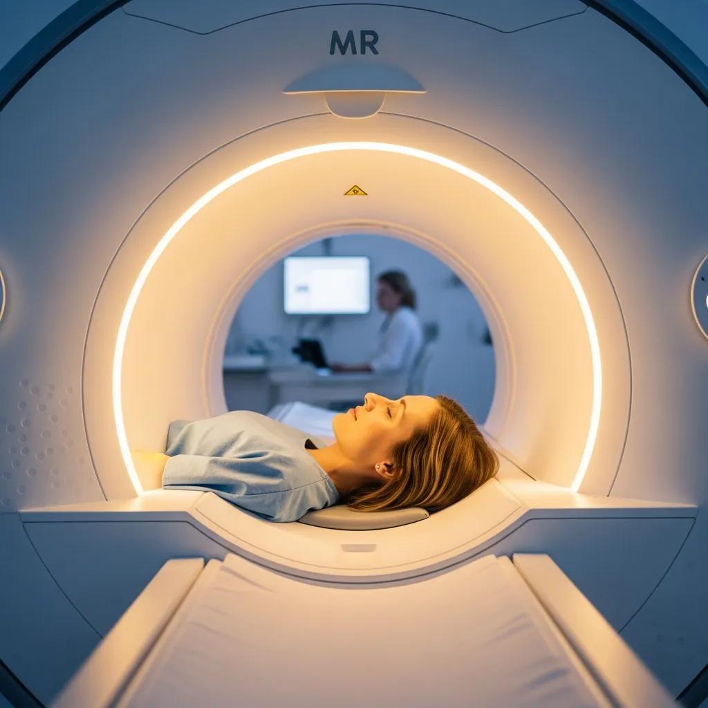
MRI produces detailed internal images by aligning hydrogen atoms in the body with a powerful magnetic field and then briefly disturbing that alignment with radiofrequency pulses. The signals returned are reconstructed into images that highlight soft tissue contrast. That makes MRI especially valuable for neurological conditions (for example, stroke and tumours), spinal assessments, joint and soft tissue injuries, and many cancer indications. MRI can be performed with or without intravenous gadolinium‑based contrast when vascular detail or lesion characterisation is needed; contrast use requires consideration of kidney function and informed consent. Patients with certain implants, pacemakers or severe claustrophobia need assessment before MRI; modern scanners, safety checks and open designs help reduce anxiety and allow many patients to be scanned safely. MRI’s soft‑tissue detail explains why it’s often the preferred test when detailed anatomy and tissue character are required.
Contrast Mechanisms in MR Imaging Explained
This paper gives an accessible overview of tissue‑specific parameters and how MR sequences use those parameters to distinguish normal and pathological tissue. It explains key factors such as proton density and relaxation times (T1, T2 and T2*), and how sequence choices — including magnetisation preparation, fat suppression and diffusion weighting — influence image contrast. The review also touches on advanced approaches like magnetisation transfer imaging and the role of functional parameters such as diffusion and perfusion in clinical studies.
Contrast mechanisms in MR imaging, 1999
What Are the Benefits and Risks of CT Scans in Hospital Imaging?
CT uses a rotating X‑ray tube and detector array to produce fast cross‑sectional images, making it extremely useful in acute settings such as trauma, suspected internal bleeding, lung disease assessment and detailed bone imaging. Cardiac CT angiography is a specialised application that visualises coronary arteries and cardiac anatomy to assist cardiology teams. The main risks are exposure to ionising radiation and potential reactions to iodinated contrast. Modern scanners and protocols — including ultra‑low dose CT techniques — significantly reduce radiation while maintaining diagnostic quality. Patients with contrast allergies or impaired kidney function may need tailored protocols, pre‑medication or alternative imaging. These safety measures explain CT’s central role in rapid diagnosis and why dose‑reduction advances are important.
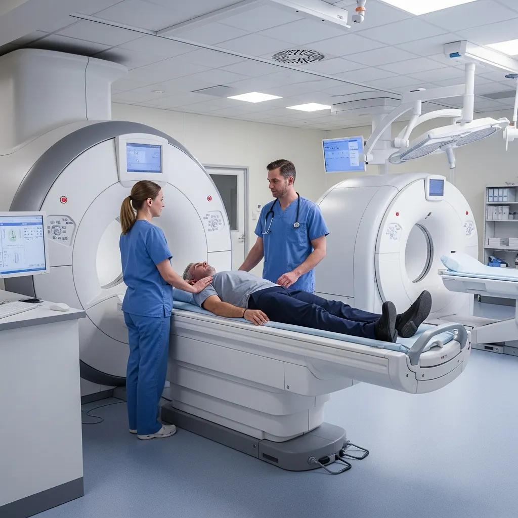
How Can Patients Prepare for Common Imaging Procedures?
Good preparation improves image quality, reduces repeat scans and speeds clinical care. Preparation varies by modality: MRI needs implant checks and removal of metal; CT may require fasting if contrast is planned; ultrasound often depends on bladder fullness or fasting for abdominal studies; most X‑rays need only removal of metal and loose clothing; and DEXA usually requires avoiding calcium supplements before the scan. Also bring required documents and any prior imaging, and tell staff if you are pregnant or have kidney concerns. The sections below provide step‑by‑step checklists for MRI/CT and ultrasound/X‑ray so you can arrive prepared and relaxed.
Bring these items to your appointment to help the visit run smoothly:
- Your referral from the referring clinician and any prior imaging reports.
- Photo identification and payment or insurance details if needed.
- A list of current medications and any known allergies, especially to contrast agents.
- Comfortable, metal‑free clothing and removable jewellery.
Having these ready shortens check‑in and helps staff confirm safety requirements before scanning.
This table is a quick reference — always follow the specific instructions supplied by your imaging provider to avoid delays and ensure the best image quality.
What Is the Step-by-Step Preparation for MRI and CT Scans?
Preparing for MRI and CT follows a short checklist that protects safety and image quality. First, confirm your referral, list any implants or devices and complete the safety questionnaire — this determines suitability and scanner choice. Second, follow fasting instructions if contrast is planned and check medication advice with your GP (for example, regarding metformin). Third, remove all metal objects and wear comfortable clothing; tell staff if you have claustrophobia so they can offer support. Finally, bring prior imaging or reports to help the radiologist compare studies and provide a more accurate interpretation. These steps reduce the chance of cancellations and improve diagnostic yield.
How Should Patients Prepare for Ultrasound and X-ray Imaging?
Ultrasound and X‑ray preparation is usually simple but differs by exam. For abdominal ultrasound, fasting (typically 6–8 hours) reduces bowel gas and improves organ visualisation. For pelvic and obstetric scans, a comfortably full bladder provides a better acoustic window. X‑rays rarely need special preparation beyond removing metal and wearing loose clothing, though chest X‑rays may require a deep inhalation during the image. For children, preparation focuses on distraction, parent involvement where safe, and age‑appropriate explanation to minimise movement. Clear communication with staff helps ensure fast, accurate exams and reduces the need for repeats.
What Specialized Hospital Imaging Services Are Offered for Children and Women?
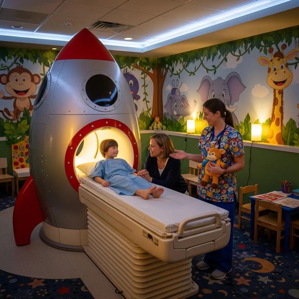
Specialised imaging for children and women uses adapted techniques, equipment and staff training to deliver safe, accurate care. Paediatric imaging prioritises dose reduction, child‑friendly communication and minimising distress through distraction or, when necessary, sedation; ultrasound and low‑dose CT protocols are commonly used. Women’s imaging includes obstetric 3D/4D scans, diagnostic breast ultrasound and gynaecological pelvic scans to assess conditions such as fibroids and ovarian cysts. These services combine technical protocol adjustments with staff skilled in communicating with patients and carers, ensuring sensitive questions are answered while safety is maintained.
Paediatric imaging teams focus on radiation safety and compassionate approaches to support families. Technologists use paediatric dose protocols and choose non‑ionising modalities like ultrasound or MRI when clinically appropriate. Communication strategies include age‑appropriate explanation, parental involvement during the scan and distraction tools to avoid sedation where possible. Common paediatric studies include head ultrasound in infants, abdominal ultrasound and, when necessary, trauma CT performed with strict dose‑reduction measures. These child‑centred practices improve cooperation, reduce anxiety and preserve diagnostic quality.
Women’s imaging services cover routine obstetric monitoring through to targeted breast and gynaecological assessments tailored to clinical need. 3D/4D obstetric imaging can supplement standard scans by offering volumetric views for parental bonding and selected anomaly assessments at recommended timepoints. Breast ultrasound is a key diagnostic tool that complements clinical examination and mammography. Gynaecological ultrasound evaluates conditions such as fibroids, ovarian cysts and suspected endometriosis using transabdominal or transvaginal approaches as appropriate. Staff trained in women’s health imaging ensure sensitivity, technical quality and clear communication of results.
How Does Hospital Imaging Support Diagnosis and Treatment Monitoring?
Imaging plays a central role in diagnosis and ongoing care — from confirming acute injury to staging cancer and tracking treatment response. Images give clinicians objective information to confirm or rule out conditions, plan surgery or radiotherapy, and monitor how disease evolves. Imaging also enables minimally invasive interventions such as image‑guided biopsies and injections, which can diagnose and treat with less morbidity than open surgery. The sections that follow explain radiologist reporting and common interventional procedures so you can see how imaging informs clinical decisions.
Radiologists interpret images in the context of clinical history and prior studies to produce a structured report that guides the treating clinician. A typical report lists findings, compares with any earlier imaging, and finishes with an impression summarising likely diagnoses and recommended follow‑up. Reports are prioritised by urgency and communicated to referrers so management decisions can proceed promptly. Providing a clear clinical history and previous imaging helps radiologists give the most accurate interpretation.
Image‑guided procedures use real‑time imaging to target tissues for biopsy, aspiration or therapeutic injection, reducing the need for open surgery. Common procedures include ultrasound or CT‑guided biopsies for pathology, spinal and joint injections for pain relief, and aspiration of fluid collections. These procedures usually require focused preparation, local anaesthesia and informed consent. Their advantages include shorter recovery times and precise targeting of diseased tissue, making them central to many diagnostic and therapeutic pathways.
What Are the Costs and Booking Options for Private MRI and Other Imaging Services?
Private imaging costs depend on several factors: the type and complexity of the scan, whether contrast or specialised sequences are required, professional reporting fees, and whether rebates or private insurance apply. Rather than list fixed prices, it helps to understand the cost components so you can get an accurate estimate for your situation. To obtain a personalised quote, contact the provider with the requested scan type and clinical details so staff can confirm if contrast, specialised protocols or extended reporting are needed.
Private imaging fees typically include a technical/service charge for the scan, a radiologist’s report fee, and possible extra charges for contrast agents or interventional procedures. Insurance and Medicare rebate eligibility depend on the clinical indication and provider billing practice; check your cover and ask the clinic for an itemised estimate that clarifies rebateable components. Many clinics can discuss concessions or payment plans if needed. Understanding these components lets patients request clear pricing information and avoid unexpected costs.
Patients can book private imaging via online booking systems, over the phone or by presenting a valid referral from a GP or specialist. Referrers can often use eReferral workflows; images and reports are returned directly to the referrer to speed clinical care. For practical assistance, contact the local imaging provider by phone or use their online booking tool to choose a convenient clinic and appointment time. Bringing your referral, identification and any prior imaging on the day helps ensure a smooth check‑in and accurate reporting.
Locally, Life Medical Imaging Central Coast operates multiple clinics across the region and offers online appointment booking and phone enquiries. To get a cost estimate or discuss referral requirements, call the clinic and provide the scan type and clinical indication so staff can outline likely fees and rebate considerations. Ask for pre‑appointment preparation guidance and confirmation of required documentation to avoid delays and make sure the appropriate protocol is scheduled.
How Is MRI Scan Cost Structured in Private Hospital Imaging?
MRI fees reflect equipment use, technologist time and the radiologist’s professional interpretation; contrast studies add material and monitoring costs. Insurance rebates and Medicare eligibility vary by indication, so your out‑of‑pocket amount depends on clinical necessity and billing classification. Ask the imaging provider for an itemised quote that separates technical and professional fees and lists any additional charges for contrast or specialised reporting. This breakdown helps you compare options, check your cover and plan for private imaging costs.
How Can Patients Book Appointments and Obtain Referrals for Imaging?
Booking normally requires a valid referral from a GP or specialist and can be completed online or by telephone. For referring clinicians, eReferral pathways are commonly accepted — include clinical history and the specific scan requested to avoid delays. When booking, confirm preparation instructions, estimated duration, arrival time and any fasting or medication guidance. After your scan, reports are usually sent to the referrer and your clinician will discuss results and next steps with you.
Life Medical Imaging Central Coast accepts online bookings and phone enquiries for appointments across its Central Coast clinics, including Bateau Bay, Killarney Vale, Umina Beach and Erina. Call the clinic to confirm referral requirements and to request a cost estimate tailored to the specific scan and any contrast or intervention required.
Why Choose Accredited Hospital Imaging Services on the Central Coast?
Accreditation and modern equipment directly affect diagnostic accuracy, safety and consistency. NATA accreditation shows adherence to quality assurance, equipment calibration and staff competency standards — factors that reduce variability and increase confidence in reported findings. Advanced technology such as ultra‑low dose CT protocols and 3D/4D ultrasound improves image clarity while lowering patient risk, and specialist services for women’s and cardiac imaging add focused expertise for complex cases. Below are tangible benefits patients and referrers should expect from accredited, well‑equipped providers.
- Consistent image quality through routine calibration and quality assurance checks.
- Credentialed staff ensuring skilled image acquisition and reporting.
- Documented processes for safety, radiation dose tracking and incident management.
These quality features lead to more reliable diagnoses and smoother care pathways, and they explain why accreditation matters to patients and clinicians.
What Does NATA Accreditation Mean for Imaging Quality and Safety?
NATA accreditation evaluates technical competence, equipment calibration, quality systems and staff training to ensure diagnostic services meet national standards. For patients, this means protocols are audited, machines are maintained to consistent specifications and staff undergo competency assessments — all of which reduce the risk of technical error and inconsistent image quality. Accreditation also requires documented procedures for reporting accuracy and incident management, giving extra reassurance for clinicians and patients who rely on imaging for important decisions. Choosing an accredited facility supports reproducible, safe imaging results.
How Does Advanced Technology Enhance Diagnostic Accuracy and Patient Care?
Advanced imaging improves diagnostic confidence by increasing spatial resolution, reducing noise and lowering radiation exposure where possible. Ultra‑low dose CT protocols preserve diagnostic quality while minimising exposure, and 3D/4D obstetric ultrasound adds volumetric detail to standard monitoring. Digital detectors speed image acquisition and improve post‑processing, and emerging AI‑assisted tools can streamline workflow and highlight findings for radiologist review. These technologies shorten appointment times, reduce repeat scans and support earlier detection — all contributing to better patient journeys and outcomes.
For patients seeking accredited, specialist imaging on the Central Coast, Life Medical Imaging Central Coast highlights NATA accreditation, a focus on women’s and cardiac imaging, and modern scanners across multiple clinics. These features support accurate, safe imaging in a patient‑centred environment, with options for online appointment booking and phone enquiries for convenience.
- Checklist: What to bring and confirm before your imaging appointment
A valid referral from your GP or specialist.Photo identification and any insurance details.Prior imaging reports and a list of current medications.Disclosure of allergies, pregnancy status and any implanted devices. - When to contact the imaging provider by phoneTo request a personalised cost estimate for a private scan.If you have implants, a pacemaker, severe claustrophobia or kidney issues.To clarify fasting or medication instructions for contrast studies.To arrange child‑specific support or interpreter services if needed.
- Key takeaways about hospital imaging on the Central CoastDifferent modalities answer different clinical questions — MRI, CT, ultrasound, X‑ray and DEXA each have specific strengths.Modality‑specific preparation improves image quality and reduces repeat scans.Accredited services and modern equipment enhance diagnostic accuracy and patient safety.For private imaging, request an itemised cost estimate and confirm referral and preparation requirements when booking.
This guide has covered imaging modalities, preparation, specialised services, clinical roles, cost structure and accreditation to help patients and referrers make informed choices about hospital imaging on the Central Coast. For appointments, referrals or cost enquiries, contact your local imaging provider by phone to get the most accurate, up‑to‑date guidance tailored to your clinical needs.
Frequently Asked Questions
What should I expect during my imaging appointment?
When you arrive, check in at reception with your referral and ID. A technologist will explain the procedure, answer questions and accompany you to the imaging room. You may need to change into a gown and remove metal items. Most scans are quick and you’ll be supported and monitored for comfort throughout. Afterwards you may wait briefly while images are reviewed before leaving.
Are there any specific risks associated with imaging procedures?
Imaging is generally safe, but some risks exist. CT exposes you to ionising radiation, which adds small cumulative risk over time, especially with repeated scans. Some people can react to contrast agents used in CT or MRI. MRI is safe for most, but people with certain implants or devices need assessment beforehand. Always discuss your medical history and concerns with your referrer and the imaging team.
How long does it typically take to receive imaging results?
Timing varies by scan type and urgency. Radiologists generally aim to report routine studies within 24–48 hours, while urgent exams are prioritised for faster reporting. Reports are sent to your referring clinician, who will discuss the results and next steps with you. If you’re concerned about timing, ask the facility at the time of your appointment.
Can I bring someone with me to my imaging appointment?
Yes — companions are usually welcome for support. Some procedures have safety or space limits; for example, companions may need to wait outside during MRI due to magnetic field safety. Check the clinic’s policy before your appointment if you plan to bring someone.
What should I do if I have claustrophobia and need an MRI?
If you have claustrophobia, tell the imaging staff in advance. Many centres offer open or wider‑bore MRI scanners for a less confined feel, and staff can suggest relaxation techniques or sedation options when needed. Discuss options with your referrer or the clinic to plan a comfortable scan.
What happens if I need a follow-up imaging procedure?
If follow‑up imaging is required, your referring clinician will explain why and provide a new referral with any preparation notes. Follow‑up scans may monitor a condition, assess treatment response or investigate new symptoms. Keep in touch with your clinician about any health changes after your initial imaging.
How can I ensure my imaging results are accurate?
Follow all preparation instructions carefully — fasting, medication guidance and appropriate clothing all help. Provide a full medical history and any prior imaging to the radiology team so the radiologist can compare studies. Choosing an accredited facility with experienced staff and modern equipment also improves reliability.
Conclusion
Knowing the range of hospital imaging services available on the Central Coast helps patients make informed choices about their care. By understanding each modality’s strengths and following preparation instructions, you can improve the diagnostic experience and outcomes. Choosing accredited providers with modern equipment supports safety and accuracy. For personalised help with bookings or cost enquiries, contact your local imaging provider today.