Medical Imaging Central Coast — Comprehensive diagnostic care and services
Medical imaging uses specialised scans to create clear pictures of the body’s internal structures. These images help clinicians reach faster, more accurate diagnoses and guide treatment — from routine general practice referrals to specialist care. This guide describes how diagnostic imaging on the Central Coast improves outcomes, matches common conditions to the most appropriate modality, and offers practical advice on preparation, safety and billing so patients and referrers can book and attend with confidence. Many people arrive at imaging appointments unsure which test is needed, how to prepare or what costs to expect; clear pathways cut delays and reduce repeat tests. Below we outline local imaging services, how imaging supports care across NSW, step-by-step preparation checklists for common scans, Medicare and private insurance considerations, clinic locations and booking options, and recent technology trends that affect accuracy and access. Where helpful, we include locality-specific details for Life Medical Imaging Central Coast to make booking and enquiries straightforward — clinical education remains our focus.
Medical Imaging Central Coast: Comprehensive Healthcare and Diagnostic Services
This guide explains how diagnostic imaging on the Central Coast improves clinical outcomes, links each modality to common conditions, and gives clear, practical advice on preparation, safety and billing so patients and referrers can proceed with confidence. Many people attend imaging appointments unsure which scan is right, how to prepare, or what costs to expect; well-defined pathways reduce delays in diagnosis and treatment. Below we summarise the imaging services available locally and how they support timely care.
Evaluation of AI-assisted medical image reconstruction: More than meets the eye?, ME Klontzas, 2024
What Medical Imaging Services Are Available on the Central Coast?
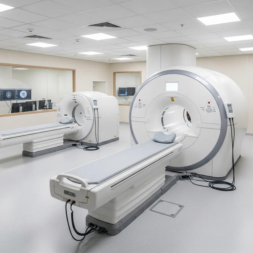
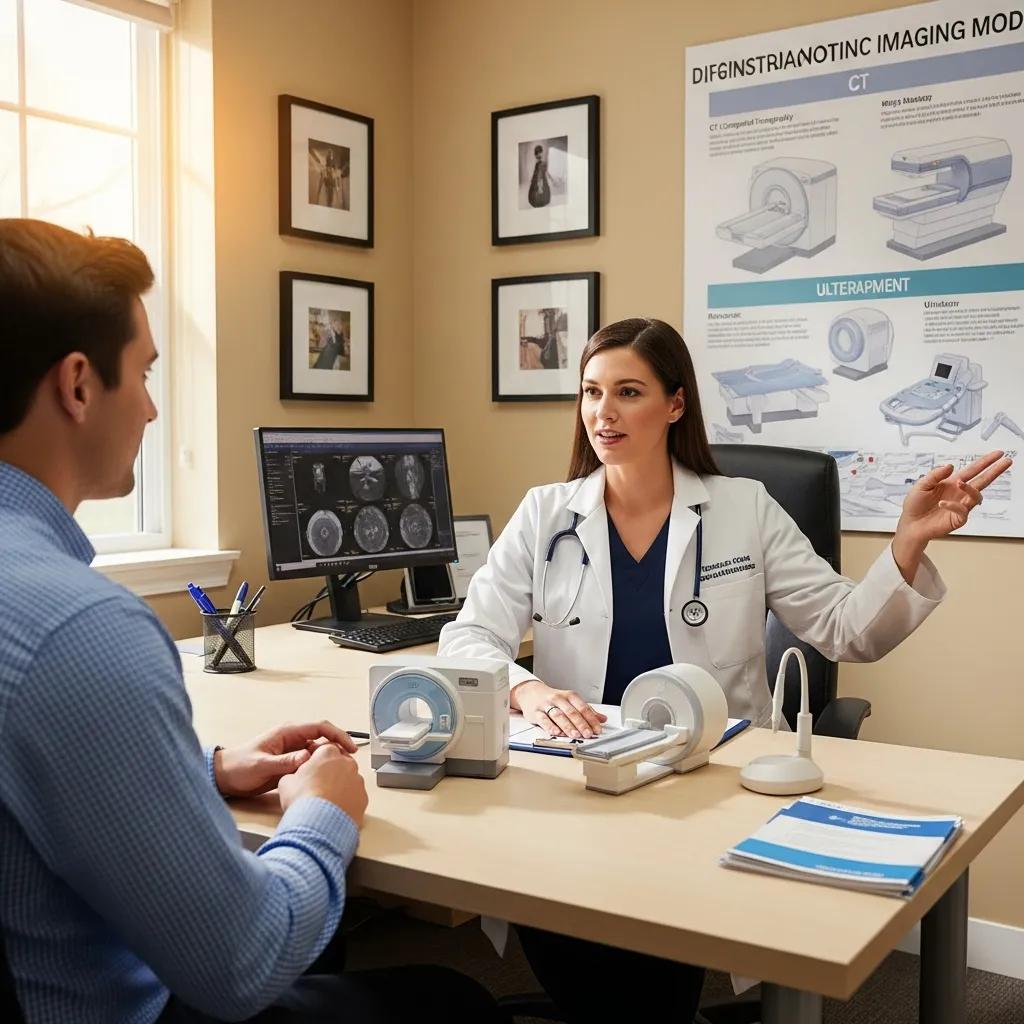
The Central Coast offers a full range of imaging modalities, each suited to different clinical questions: CT delivers fast, cross-sectional detail for trauma and chest or abdominal concerns; MRI provides excellent soft-tissue contrast for neurological and musculoskeletal issues; ultrasound gives real‑time assessment for pregnancy, vascular and abdominal conditions; X‑ray offers quick bone and chest checks; and DEXA measures bone density. These techniques operate on different physical principles — X‑rays for density contrast, ultrasound for reflected sound waves, CT for computed X‑ray slices and MRI for magnetic resonance — which is why clinicians select one test over another. The benefit to patients is quicker diagnosis, more accurate disease staging and better-targeted treatment plans based on high-quality images. Knowing what each modality does helps patients and clinicians pick the right scan and follow the correct preparation, reducing the chance of repeat imaging.
The table below is a quick reference that links common imaging modalities with their typical uses to help clinicians and patients match symptoms to likely investigations.
This table shows how each modality answers different clinical questions and why a referrer might choose one scan over another. The next section outlines which of these services Life Medical Imaging Central Coast offers locally and how that improves access to diagnostics.
Which Types of Diagnostic Imaging Does Life Medical Imaging Provide?
Life Medical Imaging Central Coast is an accredited independent radiology service offering a broad range of tests to meet common and specialised needs. Our clinic provides General CT and Cardiac CT, Dental imaging for oral and maxillofacial assessment, and Digital X‑ray for quick bone and chest checks. Ultrasound services include General, Vascular, Musculoskeletal, Obstetric and Gynaecological examinations. We also perform image‑guided interventions such as spinal and joint injections, biopsies and aspirations. Additional services include Paediatric imaging, Body Composition DEXA, Bone Mineral Densitometry and Platelet‑Rich Plasma (PRP) injections — enabling many patient pathways to be completed within the same service network on the Central Coast.
Having this breadth of services means patients often have investigations and image‑guided procedures coordinated in one network, reducing handovers and improving continuity of care. That leads to faster diagnostic work‑ups and smoother referrals back to treating clinicians. Sub‑specialist reporting in Women’s and Cardiac imaging further improves diagnostic confidence for complex cases and supports better management decisions.
How Do These Imaging Services Support Early Detection and Treatment Planning?
Imaging helps detect structural or functional problems before symptoms worsen, allowing earlier intervention and often better outcomes. For example, scans can reveal fractures not seen on exam, detect tumours at a surgically treatable stage, or confirm vascular problems that need urgent care — each finding changes the clinical pathway. Imaging also stages disease, such as identifying local invasion or spread in cancer, which informs surgery, radiotherapy planning and systemic treatment choices. Fast, accurate imaging reduces uncertainty and helps multidisciplinary teams prioritise care.
One typical pathway illustrates this: a patient with ongoing abdominal pain has an ultrasound that finds a suspicious mass, followed by CT to define extent and then an image‑guided biopsy for tissue diagnosis — together these steps produce a definitive diagnosis and a clear treatment plan. Image‑guided procedures, like joint injections or biopsies, can sometimes offer diagnostic information and therapeutic benefit in a single visit, shortening the time to effective treatment. Recognising these pathways explains why timely imaging and correct preparation matter; the next section gives practical preparation steps for common scans.
How Does Medical Imaging Improve Patient Care in Radiology Services NSW?
Across NSW, medical imaging raises diagnostic accuracy, shortens time to diagnosis and enables targeted treatments that can avoid more invasive procedures. High‑quality images and specialist radiology reports inform decisions from general practice to tertiary surgical teams, and imaging often determines whether conservative care or urgent intervention is needed. Central Coast imaging services strengthen regional care by offering local access to diagnostics that might otherwise require travel to metropolitan centres, supporting continuity and timeliness for patients in the area.
National and professional data show growing, evidence‑based use of imaging; current guidance stresses choosing the right test at the right time to avoid overuse while maximising benefit. Collaborative working between radiologists, sonographers and referrers ensures imaging is part of an integrated care plan. Timely report turnaround and clear communication of urgent results are essential to turn imaging findings into prompt clinical action, helping treating teams prioritise care.
What Conditions Can Be Diagnosed with CT, MRI, Ultrasound, and X-ray?
Each modality has strengths that make it best for particular conditions: CT is preferred for acute trauma and most chest or abdominal emergencies thanks to speed and spatial resolution; MRI is ideal for neurological, spinal and soft‑tissue problems because of superior contrast; ultrasound is the first choice in pregnancy, vascular assessment and dynamic musculoskeletal scans; and X‑ray remains the go‑to for suspected fractures and initial chest assessments. Choosing the right modality improves diagnostic yield and patient safety by avoiding unnecessary tests or exposures.
- CT: acute head trauma, pulmonary embolism, suspected abdominal perforation
- MRI: stroke assessment, spinal cord pathology, soft‑tissue tumours
- Ultrasound: obstetric follow‑up, deep vein thrombosis, gallbladder disease
- X‑ray: long‑bone fractures, chest infection screening
Picking the correct test reduces delays and aligns subsequent care to the most likely diagnosis, which leads naturally into how interventional procedures are used to diagnose and treat conditions.
How Do Interventional Procedures Enhance Pain Management and Diagnosis?
Interventional radiology uses image guidance to place needles or catheters precisely for diagnostic sampling or targeted therapy, improving accuracy and safety. Procedures like spinal or joint injections can both relieve pain and help confirm the pain source, while image‑guided biopsies and aspirations obtain tissue or fluid with minimal invasiveness. These approaches often avoid open surgery, shorten recovery and let disease‑specific treatment start sooner once pathology is confirmed.
Robust safety protocols and imaging guidance reduce procedure risks, and patients generally recover faster compared with surgical alternatives. Interventional techniques therefore bridge diagnostic imaging and definitive treatment. Clear referral pathways, pre‑procedure checks and coordinated post‑procedure reporting ensure appropriate indications, informed consent and reliable follow‑up. Understanding these roles helps clinicians choose the most efficient route for patients with pain or suspicious lesions.
What Should Patients Know About Preparing for Medical Imaging Scans?
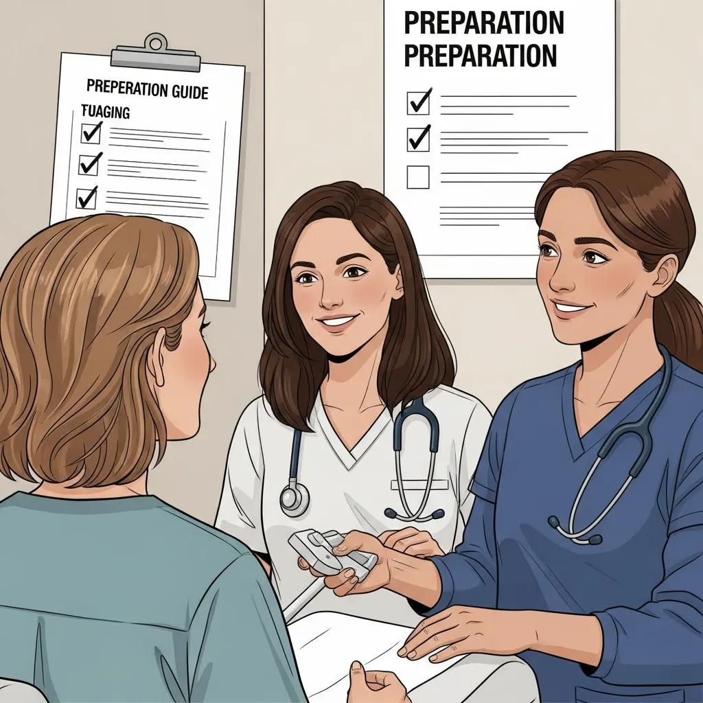
Good preparation improves image quality and reduces the chance of repeat scans, directly affecting diagnosis and treatment speed. Preparation depends on the modality: MRI needs screening for metal implants and may require strategies for claustrophobia; CT can require fasting or hydration protocols when contrast is used; ultrasound sometimes needs a full bladder or fasting depending on the exam. Clear pre‑scan instructions reduce anxiety and help technologists capture diagnostic images on the first visit.
Please bring your referral, a list of current medications and any recent imaging reports if available, and tell staff about pregnancy, allergies or implants that could affect safety. If contrast is planned, staff will check for allergies and kidney function when needed — knowing this beforehand helps you prepare and ask informed questions. The checklist below summarises practical steps for common scans so you arrive ready for an efficient appointment.
- MRI preparation checklist:
Tell staff about any implants, metal fragments or devices and complete the screening form.
Remove jewellery and clothing with metal; wear comfortable, metal‑free clothes.
If claustrophobic, discuss options ahead of time — we can plan open‑bore scanners or mild anxiolytics where appropriate. - CT preparation checklist:
Follow fasting instructions if contrast is required and disclose any allergies.
Stay well hydrated unless otherwise advised — hydration helps contrast clearance.
Bring a current medication list and recent renal function results if available. - Ultrasound preparation checklist:
For abdominal scans, follow fasting guidance; for pelvic scans, a full bladder may be requested.
Wear loose clothing for easy access to the area being examined.
For obstetric scans, bring pregnancy details and prior ultrasound reports if possible.
Following these steps reduces the risk of non‑diagnostic studies and improves comfort. If you’re unsure about anything, contact the imaging service before your appointment — Life Medical Imaging Central Coast supports online booking and eReferral and can answer preparation questions by phone on 02 4326 7000 to ensure you’re fully prepared.
How Do You Prepare for Common Scans Like MRI, CT, and Ultrasound?
Effective preparation starts with understanding why the scan is needed and following any instructions from the referrer or imaging service. For MRI, a thorough implant and metal screening is essential and some patients may need mild sedatives or access to an open bore scanner to manage anxiety; discuss this before your appointment so staff can accommodate you. For CT, fasting for a few hours and confirming contrast safety helps avoid complications. Ultrasound preparation focuses on bladder or fasting requirements specific to the organ examined. Sharing relevant medical history, allergies and pregnancy status ensures staff select the safest protocol and capture diagnostic images efficiently.
If you have mobility limitations or are bringing a child, tell the clinic when booking so appropriate supports can be arranged. Bringing previous imaging and clinical notes helps radiologists compare studies and improves diagnostic accuracy. Preparation that reduces movement and provides clinical context ultimately enhances image quality and shortens the pathway to treatment. The next section outlines safety measures imaging services use to protect patients.
What Safety Measures and Technologies Ensure Patient Wellbeing?
Imaging services follow multiple safety measures: radiation dose optimisation for CT and X‑ray, equipment accreditation and maintenance, staff credentialling and continuing education, and infection control procedures. Dose‑reduction strategies such as low‑dose CT protocols aim to keep exposure as low as reasonably achievable while maintaining diagnostic quality. Regular quality assurance checks ensure scanners perform reliably, and specialist radiologist oversight supports safe reporting of complex cases.
In addition to technical safeguards, services screen for contrast allergies and renal impairment, follow clear consent processes for interventional procedures, and offer tailored pathways for paediatric and pregnant patients to minimise risk. These systems ensure appropriate, high‑quality imaging. Emerging technologies — for example AI‑assisted image reconstruction and dose‑efficient protocols — are further improving safety and diagnostic confidence, as discussed later in this guide.
How Are Medical Imaging Costs Managed: Medicare Rebates and Private Health Insurance?
Knowing likely costs helps you plan care and avoid surprises. In Australia, many diagnostic imaging procedures attract Medicare rebates when the referral and clinical indication meet MBS criteria; rebate eligibility depends on the referring practitioner’s specialty and the documented clinical justification. Private health insurance may cover additional costs depending on your policy and whether the scan is inpatient, outpatient or part of a private admission. Check rebate rules and your insurance details before booking to estimate any gap payments.
The table below summarises common procedures, typical rebate eligibility and notes on expected out‑of‑pocket ranges so patients have a planning guide. Exact amounts vary by MBS item number and clinical context.
This table is a guide — patient‑specific factors and MBS item numbers determine actual rebates and gaps. Confirm details before your appointment to reduce uncertainty. For imaging at Life Medical Imaging Central Coast, our team can outline billing policies and help estimate out‑of‑pocket costs; call 02 4326 7000 for assistance with eligibility checks and whether an eReferral or specialist referral is needed for a Medicare rebate.
What Medicare Rebates Are Available for MRI and Other Diagnostic Scans?
Medicare rebates are linked to specific MBS item numbers and apply when clinical criteria are met. Eligibility usually depends on the documented clinical indication from the referrer and whether the referral fulfills the item‑number requirements; some MRI item numbers require a specialist referral or prior imaging. Ask your referrer to include clear clinical indications and, where relevant, the MBS item number to speed processing and reduce billing delays.
Because rebate rules can be detailed, request an estimate of likely rebates and gap fees from the imaging provider before booking. When imaging services and referrers work together to provide thorough documentation, rebate claims are processed more smoothly and patients avoid unexpected costs.
How Can Patients Navigate Out-of-Pocket Costs and Insurance Coverage?
To reduce surprise bills, take three simple steps: confirm with your referrer that the referral matches rebate criteria; contact your private health insurer to check cover and any excesses for diagnostic imaging; and ask the imaging provider for an itemised estimate before the appointment. Bring your Medicare card, proof of insurance and referral paperwork to speed billing and claims. If cost is a barrier, discuss options with the imaging service — many clinics can provide estimates, explain rebate scenarios and advise whether private admission or outpatient billing will lower gaps.
Clear communication between you, your referrer and the imaging provider minimises billing errors and helps choose the most cost‑effective pathway. Life Medical Imaging Central Coast offers phone support for billing enquiries and out‑of‑pocket estimates; our staff can explain eReferral requirements and documentation needed to support Medicare rebate claims — call 02 4326 7000 for guidance.
Where Are Life Medical Imaging Central Coast Clinics Located and How Can You Book?
Life Medical Imaging Central Coast operates multiple clinics across the region to improve access to diagnostic imaging and interventional services. Each site offers specific services to match local demand. Clinics are listed below with the services they provide and the booking methods available so patients and referrers can pick the most convenient location. Booking options include online booking, eReferral submission and phone scheduling to suit both patients and referring clinicians.
This location table helps you find the site that offers the service you need and shows booking can be completed online, by eReferral or by phone. To book or check which site is best for a particular scan, call 02 4326 7000 for help with scheduling and referral requirements.
What Services Are Offered at Each Central Coast Location?
Each clinic focuses on a subset of services so staff and equipment match local clinical needs. For example, Bateau Bay commonly provides General CT, Digital X‑ray and a wide range of ultrasound services suited to obstetric and general diagnostic work. Erina offers sub‑specialist services including Cardiac CT and Women’s Imaging. Umina Beach is set up for paediatric imaging and DEXA scans, while Killarney Vale provides complex imaging and image‑guided interventions when required. This distribution keeps specialised equipment and expertise where they’re most needed while keeping services local for patients.
Choosing the right site reduces travel and ensures you arrive at a facility prepared for your specific modality, which helps avoid delays and improves imaging outcomes. If you’re unsure which location is appropriate, call the clinic and staff will advise on site selection and pre‑scan preparation.
How Do You Book Appointments and Submit Referrals Online?
Booking is simple for patients and referring clinicians: first, confirm the required imaging modality and referral details with your referrer; second, choose the preferred clinic based on service availability; third, submit an eReferral or call the clinic to schedule, providing referral documentation and patient details. For clinicians, eReferral streamlines information transfer and reduces admin by sending imaging requests and clinical notes electronically. Patients who prefer personal assistance can phone the clinic to discuss preparation, insurance or appointment times that suit mobility or paediatric needs.
Clear referral indications and clinical notes shorten wait times and ensure the correct protocols are booked. If you have logistical questions — for example about mobility support — calling 02 4326 7000 before the appointment helps the clinic prepare and ensures a smoother visit. These booking pathways support efficient coordination between referrers, patients and imaging staff.
What Are the Latest Advances and Trends in Medical Imaging Technology?
Recent advances focus on improving diagnostic accuracy, lowering patient exposure and increasing access through portable and AI‑enabled tools. Artificial intelligence is assisting with image reconstruction, lesion detection and workflow triage by flagging urgent findings and prioritising reporting, which helps radiologists deliver faster, more consistent interpretations. Low‑dose CT protocols and improved detector technology reduce radiation while keeping diagnostic quality high — beneficial for patients needing repeat imaging.
Portable imaging and telemedicine links extend specialist input into community and remote settings, allowing images to be captured locally and reviewed by specialists remotely for faster decision making. These trends lead to better outcomes by shortening time to diagnosis and enabling earlier, targeted interventions. Understanding how these technologies are used helps clinicians and patients appreciate the steady improvements in safety and diagnostic performance; the next sections cover AI and other emerging tools in more detail.
How Is Artificial Intelligence Enhancing Diagnostic Accuracy?
AI supports radiologists by automating routine tasks such as image segmentation, measurements and detection of potential abnormalities like nodules or fractures. This increases efficiency and helps catch subtle findings earlier. When AI flags a possible urgent abnormality, workflows can prioritise that case so a radiologist reviews it sooner, reducing delays for critical results. AI is a decision‑support tool — radiologist oversight remains central to ensure clinical context guides interpretation and patient safety is preserved.
Clinical validation and governance frameworks guide safe AI use, and imaging services incorporate AI outputs into reporting only when accuracy and reliability meet clinical standards. The model is collaborative — AI suggests, clinicians confirm — and this improves throughput while maintaining report quality. These workflow gains help radiology services meet rising demand without compromising diagnostic standards across the Central Coast.
What New Imaging Technologies Improve Patient Access and Care?
Emerging tools such as portable ultrasound units, low‑dose CT scanners and advanced 3D/4D obstetric imaging improve access and patient experience by enabling bedside scans, lowering radiation risk and enhancing visualisation for counselling. Telehealth‑enabled reporting and virtual consultations let clinicians and patients discuss results without an extra in‑person visit, improving convenience and continuity of care. These innovations expand specialist reach into community clinics and streamline the path from imaging to treatment.
As these technologies spread into regional services, patients benefit from faster diagnosis, less travel and better communication with treating teams. Local providers adopting these tools contribute to fairer access and a better patient experience — completing the cycle from improved diagnostics to timelier, effective care across the Central Coast.
A Comparison of CT, MRI, and PET/CT for the Diagnosis of Mediastinal Masses
Each imaging modality has strengths and limitations when assessing the mediastinum. CT offers high spatial resolution but uses ionising radiation and provides less soft‑tissue contrast. MRI gives superior soft‑tissue characterisation, though at higher cost and longer scan times. Ultrasound delivers real‑time imaging but is operator‑dependent and limited by tissue windows. FDG PET/CT adds functional information but has lower spatial resolution. Recent work has focused on using MRI to better characterise cystic mediastinal lesions when CT is inconclusive.
Imaging modalities (MRI, CT, PET/CT), indications, differential diagnosis and imaging characteristics of cystic mediastinal masses: a review, A Shah, 2022
Frequently Asked Questions
What should I expect during my first medical imaging appointment?
When you arrive, a member of our team will welcome you and check your referral and medical history. The technologist will explain the procedure and any preparation (for example fasting or clothing). Depending on the scan you may need to lie still, hold your breath briefly, or follow simple instructions while images are taken. We aim to make the visit as comfortable and efficient as possible and will answer any questions before you start.
Are there any risks associated with medical imaging procedures?
Most imaging tests are safe, but there are some considerations. X‑rays and CT scans use ionising radiation, which carries a small long‑term risk that is weighed against the clinical benefit. MRI uses strong magnetic fields and is not suitable for some implants or devices. Contrast agents can cause allergic reactions in rare cases and require kidney‑function checks when appropriate. Your referrer and our staff will discuss risks and benefits related to your individual situation.
How can I ensure the best quality images during my scan?
Follow the preparation instructions given by your referrer or the imaging centre — for example fasting or bladder filling. Arrive on time and wear comfortable, metal‑free clothing. Let staff know about movement limitations, recent procedures or prior imaging. Clear communication and correct preparation help technologists capture diagnostic images and reduce the chance of repeat scans.
What should I do if I have claustrophobia and need an MRI?
If you have claustrophobia, tell the clinic when you book. Options may include an open‑bore scanner, scheduling a time with more support, or discussing mild sedative measures with your referrer. Technologists will also explain techniques to help you relax. Letting us know in advance allows us to plan and make the scan as comfortable as possible.
How long does it typically take to receive results from my imaging?
Timing varies by scan and clinical urgency. Preliminary results may be available within hours to a couple of days, while final radiology reports are typically returned within 24–72 hours. Urgent findings are communicated to your referrer immediately. Your treating clinician will discuss the results and next steps with you when the report is ready.
Can I bring someone with me to my imaging appointment?
Yes — you may bring a support person, but check with the clinic beforehand as some procedures or areas may have visitor limits. A companion can help with transport or emotional support, particularly for children or patients needing assistance. If sedation or contrast is used, plan for someone to drive you home if advised.
What should I do if I have questions about my insurance coverage for imaging services?
Start by contacting your insurer to confirm cover and any excesses for diagnostic imaging. You can also speak with the clinic’s billing team, who can provide an itemised estimate and explain likely Medicare rebates and gaps. Bringing your referral, Medicare card and insurance details to the appointment speeds the process and helps avoid surprises.
Conclusion
Imaging services on the Central Coast play a vital role in diagnosis and treatment planning by delivering timely, accurate assessments. When patients and referrers understand the different modalities and how to prepare, the pathway from imaging to treatment is faster and more reliable. Life Medical Imaging Central Coast provides a comprehensive range of services tailored to local needs, prioritising access, safety and continuity of care. For more information or to book an appointment, please call us on 02 4326 7000.