Frequently Asked Questions
What should I expect during a medical imaging appointment?
When you arrive, check in at reception with your referral and photo ID. A member of our team will then take you to the imaging room, explain the procedure and make sure you’re comfortable. Some exams require changing into a gown and removing metal items. Scan times vary by test, but the staff will keep you informed at every step to help ease any concerns.
Are there any alternatives to traditional imaging methods?
Yes. Depending on your situation, alternatives to X‑rays and CT include ultrasound, which uses sound waves and no radiation, and MRI, which uses magnetic fields and radiofrequency pulses instead of ionising radiation. These options are often preferred for soft tissue assessment and for patients who are pregnant. Your referrer will recommend the most appropriate modality for your needs.
How can I ensure the best quality images during my scan?
Follow the preparation instructions we give you — for example fasting, drinking water or pausing certain medications — and arrive on time. Wear comfortable, metal‑free clothing. Tell the staff in advance if you have claustrophobia, anxiety or other concerns so they can support you and help obtain the best images.
What happens if I have an allergic reaction to contrast material?
If you experience an allergic reaction to contrast, tell the imaging staff immediately. Our teams are trained to manage reactions and will provide appropriate treatment, such as antihistamines, based on the severity. Always disclose any known allergies to contrast agents during pre‑scan checks so we can plan and reduce risk.
Can I bring someone with me to my imaging appointment?
In most cases, yes — a friend or family member can come with you for support. Some procedures have space or safety limits on who can be in the imaging room, so check with us ahead of time if you plan to bring someone.
What should I do if I have questions after receiving my imaging results?
If anything in your report is unclear, contact the clinician who referred you. They can explain the findings in context and discuss next steps. Imaging reports can be technical, so your referrer is best placed to interpret results and recommend further action. Keep a copy of the report for your records.
How often should I have medical imaging done?
Frequency depends on your health situation and the condition being monitored. Some people need routine imaging to track chronic issues, while others only have scans when symptoms appear. Follow your healthcare provider’s advice, which will balance clinical need and safety.
What Are the Main Types of Medical Imaging and How Do They Work?
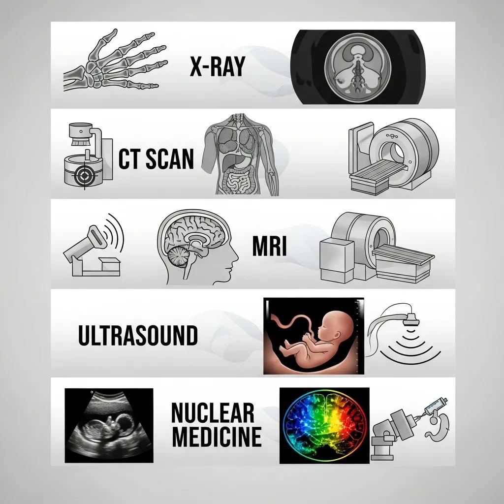
Medical imaging uses several different technologies to create pictures of the body’s structure and function. The main modalities — X‑ray, CT, MRI, ultrasound and nuclear medicine — rely on different physical principles and are chosen according to the clinical question. Knowing the differences helps patients understand preparation, expected duration and safety considerations. Below we summarise how each modality works, common uses, basic preparation and typical exam length.
The table that follows compares each modality’s mechanism, usual indications, preparation and typical duration to help guide decisions and referrals.
This comparison shows how each technique’s mechanism and clinical role determine preparation and exam length. The sections below describe X‑ray and CT in more patient‑friendly detail.
What Is an X-ray and When Is It Used?
An X‑ray is a rapid projection technique that uses ionising radiation to image dense structures such as bone and the chest. It’s commonly used to assess fractures, detect chest infections and for routine dental checks. Preparation is minimal — remove jewellery and metal items and wear comfortable clothing. If you are, or might be, pregnant, tell staff so we can consider alternatives. A single X‑ray carries a low radiation dose compared with many CT exams, and every X‑ray is performed only when clinically justified.
X‑ray is often the first‑line test; when more detailed cross‑sectional views are needed, a CT scan may be recommended.
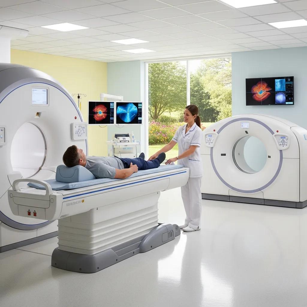
How Does a CT Scan Work and What Should Patients Expect?
CT acquires multiple X‑ray measurements around the body and reconstructs them into detailed cross‑sectional images. It’s particularly useful for complex fractures, lung disease and acute abdominal conditions. During the scan you lie on a motorised table that moves through the scanner gantry while the tube rotates. Many CT exams are quick, though contrast‑enhanced studies can take longer because of preparation and observation. Contrast may be given orally or intravenously to improve visualization — always tell staff about allergies and kidney problems so we can manage risk. Modern techniques such as ultra‑low dose CT help reduce radiation while maintaining image quality.
Because CT is more sensitive for certain injuries, it’s chosen when X‑ray is insufficient — MRI preparation and considerations differ and are described next.
How Should Patients Prepare for Different Medical Imaging Procedures?
Good preparation improves image quality and reduces delays. While some instructions are modality‑specific, most scans share basic requirements such as the documents to bring, clothing choices, medication guidance and arrival time. The list below summarises essential items to consider before your appointment to help ensure a smooth visit and avoid rescheduling.
- Bring referral and photo ID: Have your referral letter and identification ready at reception.
- Wear comfortable clothing: Loose, metal‑free clothes help prevent interference with scans.
- Inform staff of pregnancy, allergies and implants: Tell us if you are pregnant, have allergies or any implanted devices before booking.
These general steps prepare you for specific instructions for each modality and for contacting the clinic with any questions.
What Are the General Preparation Guidelines for Medical Scans?
General preparation focuses on documentation, safety declarations and simple practicalities that apply across most modalities. Bring your referral, Medicare or insurance details where relevant, and any prior imaging on disc or printed reports — these help the radiologist compare studies. Remove jewellery, metal fastenings and electronic devices, and arrive a little early to complete safety forms. If you have claustrophobia, implants or known contrast allergies, contact us before your appointment so the team can arrange appropriate support.
Clear preparation reduces the need for repeat scans and improves diagnostic accuracy. Specific rules for MRI follow below.
How Do You Prepare Specifically for an MRI Scan?
MRI uses a powerful magnetic field, so preparation centres on screening for metal implants, foreign bodies and devices that could interact with the scanner. Complete the MRI safety questionnaire fully and bring any implant documentation, including manufacturer details where available. Tell staff if you experience claustrophobia — sedation or open MRI options may be possible. Remove all metal, watches and electronic items. Medication changes are rarely required, but follow any instructions we provide if contrast is planned. If you’re unsure about device compatibility, discuss it with your referrer and the imaging team before the appointment.
Careful MRI screening helps ensure the scan is safe and successful, and connects directly to the MRI safety measures outlined next.
What Are the Safety Considerations and Risks Associated with Medical Imaging?
Safety in imaging covers radiation from X‑ray and CT, MRI implant compatibility, contrast reactions and sedation risks where relevant. Modern practice follows two core principles: justification — only perform scans when the expected benefit outweighs risk — and optimisation — use the lowest effective doses and best protocols. Facilities using ultra‑low dose CT and subspecialist reporting aim to improve diagnostic confidence while reducing exposure and error. The Q&A subsections that follow address common patient safety questions with clear, practical answers.
Before the modality‑specific notes, the table below summarises typical risks and recommended precautions to help you discuss options with your referrer or imaging team.
This overview highlights relative risks and precautions so you can raise any concerns with your referrer or our team.
Are X-rays and CT Scans Safe Regarding Radiation Exposure?
X‑rays and CT use ionising radiation. Safety depends on the dose, clinical justification and optimisation of protocols. To put dose in perspective, clinicians often compare it with natural background radiation, and where appropriate we use dose‑reduction techniques such as ultra‑low dose CT. If you are pregnant or planning pregnancy, tell your clinician so alternative tests can be considered. If you have any questions, ask your referrer or the imaging provider about dose‑reduction strategies and why the scan is recommended.
Questions about radiation naturally lead to MRI safety when implants are present, which is covered next.
What Safety Measures Are Important for MRI Scans, Especially with Implants?
MRI safety focuses on the magnetic environment and device compatibility. Implants are classified as MRI‑safe, MRI‑conditional or contraindicated based on manufacturer information. Disclose all implants, metal tattoos or any history of metal fragments. Our screening forms and device documentation let the imaging team determine whether an MRI can proceed safely. If manufacturer information is unavailable, staff may recommend alternative imaging or specialist advice; some conditional implants allow specific scan parameters. When MRI is not suitable, ultrasound, CT or nuclear medicine often provide useful diagnostic alternatives.
Knowing implant protocols helps set expectations for reporting timelines and interpretation, which the next section explains.
How Can Patients Understand Their Medical Imaging Results and Next Steps?
Imaging reports present findings and the radiologist’s diagnostic impression. Understanding the report’s structure and expected timelines helps you ask the right follow‑up questions. Reports usually separate an objective Findings section from an interpretive Impression or Conclusion; this distinction shows what is descriptive and what is diagnostic. Turnaround times vary by modality and urgency — the list below outlines typical pathways and suggested actions after you receive a report. Clear expectations reduce anxiety and support coordinated care with your referrer.
- Typical report sections: Findings (descriptive) and Impression (diagnostic summary).
- Result timelines: Routine versus urgent reporting differs — ask the clinic about expected turnaround.
- Next steps: Discuss the Impression with your referring clinician to plan treatment.
These points lead into more specific timelines and interpretation guidance in the subsections below.
How Long Does It Take to Receive Scan Results?
Turnaround depends on modality, clinical urgency and the facility’s workflow. Routine outpatient reports commonly take a few days, while urgent or inpatient studies can be communicated within hours. For many standard exams expect results within 24–72 hours; complex studies or those needing subspecialty review may take longer. Usually the referring clinician receives the formal report and will advise you. Some providers offer patient portals or direct image access, while others send results via the referrer — confirm the process when you book. If a report is delayed beyond the advised timeframe, contact the imaging provider or your referrer for an update.
Understanding timelines makes it easier to interpret the report format, which we explain next.
How Do You Interpret a Radiology Report?
A radiology report starts with objective Findings and finishes with the radiologist’s Impression, which synthesises likely diagnoses in the clinical context. Terms such as “attenuation”, “contrast enhancement”, “nodule” or “effusion” have specific meanings; a short glossary can help, but clinical interpretation requires input from your referrer who knows your history. If a report uses cautious language (for example, “suggests” or “consistent with”), ask your referrer what further tests or management steps are recommended. Keep a copy of the report and key images to support follow‑up consultations or second opinions when needed.
Knowing how reports are delivered and understood transitions naturally to booking and cost information covered next.
How Do Patients Book Appointments and What Are the Costs Involved?
Booking an imaging appointment involves selecting a booking route, preparing required documentation and understanding likely costs. Options include GP referral, private self‑referral where permitted, or direct phone/clinic booking. The table below compares routes, documentation needs and typical cost or insurance considerations so you can choose the best pathway. After the table we outline simple, step‑by‑step booking instructions.
This comparison clarifies pathways and leads to a straightforward booking workflow patients can follow.
What Is the Process for Booking a Medical Imaging Appointment?
Booking typically follows three steps: obtain the appropriate referral; contact the imaging provider by phone or use online booking; and follow any pre‑scan instructions sent with your appointment confirmation. Have your referral, ID and clinical details ready when you call to speed up scheduling — urgent referrals are triaged differently to routine outpatient scans. If you have questions about preparation, implants or contrast allergies, contact the imaging team before your appointment to confirm instructions and any special arrangements. For Life Medical Imaging Central Coast patients, phone enquiries can clarify booking options and referral requirements.
Clear booking steps also help you understand likely costs, addressed in the next subsection.
What Are the Typical Costs and Insurance Options for Private Scans?
Private scan costs vary with modality, complexity and whether a report attracts a Medicare rebate. Factors that affect price include use of contrast, scan duration and whether specialist reporting is required. Check with your insurer about rebate eligibility and ask clinic billing staff for an exact quote before booking. Some providers offer self‑pay pricing and can help with preauthorisation. Typical private‑scan considerations include out‑of‑pocket fees for non‑rebated studies and possible gap payments after insurer rebates. Contact the imaging provider’s billing team at booking for a precise cost estimate.
With booking and costs covered, the following sections address specialised imaging for women, children and cardiac patients.
What Are the Frequently Asked Questions About Specialised Imaging Services?
Specialised imaging areas — Women’s imaging, Paediatric imaging and Cardiac imaging — have tailored protocols for preparation, safety and reporting to meet specific patient needs. Women’s imaging commonly combines mammography and ultrasound and prioritises privacy and pregnancy‑safe options. Paediatric imaging uses child‑friendly techniques and strict dose reduction. Cardiac imaging employs modality‑specific protocols such as cardiac CT and MRI to assess anatomy and function. The sections below explain what to expect, how specialised reporting supports care and where to seek follow‑up after a focused study.
The summary list highlights the main considerations for each specialised service to help patients and referrers choose the appropriate pathway.
- Women’s imaging: choosing the right modality, pregnancy precautions and privacy measures.
- Paediatric imaging: child‑friendly approaches, sedation policies and dose reduction.
- Cardiac imaging: indications, preparation and specialist reporting requirements.
These points prepare you for the detailed subsections that follow.
What Should Women Know About Women’s Imaging Services?
Women’s imaging typically uses mammography and ultrasound to investigate breast concerns, with modality choice guided by age, symptoms and pregnancy status. If pregnancy is possible, staff will consider non‑ionising options such as ultrasound where appropriate; mammography is performed only when the clinical benefit outweighs the small risk, with shielding and exposure minimisation. Our service emphasises privacy, comfort and subspecialist reporting to ensure accurate, sensitive communication. If timing (for example around your menstrual cycle) or additional support matters to you, let booking staff know so we can accommodate your needs.
Information about women’s imaging leads into the paediatric and cardiac imaging considerations described next.
What Are the Key Concerns for Paediatric and Cardiac Imaging?
Paediatric imaging focuses on minimising radiation exposure, communicating in a child‑friendly way and using sedation only when necessary; protocols are adjusted for the child’s size and clinical question to reduce dose. Cardiac imaging includes echocardiography, cardiac CT and cardiac MRI to evaluate heart structure and function; preparation varies — some cardiac CTs require heart‑rate control or fasting. Both areas benefit from subspecialist reporting to ensure precise interpretation and clear follow‑up advice. Parents and patients should discuss sedation risks, pre‑scan medication instructions and expected result timelines with the clinic when booking.
If you need more information or want to make an appointment, contact Life Medical Imaging Central Coast by phone. Our accredited clinic uses advanced technology, including ultra‑low dose CT and subspecialist reporting, to support safe, accurate diagnostics. Phone enquiries allow staff to advise on suitable modalities and provide cost guidance.
- Call to clarify referral and booking options: Have your referral and clinical details ready.
- Ask about preparation and safety: Mention implants, pregnancy, allergies or claustrophobia.
- Request a cost estimate: Confirm private billing or rebate questions before attendance.
Medical Imaging Modalities: Trends and Applications in Disease Diagnosis
Medical imaging is central to modern healthcare — diagnosing disease, guiding treatment and monitoring progress. Techniques such as X‑ray, PET, CT, MRI and ultrasound provide non‑invasive views of internal structures. Advances in medical image processing have improved detection and analysis and are now being combined with machine learning and deep learning to support clinical decision making. While automated tools can assist interpretation, high accuracy and careful clinical oversight remain essential to deliver safe, effective care. This review summarises how different imaging modalities work, their benefits and limitations, and emerging trends shaping the field.
Ultra-Low-Dose CT vs. Standard-Dose CT for Fracture Diagnosis
This study compared ultra‑low‑dose CT (ULD‑CT) with standard‑dose CT (SD‑CT) for diagnosing non‑displaced fractures of shoulder, knee, ankle and wrist. Ninety‑two patients had SD‑CT followed by ULD‑CT about nine days later. While SD‑CT showed somewhat better objective and subjective image quality, diagnostic performance for detecting fractures was similar: SD‑CT sensitivity 95.35% and ULD‑CT 90.70%, with both methods demonstrating high specificity and accuracy. The effective radiation dose was significantly lower with ULD‑CT. The authors conclude ULD‑CT can be useful for fracture diagnosis and supports clinical decision making where dose reduction is important.
Conclusion
Knowing the strengths and requirements of different imaging tests helps you make informed choices with your referrer. From quick X‑rays to detailed MRI studies, each modality has specific benefits and preparation steps. Following the guidance provided and discussing any safety concerns with the imaging team will improve your experience and the quality of the exam. For help or to book an appointment, contact our team today.