Diagnostic Ultrasound Guide: Scan Types, Preparation, Safety & Costs
Diagnostic ultrasound is a safe, non‑invasive imaging method that uses high‑frequency sound waves to produce real‑time images of organs, blood flow and developing babies — all without ionising radiation. This guide describes how ultrasound works, summarises common scan types and when they’re used, and gives clear, practical advice on how to prepare and what to expect. We also explain safety considerations, how reports are produced, and what private patients can expect to pay. Use the scan checklists and cost guidance to plan your appointment and know who to contact if you have questions.
What are the different types of ultrasound scans offered?

Ultrasound covers a range of specialised examinations tailored to particular body systems and clinical questions. Using different transducers and Doppler techniques, we can image anatomy and measure blood flow in real time. Each scan has its own indications, preparation needs and reporting considerations. Knowing the difference between obstetric, abdominal, vascular, musculoskeletal, gynaecological, breast and 3D/4D obstetric scans helps referrers and patients choose the right test and plan any follow‑up.
The table below gives a quick comparison of common ultrasound types, their main uses and typical indications to help with selection and referral.
This overview helps you identify which modality best suits a given clinical situation. Detailed sections below explain each area in more depth.
How does obstetric ultrasound monitor fetal development?
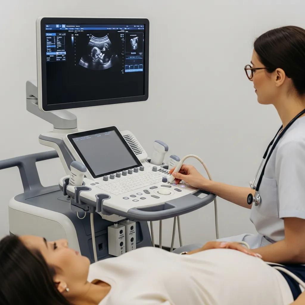
Obstetric ultrasound uses a series of timed scans to date the pregnancy, check fetal anatomy and follow growth. Early dating scans give an accurate gestational age, the mid‑trimester anatomy scan examines organ development and screens for structural anomalies, and later growth scans assess fetal size, amniotic fluid and placental function. 3D and 4D imaging are useful extras for surface detail and parental keepsakes, but they supplement — not replace — standard 2D diagnostic scans. Understanding the purpose and timing of each scan helps parents and clinicians plan appropriate follow‑up and spot when specialist referral is needed.
3D/4D ultrasound can provide additional surface detail for diagnostic questions and parental information.
Advanced 3D/4D ultrasound for prenatal diagnosis and fetal anatomy visualisation
3D and 4D ultrasound techniques now support routine clinical use by offering views that are difficult to obtain with conventional 2D imaging. These modes can improve detection and assessment of certain fetal features, assist with counselling, and allow digital storage of volumes for later review and teaching. While valuable, they are best used alongside standard 2D scans performed for diagnostic purposes.
Next we look at how ultrasound detects abdominal and vascular conditions in patients who are not pregnant, and when Doppler studies are indicated.
What conditions can abdominal and vascular ultrasounds detect?
Abdominal ultrasound is an excellent first‑line test for problems such as gallstones, fatty liver changes, cirrhosis features, kidney stones and biliary dilation, and it’s commonly used for unexplained abdominal pain or abnormal liver blood tests. Vascular Doppler studies assess blood flow to identify deep vein thrombosis, carotid narrowing and peripheral arterial disease, and they measure flow velocities to quantify clinically important stenosis. Ultrasound has limitations — for example small pancreatic lesions or bowel wall disease can be hard to see — and sometimes CT or MRI are needed for further characterisation. When findings are unclear, clinicians often request cross‑sectional imaging or specialist input to guide management.
Good preparation helps each study deliver its maximum diagnostic value, which is covered in the next section.
How should patients prepare for their ultrasound scan?

Proper preparation improves image quality and reduces the chance of needing a repeat scan. Requirements depend on the scan type: abdominal scans usually need fasting to reduce bowel gas, pelvic or early pregnancy scans commonly require a full bladder to act as an acoustic window, and vascular or musculoskeletal scans generally need only comfortable clothing. Clear instructions make the appointment smoother for you and help our sonographers capture accurate diagnostic images. The checklist below covers the most common preparation steps.
- Bring your referral and a list of current medications when you arrive at reception.
- Choose comfortable, loose clothing that allows easy access to the area being examined.
- Follow any fasting or bladder instructions included with your appointment details.
- Tell staff if you are pregnant, have diabetes or have mobility needs so we can make appropriate arrangements.
Following these steps reduces delays and helps the imaging team acquire the views needed for diagnosis. The next section gives specific fasting and bladder timing for abdominal and pelvic scans.
What are the preparation requirements for abdominal and pelvic ultrasounds?
For most abdominal scans we recommend fasting for 6–8 hours beforehand to reduce bowel gas and improve views of the liver, gallbladder and pancreas — water may be taken with essential medications. For transabdominal pelvic or early pregnancy scans, a comfortably full bladder helps displace bowel and creates a better acoustic window; drinking about 500–750 ml (roughly one hour before arrival) is a common instruction. Patients with diabetes or those taking regular medicines should contact the clinic or their referrer for tailored fasting advice. Good preparation reduces the need for repeat imaging and supports accurate assessment.
Next, a short note on what to wear and what to expect on the day of your appointment.
What should you wear and expect on the day of your ultrasound appointment?
Wear loose clothing and avoid tight belts or garments; you may be given a gown for modesty and easier access during pelvic or breast scans. Please arrive 10–15 minutes early to complete registration and confirm preparation details. Most scans last between 15 and 45 minutes depending on complexity. During the examination, a sonographer will apply warm gel and move a handheld transducer over the area while explaining steps; most people feel little or no discomfort and privacy is maintained throughout. Knowing what to expect helps reduce anxiety and ensures the appointment goes smoothly.
With practicalities covered, many patients understandably ask about safety — why ultrasound is widely used and how we apply safety standards in practice.
Is diagnostic ultrasound safe? Understanding ultrasound safety guidelines
Yes. When used appropriately, diagnostic ultrasound is considered safe and does not use ionising radiation, making it suitable for pregnancy and general medical imaging. Safety relies on clinical justification, skilled operators and adherence to principles such as ALARA (as low as reasonably achievable) to keep exposure times and acoustic output to the minimum needed for a diagnostic result. Accreditation, specialist reporting and compliance with local and international guidelines further support safe practice. New tools like elastography and validated AI aids can enhance diagnosis when used by trained teams and within approved pathways.
The ALARA principle underpins responsible ultrasound practice by keeping exposure as low as needed for clinical quality.
Ensuring diagnostic ultrasound safety with the ALARA principle
Users are responsible for prudent ultrasound use and for reducing output levels when indices exceed recommended thresholds. Applying the ALARA principle — using the lowest exposure consistent with diagnostic quality — is central to ultrasound safety.
Next we explain why ultrasound is the preferred imaging method in pregnancy and how operator skill adds to safety.
Why is ultrasound considered safe for pregnant women and general use?
Because ultrasound does not use ionising radiation, professional bodies endorse its use in pregnancy when clinically indicated. Following ALARA, trained sonographers and radiologists use appropriate settings and limit scan time to answer the clinical question. Facility accreditation and operator experience are useful indicators of quality — skilled operators select settings that optimise images while avoiding unnecessary exposure. Taken together, these safeguards make ultrasound a reliable tool for monitoring fetal and maternal health as well as general diagnostic imaging.
Understanding safety also means tracking technological developments; the next section summarises recent advances expanding ultrasound’s clinical role.
What are the latest technological advances in ultrasound imaging?
Recent innovations include 3D/4D imaging for improved obstetric visualisation, elastography to assess tissue stiffness non‑invasively (useful in liver and breast workups), and AI tools that support image acquisition, quality checks and consistent reporting. Enhanced Doppler sensitivity improves detection of subtle blood‑flow changes, aiding vascular and fetal surveillance. These advances increase diagnostic accuracy and patient experience when they are validated and used within established clinical pathways by trained specialists. Ongoing research and guideline updates continue to define best practice for these technologies.
Artificial intelligence is increasingly used to support ultrasound acquisition and interpretation, improving consistency and workflow.
AI in diagnostic ultrasound: enhancing accuracy and workflow
AI and deep learning methods are being applied to ultrasound to improve image acquisition, automate quality assessment and assist in lesion detection. When validated, these tools can support clinicians by streamlining workflows and reducing observer variability, contributing to more consistent diagnostic performance.
With safety and technology in mind, many patients then want to understand likely costs and how private insurance can affect billing.
How much does a private ultrasound cost in the UK and what are the insurance options?
Private ultrasound prices vary with scan complexity, the need for specialist reporting and local market factors. Insurance policies differ too: some cover imaging fully, others offer partial rebates or require a GP referral or pre‑authorisation. Some private clinics accept self‑referrals, but it’s wise to check your insurer’s requirements before booking to avoid unexpected fees. The table below shows representative private cost ranges for common scans and key insurance notes to help with planning.
We recommend calling your insurer to confirm cover, excesses and whether pre‑authorisation is needed. Asking the clinic for an itemised estimate at booking helps avoid surprise bills and speeds up claims processing.
What are the typical costs for common ultrasound scans at private clinics?
Typical costs reflect scan complexity and reporting level: routine abdominal or pelvic scans are usually toward the lower end of the range, while combined or specialist Doppler studies sit at the higher end. Extra fees may apply for procedures such as image‑guided biopsy or extended specialist reporting — please confirm these when you book. Because prices vary by clinic and location, ask for a written estimate and whether radiologist reporting is included to make an informed decision.
The next section explains practical steps to check your insurance before a private scan.
How can private health insurance affect ultrasound scan costs?
Whether insurance covers an ultrasound depends on your policy. Some insurers cover diagnostic imaging fully, others apply an excess or limit outpatient imaging benefits, and many require a GP or consultant referral or pre‑authorisation. Practical steps: check your insurer’s outpatient imaging benefits, confirm pre‑authorisation requirements, and note any excesses or annual caps. Keep an itemised receipt and the radiology report to support claims. If your policy won’t cover the scan, clinics usually accept self‑payment and can provide invoices and reports for later reimbursement.
With costs and insurance covered, the following section describes what happens during and after your scan.
What happens during an ultrasound scan and how are results interpreted?
Your appointment typically follows a clear patient journey: check‑in and confirmation of preparation, image acquisition by a sonographer, and formal radiologist reporting returned to the referrer within an agreed timeframe. The sonographer acquires diagnostic images and measurements; a radiologist or sub‑specialist then reviews those images to produce the clinical report that is sent to the referring clinician. Routine reports are usually available within a few days, while urgent results are relayed promptly. Knowing who performs and reports your scan clarifies next steps and expected timelines.
The sonographer and radiologist perform complementary roles to ensure technical quality and expert interpretation, as explained below.
What is the role of the sonographer and radiologist in your ultrasound?
The sonographer carries out the scan, captures high‑quality images and records measurements. The radiologist provides the specialist interpretation and issues the formal diagnostic report. Sonographers apply clinical judgement during the study to capture relevant views; the radiologist combines those images with clinical information to reach a diagnosis and recommend further testing when needed. In complex cases, sub‑specialist radiologists (for example in women’s or cardiac imaging) add extra expertise to improve diagnostic accuracy. This team approach supports reliable, actionable reports for clinicians and patients.
Understanding who prepares the report explains how results are shared — the next subsection covers that.
How will you receive and understand your ultrasound results?
After the radiologist finalises the report, results are usually sent to your referring clinician who will discuss findings and next steps with you; some clinics also provide direct patient access to reports on request. Reports contain descriptive findings, measurements and a clinical impression that indicates whether results are benign, indeterminate or warrant further investigation. If an urgent or unexpected finding arises, the clinic notifies the referrer immediately and may arrange follow‑up imaging or specialist referral. We encourage patients to review reports with their referrer to understand the clinical context and agree a management plan.
Now that you know how results are handled, here’s how to book an appointment with Life Medical Imaging Central Coast.
How can you book a diagnostic ultrasound appointment at Life Medical Imaging Central Coast?
To book with Life Medical Imaging Central Coast, have your referral and preferred site ready, then call the clinic or use the online booking link provided in your appointment materials to check availability. We are a NATA‑accredited independent radiology provider offering a full range of ultrasound services, including sub‑specialist women’s and cardiac imaging across Central Coast locations. When you book, tell staff about any preparation needs — fasting, diabetes management or mobility support — so we can provide tailored instructions and the right appointment length. Keep your referral and Medicare or insurance details handy when you call.
- Have your referral: Provide the referring clinician’s details and the reason for the scan.
- Be ready with personal details: Give full name, date of birth and a contact number when booking.
- Mention special needs: Let staff know about mobility issues, diabetes, pregnancy or interpreter requirements.
- Confirm preparation: Ask for specific fasting or bladder instructions and note the recommended arrival time.
Following these steps helps reception allocate the correct appointment type and ensures you receive clear preparation instructions.
What locations and services are available on the Central Coast?
Life Medical Imaging Central Coast operates multiple clinics across the region — including Bateau Bay, Killarney Vale, Umina Beach and Erina — offering core ultrasound services such as general abdominal, vascular Doppler, musculoskeletal, obstetric and gynaecological scans, breast ultrasound and 3D/4D obstetric imaging. Availability of sub‑specialist clinics and advanced techniques may vary by site, so please confirm the exact service at your chosen location when booking. Our NATA accreditation and specialist reporting support consistent quality and clinical governance across all sites. For questions about accessibility, parking or site‑specific details, contact the clinic reception when you arrange your appointment.
This listing clarifies where services are offered and highlights the importance of checking availability; the next subsection explains the booking flow and patient support options.
How does online booking and patient support work?
Online booking usually involves choosing a service type, selecting a site and time, and entering basic personal and referral details before confirming the appointment. If you’re unsure about preparation or need special assistance, phone support is available. When booking by phone, staff will confirm preparation instructions, advise arrival time and note any mobility or clinical needs to ensure suitable scheduling. To change or cancel an appointment, contact the clinic as early as possible so we can reallocate the slot. If you need help with insurance pre‑authorisation or paperwork, reception staff can advise on the documents required.
This process ensures a patient‑centred approach to scheduling and helps you arrive prepared for your scan.
- Contact: Call the clinic number if you prefer phone support and have your referral details ready.
- Modify: Request changes or cancellations early to allow rebooking flexibility.
- Support: Ask reception for help with insurance paperwork and pre‑authorisation where needed.
Following these steps makes it simpler to access the right ultrasound service at the right time.
This guide has used tables and lists to make preparation, costs and service options easy to scan. It covers major ultrasound modalities, preparation tips, safety advice and practical booking information for Life Medical Imaging Central Coast. If you need personalised assistance with booking or preparation, please call the clinic and our team will be happy to help and confirm site‑specific details.
Frequently asked questions
What is the difference between 2D, 3D and 4D ultrasound imaging?
2D ultrasound produces flat, cross‑sectional images and is the mainstay of diagnostic work. 3D ultrasound reconstructs a three‑dimensional view, which can help with certain anatomical assessments — especially in obstetrics. 4D adds real‑time motion to 3D, showing moving images such as fetal movements. While 3D/4D add value for specific questions and parental visualisation, they are adjuncts to — not replacements for — standard 2D diagnostic scans.
How long does an ultrasound scan typically take?
Scan length varies with the examination. Most routine scans take about 15–45 minutes. Simpler studies (routine abdominal or pelvic scans) are often shorter; more complex vascular Doppler or 3D/4D obstetric scans may take longer. Allow extra time for registration and any discussion with staff before or after the scan.
Can I eat or drink before my ultrasound appointment?
Instructions depend on the scan type. For abdominal scans, fasting for 6–8 hours is usually advised to reduce bowel gas. For pelvic or obstetric transabdominal scans, you may be asked to drink water to have a comfortably full bladder. Follow the specific guidance given by the clinic for best results.
What should I do if I have anxiety about the ultrasound procedure?
Feeling nervous before a scan is common. Tell the sonographer or reception staff about your concerns — they can explain the process and help you feel more comfortable. Bringing a support person is often allowed. Simple relaxation techniques like deep breathing can also help. The procedure is non‑invasive and typically causes minimal discomfort.
What happens if the ultrasound results are abnormal?
If results are abnormal, the radiologist will communicate findings to the referring clinician promptly. Your clinician will explain the meaning of the results and recommended next steps, which may include further imaging, tests or specialist referral. Open discussion with your healthcare team will help you understand options and the plan going forward.
Are there any risks associated with ultrasound scans?
Diagnostic ultrasound is considered low risk and does not use ionising radiation. The main safety considerations relate to appropriate clinical use and operator training. When performed by qualified professionals following accepted protocols and the ALARA principle, ultrasound poses minimal risk.
How can I access my ultrasound results?
Results are usually sent to the referring clinician, who will discuss them with you. Some clinics can also provide direct access to reports on request. Reports include findings, measurements and a clinical impression. If you have questions about the report, review it with your clinician to understand its significance and any recommended follow up. Keeping a copy for your records is a good idea.
Conclusion
Ultrasound is a safe, non‑invasive way to get important information about many health conditions. Understanding the different scan types, how to prepare, and likely costs helps you make informed choices and reduces surprises on the day. If you need help booking or preparing for a scan, contact Life Medical Imaging Central Coast and our team will guide you through the next steps. Book your appointment today and let us support your care with clear information and professional imaging services.