Diagnostic Radiology — a practical guide to imaging, safety and patient care
Diagnostic radiology uses specialised imaging to look inside the body without surgery. It helps doctors diagnose conditions, plan treatment and monitor progress across many areas of medicine. This guide describes the main imaging types we use — MRI, CT, ultrasound, digital X-ray and bone densitometry — explains why each is chosen, and sets out what patients can expect before, during and after their scan. You’ll find clear comparisons of benefits, risks and preparation, practical checklists, safety notes about radiation and contrast, and condition-based pathways to support shared decision making. After reviewing how each modality works and when it’s used, the guide summarises safety steps, outlines ultrasound and DEXA roles, and explains how to book appointments and use eReferral pathways with Life Medical Imaging Central Coast where relevant.
What are the main types of diagnostic radiology imaging?
Diagnostic radiology includes several core modalities. Each uses different physics to highlight tissues or disease: MRI uses magnetic fields and radiofrequency to produce high-contrast soft tissue images; CT reconstructs cross-sections from X-rays for fast, detailed views of bone and lung; ultrasound uses reflected sound for real‑time, radiation‑free imaging; digital X-ray gives quick planar views for fractures and chest problems; and DEXA measures bone mineral density for osteoporosis assessment. These tests differ in radiation exposure, scan time and indications, so clinicians select the best option based on the clinical question and patient factors. The short comparison below can help you and your referrer decide which test to discuss.
Different modalities suit different clinical questions and body regions:
- MRI — best for soft tissue contrast, such as brain, spine and joint problems.
- CT — ideal for trauma, lung and abdominal emergencies where speed and detail matter.
- Ultrasound — first choice for pregnancy, vascular flow studies and dynamic musculoskeletal checks.
- Digital X-ray — commonly used for initial fracture assessment and routine chest imaging.
- DEXA — specialised for measuring bone mineral density and assessing fracture risk.
The table below summarises typical uses, expected duration and radiation considerations for each modality to support quick decisions.
Choosing a modality balances diagnostic accuracy, speed and safety. Below we explain MRI and CT in more detail and outline what patients commonly experience.
What is an MRI scan and how does it work?
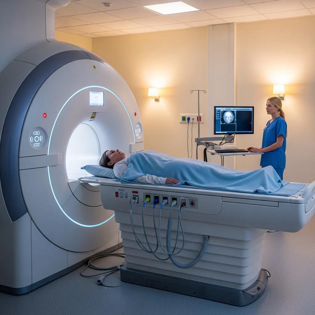
MRI (magnetic resonance imaging) creates detailed images by aligning hydrogen atoms in the body with a strong magnetic field, then using radiofrequency pulses to produce signals that are converted into images. It gives excellent soft tissue contrast without ionising radiation, so it’s very useful for neurological, spinal and musculoskeletal problems. Scans are performed with the patient lying in a bore that can be narrow and noisy; typical scan times range from 20 to 60 minutes depending on the area and protocol. Preparation usually means removing metal, completing a safety screen for implants, and fasting only when intravenous contrast is planned. Please tell your referrer and our team about implants or pregnancy before booking.
To make your visit smoother, follow these simple preparation steps.
- Leave jewellery and metallic items at home or remove them before the scan.
- Tell staff about implants, pacemakers or any metal fragments in your body.
- Only fast if you have been told contrast will be used; otherwise normal eating is fine.
These steps help avoid delays and improve image quality. You can Book Online or use Request An Appointment to arrange an MRI at Life Medical Imaging Central Coast; location‑specific guidance is provided during booking.
MRI safety and screening for children with implants
MRI is the preferred imaging tool for many paediatric conditions. While the MRI environment carries specific safety considerations, strict screening and established protocols mitigate risks and allow safe imaging in most children. Implanted medical devices can present additional challenges, so awareness of device-specific MRI safety and careful assessment are essential for safe practice.
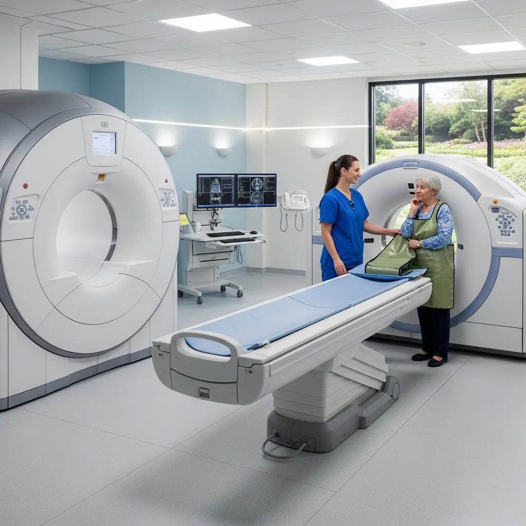
How does a CT scan work and when is it used?
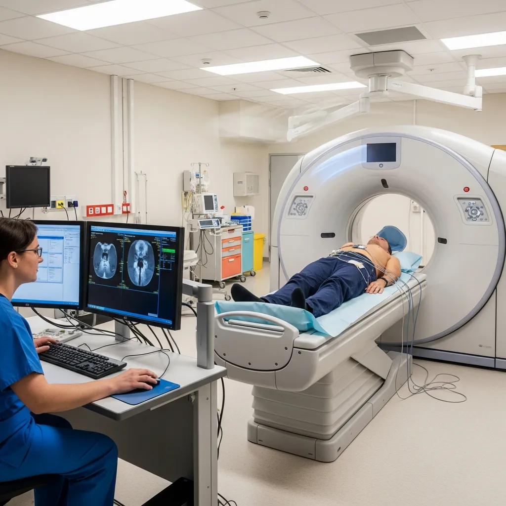
CT (computed tomography) captures multiple X‑ray projections around the body and reconstructs them into cross‑sectional images that show bone, air and soft tissues with high spatial resolution. Intravenous contrast may be used to highlight blood vessels and organs. CT is fast and commonly used in acute settings such as trauma, suspected pulmonary embolism or acute abdominal pain; specialised CT angiography assesses vascular and cardiac anatomy. Radiation exposure is an important consideration, but modern scanners offer ultra‑low dose protocols for specific indications — for example, lung screening or routine follow‑up — without compromising diagnostic quality. Most CT exams take five to twenty minutes; if contrast is planned we will check for allergies and kidney function beforehand.
Because CT uses ionising radiation, our clinicians apply dose‑optimisation strategies and consider alternatives where appropriate. That balance of benefit and risk guides consent and modality choice.
Predicting adverse reactions to CT contrast agents
Reactions to CT contrast are uncommon but can be serious. Research has looked at risk factors and scoring approaches to predict which patients are more likely to have adverse reactions to iodinated contrast, supporting safer decision making before administration.
If you need a CT, Life Medical Imaging Central Coast offers ultra‑low dose CT where suitable. Book Online or use Request An Appointment, and referrers can submit eReferrals for efficient scheduling.
Benefits and risks of common imaging modalities
Each modality brings distinct advantages and risks. Understanding these helps clinicians and patients choose the most appropriate test. MRI gives excellent soft tissue contrast without radiation; CT is fast and sensitive for acute problems; ultrasound is safe and dynamic; digital X‑ray is quick for fractures; and DEXA provides quantitative bone assessment. Main risks include ionising radiation with CT and X‑ray, MRI contraindications for some implants and claustrophobia, and rare contrast reactions for CT and MRI. We manage risk by using low‑dose protocols, choosing ultrasound when suitable, screening carefully and using child‑ and pregnancy‑friendly protocols.
The table below summarises benefits versus risks to support conversations with referrers and patients.
- Where diagnostic yield permits, consider ultrasound or MRI as radiation‑free alternatives.
- Use ultra‑low dose CT protocols for suitable indications to reduce exposure.
- Screen thoroughly for contrast allergies and renal risk before giving contrast agents.
Life Medical Imaging Central Coast is NATA‑accredited and offers low‑dose CT options as part of our safety and quality program. Patients and referrers can Book Online or Request An Appointment to discuss the most appropriate imaging plan.
What are the benefits and risks of MRI scans?
MRI’s main strength is its superior soft tissue contrast without ionising radiation, which makes it the preferred test for many neurological, musculoskeletal and soft tissue conditions. Risks centre on magnetic safety: some implants, metallic fragments and certain electronic devices may prevent MRI. Some patients find the bore confining or the scan uncomfortable if they must remain still for a long time. Gadolinium‑based contrast agents improve lesion characterisation but carry a small risk of allergic reaction and, rarely, concerns about long‑term deposition; we minimise risk by screening, choosing the lowest effective dose and using alternative sequences when possible. With correct screening and modern scanner designs, MRI is very safe, and options such as open bore scanners or sedation can help anxious patients.
Given these points, MRI is often the best choice for soft tissue evaluation unless specific contraindications or urgent clinical needs favour CT or ultrasound.
What are the benefits and risks of CT scans, including low‑dose options?
CT provides fast, high‑resolution images ideal for bony injury, lung disease and intra‑abdominal problems. CT angiography allows non‑invasive vascular assessment, including coronary vessels. The primary risk is ionising radiation; modern scanners and reconstruction algorithms, plus ultra‑low dose protocols for selected indications, substantially reduce exposure while maintaining diagnostic quality. Contrast improves lesion detection but requires screening for allergy and kidney function. Special care is taken with children and pregnant patients to avoid unnecessary radiation. Clinicians weigh urgency, diagnostic yield and patient vulnerability when choosing CT and favour low‑dose techniques where clinically appropriate.
These considerations highlight why accredited services and robust dose optimisation protocols are important — topics we cover in the provider section below.
How is ultrasound used in diagnostic radiology?
Ultrasound uses high‑frequency sound waves from a transducer to produce real‑time images, allowing dynamic assessment of organs, blood flow and musculoskeletal structures without ionising radiation. Its safety and portability make it first‑line for routine pregnancy monitoring, duplex Doppler vascular studies and musculoskeletal assessments with guided interventions. Limitations include operator dependence and reduced utility for gas‑filled or very deep structures, but the ability to image dynamically and at the bedside is a major advantage. Preparation varies by exam — for example, fasting for abdominal scans or a full bladder for pelvic studies — so following pre‑scan instructions improves diagnostic quality.
- Ultrasound is safe and radiation‑free, making it suitable for pregnancy and paediatric use.
- It provides real‑time dynamic views and Doppler assessment of blood flow.
- It can guide procedures such as injections or biopsies under direct visualisation.
Below we outline common women’s and paediatric uses and how vascular and MSK ultrasound support diagnosis and treatment.
Common uses of ultrasound in women’s and paediatric health
In women’s health, ultrasound is essential for early pregnancy dating, anatomy surveys, 3D/4D obstetric imaging and gynaecological assessment of the uterus and ovaries. Breast ultrasound complements mammography for evaluating palpable lumps and dense breasts. For children, ultrasound avoids radiation and often removes the need for sedation by using child‑friendly techniques and quicker protocols; common paediatric uses include appendicitis assessment, renal imaging and neonatal cranial scans. Timing matters in obstetrics — first‑trimester scans check viability and dating, second‑trimester scans review fetal anatomy, and third‑trimester scans monitor growth and placental position. Bring any prior imaging to your appointment and follow instructions such as keeping a full bladder for pelvic scans. You can Book Online or Request An Appointment to choose the most suitable clinic location.
Next we describe how vascular and musculoskeletal ultrasound are used in diagnosis and intervention.
How do vascular and musculoskeletal (MSK) ultrasound help diagnosis?
Vascular ultrasound uses duplex Doppler to measure flow velocity and waveform patterns, helping diagnose deep vein thrombosis, carotid stenosis and peripheral arterial disease; it is non‑invasive and suitable for repeat monitoring. MSK ultrasound visualises tendons, ligaments, bursae and small joints dynamically to detect tears, tendinopathy and fluid collections, and it guides injections and aspirations in real time. MSK ultrasound is convenient and cost‑effective for many problems, though MRI may be better for deep intra‑articular or complex soft tissue pathology. Clinicians select vascular or MSK ultrasound when dynamic information or image guidance is needed, and patients can request these tests via Book Online or Request An Appointment at Life Medical Imaging Central Coast.
What are the types and applications of X‑ray imaging?
Digital X‑ray records planar images with electronic detectors rather than film, giving faster processing, immediate review and better post‑processing tools. Common uses include chest radiographs, limb fracture series and dental panoramic (OPG) images. Digital systems reduce repeat rates, support secure archiving and speed clinical decisions in emergency and outpatient settings. Although X‑ray delivers lower radiation than CT, it is less sensitive for soft tissue pathology, so it’s often a first‑line test before escalating to CT or MRI when needed. Knowing when an X‑ray is sufficient helps streamline diagnostic pathways for patients and referrers.
- Faster image acquisition and immediate review support quicker diagnosis.
- Lower repeat rates and digital processing improve image quality and workflow.
- Digital storage and sharing streamline multidisciplinary care.
These practical advantages make X‑ray the pragmatic first step for many trauma assessments before further imaging.
Advantages of digital X‑ray versus traditional film
Digital X‑ray converts X‑ray photons to digital signals that radiographers can process to enhance contrast, take measurements and annotate instantly. Automatic exposure controls and improved detector efficiency typically reduce patient radiation compared with older film techniques. Digital storage integrates with PACS for quick comparison with prior studies, helping clinicians track healing or disease progression. For patients, this means shorter waits and fewer repeats — practical benefits that improve care and convenience.
Because of these strengths, X‑ray remains the default first test in many trauma situations before CT or MRI is used for further characterisation.
How are X‑rays used for fractures and dental conditions?
For suspected fractures, targeted X‑ray views localise cortical breaks, joint alignment and displacement to guide immediate management such as immobilisation or surgical referral. If an occult fracture or complex anatomy is suspected, CT offers complementary high‑resolution cross‑sectional detail. Dental panoramic radiographs (OPG) provide an overview of tooth roots, jaw pathology and impacted teeth to guide dental and oral surgery planning. Referrers should include a clear clinical history and the suspected region to ensure the correct imaging series and avoid unnecessary exposure. Timely reporting and follow‑up imaging instructions ensure evolving diagnoses are captured appropriately.
From here, we turn to bone densitometry, a specialised low‑dose test for assessing bone health.
Why is bone densitometry important in diagnostic radiology?
Bone densitometry (DEXA) measures bone mineral density to assess fracture risk and diagnose osteoporosis. It provides quantitative results that directly inform prevention and treatment decisions. DEXA compares values to young adult norms (T‑scores) and age‑matched standards (Z‑scores) to classify bone health and guide management such as lifestyle changes, calcium and vitamin D optimisation or pharmacologic therapy. The scan is quick, painless and uses very low radiation, so it’s suitable for baseline screening and follow‑up. Typical indications include postmenopausal women, older adults with prior fractures and conditions that increase bone loss; reports usually include T‑score interpretation and follow‑up recommendations.
The table below compares DEXA with other bone assessment methods to clarify its role.
How does bone mineral density scanning detect osteoporosis?
DEXA measures BMD at the lumbar spine and hip and reports T‑scores and Z‑scores that clinicians use to classify normal bone density, osteopenia or osteoporosis. A T‑score of −2.5 or lower typically indicates osteoporosis and increased fracture risk. By providing objective baseline values and tracking changes over time, DEXA helps guide lifestyle advice, supplementation and medical therapy. Results can be combined with clinical risk tools to estimate 10‑year fracture risk and to monitor treatment response at recommended intervals. These practical outputs mean DEXA directly influences prevention and management for patients at risk of fragility fractures.
Knowing what the test shows helps you prepare and discuss results with your clinician.
What should patients expect during a bone densitometry scan?
A DEXA scan usually takes 10–20 minutes, is painless and involves lying on a scan table while a scanning arm passes over the hip and spine. Wear loose clothing without metal and avoid recent contrast studies that might affect results. Radiation exposure is very low — a small fraction of a chest X‑ray — and no IV contrast or sedation is required. Reports typically include T‑score and Z‑score values with an interpretive comment and follow‑up recommendations from the reporting radiologist. Book Online or Request An Appointment to schedule a DEXA and discuss result implications with your referrer or our imaging clinicians.
Why choose Life Medical Imaging Central Coast for diagnostic radiology?
Life Medical Imaging Central Coast is an independent, NATA‑accredited radiology practice serving the Central Coast from four locations: Bateau Bay, Killarney Vale, Umina Beach and Erina. We specialise in women’s and cardiac imaging and offer ultra‑low dose CT, digital X‑ray, a full suite of ultrasound services (general, vascular, musculoskeletal, obstetric and gynaecological), image‑guided interventional procedures, paediatric imaging, CT angiography, DEXA bone densitometry and 3D/4D obstetric ultrasound. Patients and referrers can Book Online or Request An Appointment, and referrers may use eReferral pathways to streamline scheduling. These options support timely access while meeting our quality and safety standards.
Below we outline the expertise and technology that support our clinical care.
What expertise and technology set our radiologists apart?
Our radiologists and imaging team have strong clinical experience in women’s and cardiac imaging and use modern equipment such as ultra‑low dose CT and advanced ultrasound platforms for 3D/4D obstetric studies. NATA accreditation reflects our commitment to quality assurance and technical standards for imaging and reporting, which supports diagnostic confidence and patient safety. We offer specialised studies like CT angiography and image‑guided procedures, and we follow dose‑optimisation and screening protocols to protect patients. Together these capabilities support accurate diagnosis and seamless patient care.
How can patients book appointments and access multiple locations?
You can Book Online or use Request An Appointment to choose the most convenient clinic — Bateau Bay, Killarney Vale, Umina Beach or Erina — based on the services you need. Referrers may submit eReferrals with clinical details that help us tailor protocols and schedule appropriately. For specific preparation advice — for example MRI safety screening, CT contrast precautions, ultrasound preparation or DEXA pre‑scan instructions — please review the location‑specific notes when booking or discuss them with your referrer. Clear booking pathways reduce delays and help ensure imaging supports timely clinical care.
These practical access options close the loop between clinical need and imaging delivery, helping clinicians and patients get the answers they need.
Frequently asked questions
What should I wear for my imaging appointment?
Wear loose, comfortable clothing without metal fastenings such as zippers or large buttons, since metal can interfere with images. For MRI you will usually change into a gown provided by the facility to ensure no metal is present. For DEXA, avoid clothing with metal components. If you’re unsure, check the pre‑scan instructions or ask when you book.
How can I prepare for a CT scan?
Preparation varies by the type of CT. Commonly you may be asked to avoid food and drink for a few hours before the scan if contrast is planned. Tell the imaging team about any allergies (especially to iodine) and any medications you take. Follow any specific instructions provided at booking to ensure the best possible images and safety.
Are there any age restrictions for imaging procedures?
Most imaging procedures have no strict age limits, but we adapt protocols for children and older adults. For children we focus on minimising radiation and using child‑friendly techniques; for elderly patients we may adjust preparation and monitoring to account for other health issues. Discuss individual needs with your referrer or our team.
What are the potential side effects of contrast agents used in imaging?
Contrast agents for CT and MRI are generally safe. Mild reactions can include nausea, a warm sensation or a metallic taste. Serious allergic reactions are rare. Patients with a history of allergies or kidney problems should tell the imaging team before the procedure so we can take precautions and monitor appropriately.
How long will it take to receive my imaging results?
Timing depends on the exam and the facility’s reporting workflow. Preliminary findings may be available within hours, while final radiology reports typically arrive within one to three days. Your referrer will discuss results and next steps with you once the report is complete.
Can I bring someone with me to my appointment?
Yes — companions are usually welcome for support. For safety reasons, companions may be asked to remain outside the MRI room or scanning area. Check our location policies when you book if you need someone to accompany you.
What should I do if I have claustrophobia and need an MRI?
If you have claustrophobia, tell your referrer and our team in advance. We can offer options such as open bore scanners, ear protection and music, or sedation in selected cases to make the scan more comfortable. Discuss your concerns so we can plan the best approach for you.
Conclusion
Knowing how MRI, CT, ultrasound, X‑ray and DEXA work helps you and your clinician choose the right test for diagnosis and treatment planning. Each modality has strengths and limitations, and our team at Life Medical Imaging Central Coast is here to guide you through preparation, safety and booking. To discuss the best imaging options for your care, Book Online or Request An Appointment — your next step toward an accurate diagnosis starts with the right scan.