CT Scans: A clear guide to what they are and how they work
Computed tomography (CT) uses rotating X‑ray beams and computerized reconstruction to produce detailed cross‑sectional images of the body. This guide explains how CT scanners form images, outlines common scan types and clinical uses, and gives practical preparation steps so patients know what to expect. We also address common questions about radiation dose, contrast safety and low‑dose CT options with evidence‑based explanations and clear, actionable advice. You’ll find comparisons of scan types, checklists for contrast and pregnancy considerations, and a summary of how CT supports early diagnosis and surgical planning. Later sections cover dose‑reduction technology and local booking options on the Central Coast so the information moves from technical clarity to practical next steps. Read on to learn what a CT scan does, how to prepare, what risks and benefits to consider, and how to arrange a scan if needed.
What Is a CT Scan and How Does It Work?
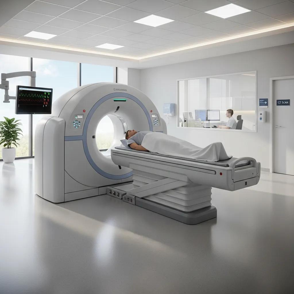
A CT scan is a diagnostic test that acquires multiple X‑ray projections around the body and uses computer algorithms to reconstruct them into cross‑sectional images. The images rely on differences in how tissues attenuate X‑rays, creating contrast between organs, bone and air. Compared with plain X‑rays, CT gives cross‑sectional detail and allows three‑dimensional reconstructions that help detect subtle disease and guide procedures. The sections that follow outline the basic physics behind computed tomography and the steps by which X‑rays and software produce diagnostically useful images.
What Is Computed Tomography and Its Basic Principles?
Computed tomography—literally “computed slicing”—collects projection data while an X‑ray source and detectors rotate around the patient to capture multiple angles. Reconstruction algorithms convert those projections into thin image slices; stacking slices yields a volumetric dataset, much like slicing a loaf of bread to inspect each piece. The key principles are attenuation (how tissues absorb X‑rays) and contrast based on density differences—bone attenuates strongly, air very little, and soft tissues in between. These properties let radiologists distinguish structures and apply windowing and tailored reconstruction kernels for specific clinical questions.
How Do X-rays and Computer Processing Create Detailed Images?
During a scan an X‑ray tube emits a fan or cone beam that passes through the body while ring‑shaped detectors record transmitted photons. Reconstruction methods—historically filtered back‑projection and now commonly iterative or model‑based techniques—process the raw measurements into voxel values representing tissue attenuation. Voxels form a 3‑D grid that can be viewed in axial, coronal or sagittal planes and rendered in 3‑D for surgical planning or vascular assessment. Improvements in detector design and reconstruction software boost spatial resolution and reduce noise, allowing clearer images at lower radiation doses and supporting better clinical decisions.
What Are the Different Types of CT Scans Available?
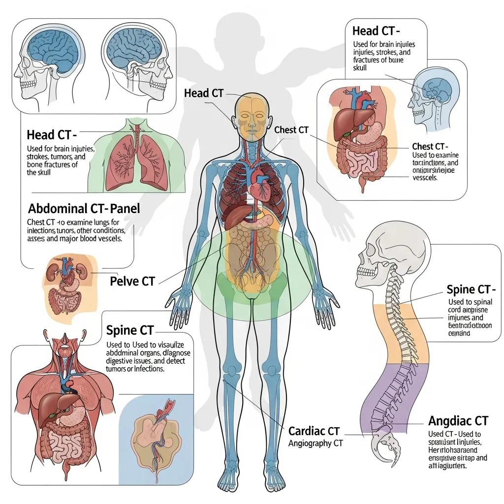
CT is used across many clinical needs, with protocols tuned to specific body regions and diagnostic aims. Common exams include head CT for stroke and trauma, chest CT for lung disease and pulmonary embolism, abdominal CT for infection or cancer staging, spinal CT for fractures, and vascular studies such as CT angiography. Cardiac CT and CT angiography use ECG‑gated techniques to reduce motion, while low‑dose CT protocols focus on minimising radiation for screening or follow‑up. The table below helps patients and referrers match indications, typical durations and contrast requirements.
Different CT modalities suit different clinical questions and patient circumstances.
This table summarises how common CT types differ by indication, typical scan time and contrast needs to guide appropriate protocol selection.
Understanding protocol differences helps ensure the right scan is ordered for each clinical question.
Different facilities offer different services; Life Medical Imaging Central Coast provides a range of CT studies including General CT, Cardiac CT and CT Angiography to meet many diagnostic needs. These options let referring clinicians choose protocols that match the clinical indication, while local patients can access specialist cardiac and vascular imaging close to home.
What Are General, Cardiac, and CT Angiography Scans?
General CT covers standard body‑region protocols—head, chest, abdomen or pelvis—used for trauma assessment, infection work‑ups and tumour detection, and it often needs minimal preparation. Cardiac CT uses ECG‑gating to synchronise imaging with the heartbeat and reduce motion artefact for clearer views of coronary arteries and cardiac chambers. CT Angiography (CTA) is timed to opacify arteries or veins after intravenous contrast, enabling non‑invasive assessment of stenosis, embolism or aneurysm. Each modality balances scan speed, spatial resolution and contrast timing to answer specific clinical questions, and selecting the correct study maximises diagnostic yield.
How Do Specific CT Scans Target Different Body Areas?
Protocol changes—slice thickness, contrast timing, patient positioning and reconstruction kernel—tune image quality to each region and problem. For example, head CT uses thin slices and bone windows for trauma, chest CT for lung parenchyma uses sharp kernels, and cardiac CT requires ECG‑gating with controlled breathing. Contrast timing matters: arterial‑phase CTA captures vessels shortly after injection, while portal‑venous abdominal scans show parenchymal enhancement. Knowing these differences helps patients follow preparation instructions and helps referrers order the most informative study.
How Should You Prepare for a CT Scan?
Good preparation improves image quality and safety, especially when intravenous contrast is planned. Key steps include fasting windows for contrast studies, medication advice (for example, metformin precautions), removing jewellery and bringing prior imaging or referrals. Screening for kidney function (eGFR) and contrast allergies is part of pre‑scan safety checks, and pregnant patients should notify their clinician so alternatives can be considered. The sections below give a practical checklist and focused guidance on contrast and pregnancy to help you prepare confidently.
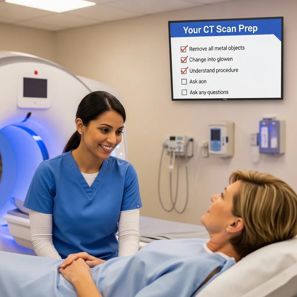
Follow these steps to prepare effectively for your CT appointment.
- Arrive on time with your referral and any prior imaging to help comparison and reporting.
- Remove metal items and wear comfortable clothing without metal fastenings.
- Follow fasting instructions—typically no food for 4 hours before contrast studies; clear fluids usually allowed unless advised otherwise.
- Inform staff about allergies, kidney disease, diabetes medications and pregnancy.
- Bring a list of current medications and a form of photo identification.
These checklist steps reduce delays, support safe contrast administration and help ensure the scan meets the clinical need.
Before the practical checklist, the following table summarises common preparation items, why they matter and required actions.
This table clarifies typical preparatory steps and helps patients complete any actions required before attendance.
If you need facility‑specific instructions or printable preparation sheets, Life Medical Imaging Central Coast provides patient resources and booking guidance. Contact the clinic in advance if you have recent kidney tests, a history of allergic reactions, or any possibility of pregnancy so staff can advise on alternatives or premedication.
What Are the Key Preparation Steps Before a CT Scan?
Key preparation reduces risk and helps achieve the diagnostic goal. Confirm your referral and bring prior images. Follow fasting guidance for contrast‑enhanced studies—usually no solid food for around four hours while staying hydrated with clear fluids unless told otherwise. Remove jewellery and metal, wear clothes that are easy to change, and bring a medication list and any recent eGFR results. Finally, tell staff about allergies or pregnancy so the team can take appropriate precautions or recommend alternative imaging if needed.
What Are the Considerations for Contrast Material and Pregnancy?
Intravenous iodinated contrast improves vessel and organ visualisation but carries small risks, including allergic reactions and potential kidney effects in vulnerable patients. Reactions are uncommon and usually mild (nausea, itching); severe reactions are rare. Pre‑scan screening and premedication protocols reduce risk for patients with prior reactions. Kidney function (eGFR) guides contrast decisions—those with impaired renal function may need alternative imaging or specialist input. Pregnant patients should inform their referrer and the imaging team because non‑ionising alternatives like ultrasound or MRI may be preferred; if CT is essential, shielding and dose‑minimised protocols will be used.
What Are the Risks and Benefits of CT Scans?
CT scans deliver rapid, detailed images that support diagnosis, staging and treatment planning, but they also involve ionising radiation and occasional contrast‑related effects. Benefits include early tumour detection, trauma assessment and non‑invasive vascular mapping that can replace invasive angiography. Radiation dose varies by study and body region; modern low‑dose techniques significantly reduce exposure while keeping diagnostic quality for many indications. The pros and cons below summarise the main points to help with informed decisions.
CT provides important clinical advantages and is best used when the expected benefit outweighs the small associated risks.
- Benefits:Fast, high‑resolution images that improve diagnostic accuracy and clinical decision‑making.Cross‑sectional and 3‑D reconstructions that guide surgery, intervention and staging.Non‑invasive vascular assessment via CT angiography often replaces more invasive tests.
- Risks:Exposure to ionising radiation—low per study but accumulative over multiple exams.Small risk of allergic reaction or kidney effects from iodinated contrast in susceptible patients.Motion artefact or suboptimal studies if preparation or timing is inadequate.
These balanced points help patients and clinicians weigh diagnostic value against potential harms and lead into quantitative comparisons and mitigation strategies.
This table presents a transparent risk‑benefit comparison and shows how mitigation strategies reduce adverse outcomes while preserving diagnostic benefit.
What Are the Radiation Exposure and Contrast Reaction Risks?
Radiation dose varies by study: a head CT generally gives a lower effective dose than an abdominal CT. Dose can be compared to average annual background radiation to provide perspective. Modern scanners and dose‑reduction technology substantially lower exposure compared with older equipment, and clinicians weigh the small individual risk against the diagnostic benefit. Contrast reactions affect a small percentage of patients and are most often mild and manageable; severe reactions are rare. Pre‑scan screening, hydration and established premedication and emergency protocols further reduce the risk of adverse events.
How Do CT Scans Aid in Early Diagnosis and Treatment Planning?
CT enables fast detection of life‑threatening conditions such as intracranial haemorrhage, major trauma, pulmonary embolism and acute abdominal emergencies, often directly changing clinical management in emergency settings. In oncology, CT staging defines tumour size, nodal involvement and distant spread to guide surgery, radiotherapy and systemic therapy. Vascular CT angiography non‑invasively maps stenoses and aneurysms to support endovascular planning and surveillance. These applications explain why timely, appropriate CT can be decisive for patient outcomes and why clinicians order CT when it will influence care.
What Is Low Dose CT Technology and Why Is It Important?
Low‑dose CT reduces ionising radiation exposure through hardware improvements, more sensitive detectors and advanced reconstruction algorithms, while keeping diagnostic image quality for many indications. Common techniques include iterative and model‑based reconstruction, tube current modulation, improved detector efficiency and AI‑assisted denoising—all of which allow lower photon counts without excessive image noise. Low‑dose CT is particularly valuable for screening programs, follow‑up imaging and younger patients where cumulative exposure is a concern; evidence supports its use in appropriately selected scenarios. The list below summarises common dose‑reduction methods used in modern practice.
- Iterative reconstruction: Repeated modelling to reduce noise and improve image quality at lower dose.
- Tube current modulation: Adjusts X‑ray output in real time based on patient size and anatomy.
- Improved detectors: Higher sensitivity detectors capture more information from fewer photons.
- AI denoising: Post‑processing algorithms reduce noise while preserving anatomical detail.
These methods work together to maintain image quality while reducing exposure, which is why centres offering low‑dose CT can deliver safer imaging without compromising diagnostic accuracy.
How Does Low Dose CT Reduce Radiation Without Compromising Quality?
Low‑dose CT takes a system‑wide approach: better hardware to capture more signal, software that suppresses noise, and reconstruction methods that extract reliable attenuation values from fewer photons. Iterative reconstruction refines image models to separate true signal from noise, enabling lower tube current settings. AI denoising and model‑based reconstructions further boost signal‑to‑noise, preserving edge detail and soft‑tissue contrast. Clinical validation and quality assurance show that for many indications—such as lung screening and routine follow‑up—low‑dose protocols deliver adequate diagnostic information while substantially lowering patient dose.
Why Choose Life Medical Imaging Central Coast for Low Dose CT Scans?
Life Medical Imaging Central Coast prioritises patient safety through ultra‑low dose, high‑definition CT equipment and maintains NATA accreditation to support quality assurance. We offer subspecialist reporting in Women’s and Cardiac Imaging, comfortable modern clinics, and convenient online booking and enquiry options to simplify access. For patients seeking low‑dose imaging, choosing a facility with advanced equipment and subspecialist reporting helps ensure the correct protocol and accurate interpretation. If you’re unsure whether low‑dose CT is suitable for your situation, contact the clinic to discuss protocol choices and preparation.
How Can You Book a CT Scan at Life Medical Imaging Central Coast?
Booking a CT scan with Life Medical Imaging Central Coast is straightforward. We accept referrals and enquiries by phone and online, with multiple Central Coast locations available. When you arrange a scan, have your referral, any prior imaging and recent kidney function results ready if contrast is planned—this helps with pre‑scan triage and safety checks. The clinic operates at sites including Bateau Bay, Killarney Vale, Umina Beach and Erina. The steps below outline the typical booking and arrival process so you know what to expect from referral to scan.
- Obtain a referral from your clinician specifying the clinical indication and requested CT type.
- Call the clinic phone number to schedule a convenient appointment time for the appropriate CT protocol.
- Provide recent blood test results (eGFR) and allergy history when booking if contrast is anticipated.
- Follow pre‑appointment instructions from the clinic and arrive with your referral and previous imaging for comparison.
These steps streamline booking and preparation. After the scan, the clinic will report findings to the referring clinician.
What Are the Clinic Locations and Contact Details for Booking?
Life Medical Imaging Central Coast serves the region from multiple locations: Bateau Bay, Killarney Vale, Umina Beach and Erina, so you can choose the most convenient site for your CT. For appointment enquiries and to discuss preparation or contrast concerns, call 02 4326 7000 and clinic staff will advise on available appointments and preparation instructions. When calling, have your referral details and any recent blood tests handy to speed booking and screening. Dedicated cardiac and women’s imaging subspecialists are available across these sites to support targeted protocols when needed.
How Does the Online Appointment Booking Process Work?
The online booking system lets you request appointments and provide preparation details securely. During booking you’ll be asked to enter referral information, the clinical question and any allergies or recent kidney function tests so we can complete pre‑scan screening. After submission, clinic staff will confirm your appointment, advise specific preparation instructions such as fasting or medication management, and advise if any additional tests are needed before your scan. This digital workflow complements phone booking and helps ensure all safety checks are in place before attendance.
Frequently Asked Questions
What should I expect during a CT scan procedure?
You’ll lie on a table that moves through a wide, doughnut‑shaped scanner. The actual imaging is usually quick—often just a few minutes—but you may be asked to hold your breath briefly to reduce motion blur. The scanner makes clicking and whirring sounds as it captures images. Stay still and follow the radiology team’s instructions to get the best quality images.
Are there any alternatives to CT scans?
Yes. Depending on the clinical situation, ultrasound or MRI may be suitable alternatives. Ultrasound is useful for many soft‑tissue and abdominal problems and uses no ionising radiation. MRI gives detailed soft‑tissue contrast and is often preferred for neurological and musculoskeletal assessments. Your clinician will recommend the best modality based on the clinical question, patient history and urgency.
How often can I safely have a CT scan?
Frequency depends on your clinical needs. CT scans are valuable diagnostic tools but involve ionising radiation, so benefits should outweigh risks. Your healthcare team will consider medical history, prior imaging and the reason for the scan when deciding how often repeat CT is appropriate. Keep an open dialogue with your referrer to make informed choices.
What happens if I have a contrast reaction during the scan?
If you have a reaction to contrast, the radiology team is trained and equipped to manage it. Mild reactions (nausea, itching) are most common and easily treated; severe reactions are rare. Staff monitor patients during and after contrast administration and follow emergency protocols if needed. Tell staff immediately if you feel unwell or notice any symptoms.
Can I eat or drink before a CT scan?
That depends on the scan. For exams requiring intravenous contrast, you’ll usually be asked to avoid solid food for about four hours beforehand; clear fluids are typically allowed. Always follow the specific instructions given by the imaging facility or your referrer to ensure a safe, effective scan.
What should I do if I am pregnant or think I might be pregnant?
If you are pregnant or may be, tell your referrer and the imaging staff before the scan. Because CT uses ionising radiation, alternative imaging—such as ultrasound or MRI—may be preferable when clinically appropriate. If CT is essential, the team will take steps to minimise fetal exposure.
How can I access my CT scan results?
After your CT, a radiologist will review the images and prepare a report that is usually sent to the referring clinician, who will discuss results with you. Some facilities also provide online patient portals to view reports and images. If you have questions about your results, contact your referring doctor for clarification and next steps.
Conclusion
CT scans give detailed, timely information that supports diagnosis and treatment planning using advanced imaging technology. Knowing the different scan types, preparation steps and associated risks helps you make informed decisions about your care. If you’re considering a CT scan, Life Medical Imaging Central Coast offers expert services tailored to individual needs. Contact us to discuss your options and how we can support your healthcare journey.