CT Scans — What They Are, How They Work, and How to Prepare
A CT (computed tomography) scan is a quick, non‑invasive imaging test that combines X‑rays and computer processing to create detailed cross‑section images of the body. This guide answers common questions: what CT scans do, how they work, the main types of CT studies, how to prepare, and what to expect before, during and after your appointment. We’ve written this to help reduce worry and give clear, practical advice about contrast, fasting and radiation. You’ll find a short technical overview, a plain‑language explanation of common CT types, a step‑by‑step preparation checklist, an honest look at benefits and risks, and a walkthrough of the patient experience and reporting process. If you’re on the Central Coast, Life Medical Imaging Central Coast is NATA accredited and uses ultra‑low dose, high‑definition CT — you can ask about appointments, eReferrals and online booking when you’re ready.
What Is a CT Scan and How Does It Work?
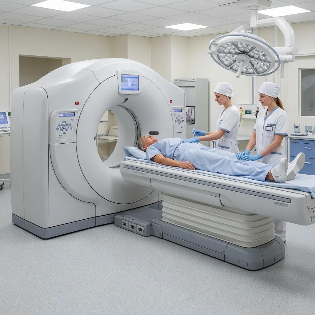
A CT scan takes many X‑ray images from different angles and uses computer software to build cross‑sectional “slices” of the body. An X‑ray tube sends beams through the body and detectors measure what comes out the other side. Reconstruction software turns those measurements into high‑contrast images that help doctors spot injuries, tumours or vascular problems. Compared with a single‑view X‑ray, CT shows bone, soft tissue and blood vessels with much more detail, which is why it’s so useful in emergencies and in planned diagnostic care. Knowing the basic parts of a CT scanner also explains why staff ask you to lie a certain way or hold your breath during some scans — these steps reduce motion and help produce clearer images.
What Does Computed Tomography Mean?
Computed tomography literally means “computerised slicing.” The scanner captures many thin X‑ray slices and the computer stacks them into a detailed image — like slicing a loaf of bread to inspect each piece. This sectional view makes it easier to find internal bleeding, fractures, tumours and other organ problems that a single X‑ray might miss. That’s why clinicians often choose CT when they need a clear picture of internal anatomy to guide treatment.
How Do X-rays and Computer Processing Create Detailed Images?
The X‑ray tube sends a rotating fan or cone of X‑rays through the body to a ring of detectors. Different tissues—bone, muscle, fat, fluid—absorb X‑rays differently, creating raw data the computer uses to reconstruct images. Reconstruction methods have evolved from filtered back‑projection to iterative and AI‑assisted techniques; these give cleaner images with lower radiation dose. The main parts you’ll hear about are the gantry (which contains the tube and detectors), the patient table, and the software that processes images and manages dose. In many studies we also use contrast medium to better distinguish blood vessels and enhance suspicious areas, which helps with diagnosis and treatment planning.
What Are the Different Types of CT Scans Offered?
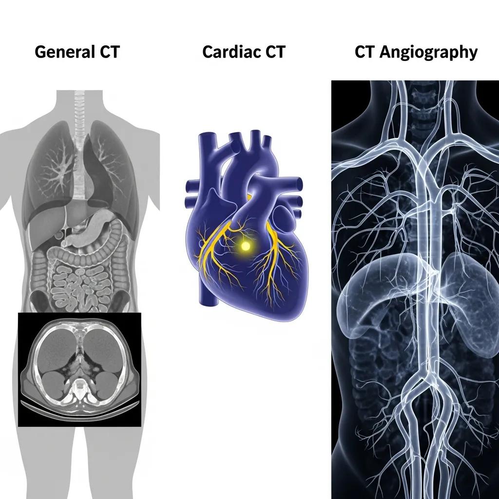
CT is a flexible tool used for different clinical questions. Common study types include General CT (head, chest, abdomen), Cardiac CT (coronary assessment and calcium scoring) and CT Angiography (CTA) for detailed vascular imaging. Each exam uses the same scanner but with different settings — for example, ECG gating for heart scans or specific timing of contrast injections for vascular studies. These differences affect how you prepare, how long the appointment takes, and the diagnostic information the scan provides. At Life Medical Imaging Central Coast we offer General CT, Cardiac CT and CTA on ultra‑low dose, high‑definition systems to balance image quality and patient safety.
Here’s a simple comparison to help you see the main differences.
What Is a General CT Scan and When Is It Used?
General CT is a fast, versatile study for major body regions. It’s routinely used in emergency departments to check for brain bleeds or stroke, to look for lung infections or nodules, and to investigate abdominal pain such as appendicitis, obstruction or organ injury. During the scan you lie on a motorised table that moves through the gantry while the scanner rotates around you; some exams need short breath‑holds to reduce motion. Knowing these typical uses helps patients understand why a clinician has ordered CT and what to expect on the day.
How Does a Cardiac CT Scan Help Assess Heart Health?
Cardiac CT images the coronary arteries and heart structures using ECG‑gated acquisition that synchronises images with the heartbeat to reduce motion blur. It can measure coronary calcium (a marker of atherosclerosis) and perform non‑invasive coronary angiography to identify significant narrowings. Some cardiac CTs require heart‑rate control medication and precise breath‑holding to get optimal images. The result is detailed anatomical information that complements stress testing and echocardiography, often without the need for invasive catheterisation.
What Is CT Angiography and How Does It Visualise Blood Vessels?
CT Angiography (CTA) uses timed intravenous contrast injections and rapid image capture to show arteries and veins in high resolution. CTA is the test of choice for suspected aneurysm, vessel narrowing, dissection or pulmonary embolus because it provides 3D views of the vascular tree quickly and non‑invasively. Contrast timing and injection rates are tailored to the area of interest, and radiologists review multiplanar images to assess vessel patency and disease extent. This clear vascular map supports surgical planning, endovascular treatment decisions and fast triage in acute settings.
How Should Patients Prepare for a CT Scan?

Preparation depends on the type of CT. Common steps include fasting for some abdominal studies, pausing or timing specific medications, staying hydrated, and removing jewellery or metal that can affect images. Good preparation improves accuracy and safety, especially when IV contrast is planned — we may need recent kidney tests and information about allergies. Your referrer or our imaging team will give clear, personalised instructions; bringing prior imaging and medical records helps radiologists compare studies and report more accurately. Life Medical Imaging Central Coast accepts eReferrals and offers online booking to make preparation simple.
Below is a short checklist of common preparation items.
- Confirm appointment and eReferral: Make sure your referral is lodged and read any pre‑test instructions from the clinic.
- Follow fasting guidelines: Observe fasting times for abdominal or contrast studies as instructed.
- Tell us about medications and allergies: Share details of medicines, allergies and recent kidney tests.
- Bring prior imaging and ID: Previous scans and identification speed up registration and reporting.
These steps help avoid delays and keep contrast procedures safe. More detailed guidance follows.
What Are the Fasting and Medication Guidelines Before a CT Scan?
For abdominal or pelvic CT we usually ask you not to eat solid food for 4–6 hours to reduce bowel gas and help contrast work as intended; clear fluids are often allowed unless we tell you otherwise. Most routine medications can be taken as normal, but drugs such as metformin may need timing adjustments when IV contrast is used — your referrer or our team will advise. If you have diabetes or complex medication needs, get personalised instructions before the appointment. Clear communication about medications and recent blood tests helps us follow safety best practice and reduces the chance of post‑contrast issues.
Pre-procedural Fasting for Contrast-Enhanced CT: Evidence and Guidelines
Recent guideline reviews (including ESUR v10.0 and ACR 2021) have questioned routine pre‑procedural fasting before standard IV iodinated contrast administration. Evidence suggests that prolonged fasting is not necessary for most patients and that non‑fasting does not increase the risk of aspiration or emetic complications in routine contrast‑enhanced CT. That said, fasting remains appropriate for certain examinations and selected patients; clinical judgement and local protocols should guide practice. These findings support more patient‑friendly preparation where safe and appropriate.
When and Why Is Contrast Medium Used in CT Scans?
Contrast medium (IV or oral) makes blood vessels, organ perfusion and abnormal tissue more visible by altering how X‑rays pass through those structures. IV contrast is routine for CTA and many contrast‑enhanced CTs to highlight vessels, inflammation or tumours; oral contrast can outline bowel loops when needed. Before giving contrast we review kidney function and allergy history to spot contraindications or to plan premedication. Mild effects such as warmth or a metallic taste are common and short‑lived; serious reactions are rare and our team is trained to manage them. Clear explanation of the purpose and safety checks helps you give informed consent.
What Are the Benefits and Risks of CT Scans?
CT offers fast, high‑resolution images that are critical for emergency care, cancer staging, vascular assessment and surgical planning. The main risks are exposure to ionising radiation and, less commonly, contrast reactions. Modern ultra‑low dose, high‑definition CT systems significantly lower radiation while keeping diagnostic quality high. Clinicians follow two key principles: justification (only scan when the benefit outweighs the risk) and optimisation (use the lowest reasonable dose). If you have concerns, ask your referrer about alternatives and about your cumulative exposure history.
How Does Ultra-Low Dose Technology Reduce Radiation Exposure?
Ultra‑low dose CT uses better detectors, smarter acquisition settings and advanced reconstruction (including iterative and AI‑assisted methods) to produce diagnostic images with fewer X‑ray photons. These computational techniques clean up image noise so clinicians can still see small findings without exposing you to the levels required by older scanners. Newer technologies like photon‑counting detectors and deep‑learning reconstruction are further improving dose efficiency and image contrast, letting us answer clinical questions with even less radiation.
What Are the Diagnostic Advantages of High-Definition CT Imaging?
High‑definition CT improves spatial resolution and contrast discrimination, helping us detect and characterise small lesions, subtle fractures and fine vascular detail. That means earlier detection of small lung nodules, clearer staging of tumours, and more precise mapping of coronary calcification or vascular disease — information that can change management. Better images also help plan surgery or interventional procedures by showing anatomy clearly, which can shorten the diagnostic pathway and support timely, targeted treatment.
What Are the Potential Risks and How Are They Managed?
The main risks are radiation exposure, contrast‑related adverse events, and incidental findings that may lead to extra tests. We manage these risks by only scanning when clinically justified, using optimised low‑dose protocols, screening for contrast risks, and providing structured reports with clear follow‑up recommendations. When incidental findings appear, radiologists and referrers work together to recommend sensible next steps that avoid unnecessary interventions while ensuring important issues are followed up. Open communication between radiology teams, referrers and patients helps balance diagnostic need with cautious, evidence‑based care.
What Happens During the CT Scan Procedure?
On the day of your scan you’ll arrive and register, confirm medical history and any allergies or kidney tests, and receive a short briefing. You’ll be positioned on the table, which moves through the gantry while the scanner rotates. You may hear mechanical noises and you might be asked to hold your breath briefly for chest or abdominal scans. Radiographers operate the scanner from a control room but maintain visual and voice contact, and they will administer contrast if required. Radiologists read and report the images after acquisition. Knowing this sequence — arrival, briefing, scanning, short observation if needed — helps reduce anxiety and sets realistic expectations for how the appointment will feel and how long it will take.
- Arrival and registration: Check in, answer screening questions and confirm consent.
- Preparation and positioning: Staff position you and explain breath‑hold instructions.
- Image acquisition: The scan itself usually takes seconds to minutes depending on the exam.
- Post‑scan observation: If contrast was used, we observe briefly to ensure you have no immediate reaction.
This predictable timeline helps you plan your day and know what to expect.
How Long Does a CT Scan Take and What Should Patients Expect?
Appointment length varies. Registration and screening often take 10–20 minutes, positioning and contrast setup another 10–20 minutes, while the image acquisition itself can be a few seconds for a simple head CT or several minutes for complex multiphase studies. Expect to lie relatively still, hear machine noises and follow breath‑hold instructions for thoracic or abdominal scans. Non‑contrast studies are usually quicker; cardiac‑gated, contrast‑enhanced or angiographic scans require more preparation and observation. Most outpatient CT visits fit into a 30–60 minute window.
What Is the Role of Radiographers and Radiologists During the Scan?
Radiographers handle patient care during the appointment: they screen, position you, operate the scanner, give instructions and administer contrast. Radiologists are the medical specialists who review the reconstructed images, compare them with earlier exams when available, apply diagnostic criteria and write a structured report for the referrer. The radiographer‑radiologist team works together to make sure the technical acquisition answers the clinical question; urgent findings are communicated quickly to the referring clinician so treatment can proceed without delay.
How Are CT Scan Results Interpreted and Communicated?
Radiologists review axial slices, multiplanar reconstructions and specialised views that match the clinical question, and they often compare current images with prior studies to look for change. Reports use clear, structured language that describes findings, gives an impression and suggests next steps or follow‑up when needed. Urgent or critical results are called through to the referrer immediately; routine outpatient reports are released according to local turnaround times. Your referring clinician will discuss the report with you and put the imaging findings into the context of your overall care plan.
- Image review: Radiologists examine multiplanar images and apply diagnostic criteria.
- Structured reporting: Reports present clear findings, an impression and recommended next steps.
- Communication: Routine reports go to the referrer; urgent findings are passed on immediately.
These steps ensure imaging results become useful, actionable parts of your care.
How Do Radiologists Analyse CT Images for Diagnosis?
Radiologists follow a systematic approach: they view axial, coronal and sagittal reconstructions, adjust windowing and use targeted reconstructions (for bone, vascular work, etc.) to characterise pathology. They combine imaging appearance with the clinical history and any prior studies to tell acute from chronic changes, and they recommend further testing or follow‑up when needed. Subspecialist reads — for example in cardiac or neurovascular imaging — provide extra expertise for complex cases and improve diagnostic confidence.
When and How Will Patients Receive Their CT Scan Reports?
Most patients receive results through their referring clinician, who reviews the radiology report with you in a follow‑up visit or phone call. Imaging centres may offer direct access in some cases depending on local arrangements. Urgent or life‑threatening findings are communicated immediately to the referrer so care can start right away. Routine outpatient reports are provided according to normal turnaround times and often include recommended next steps such as specialist referral, biopsy, surveillance imaging or conservative management.
- Urgent findings are notified immediately to the referrer.
- Routine reports are returned within standard turnaround times to the referring clinician.
- Follow‑up recommendations in reports guide the next steps in care.
These processes help CT findings translate directly into appropriate diagnosis, treatment planning and continuity of care.
Frequently Asked Questions
What should I expect during the CT scan procedure?
You’ll check in, confirm your medical history and be briefed by a radiographer. You’ll lie on the table while the scanner rotates around you — the actual image capture is usually very quick. You may hear mechanical noises and will be asked to hold your breath for short periods during chest or abdominal scans. The radiographer will stay in contact the whole time to make sure you’re comfortable.
How are CT scan results communicated to patients?
Results are generally sent to your referring clinician, who will explain the findings in a follow‑up appointment or call. In urgent cases, critical results are communicated immediately to the referrer so treatment can proceed. Your clinician will discuss what the report means and any recommended next steps.
Are there any alternatives to CT scans for imaging?
Yes. MRI and ultrasound are common alternatives. MRI is excellent for soft‑tissue detail and does not use ionising radiation, while ultrasound is useful for real‑time imaging of organs and blood flow. The best choice depends on the clinical question, your history and any contraindications — discuss options with your clinician.
What happens if I have an allergic reaction to the contrast medium?
Medical staff are trained to manage allergic reactions. Mild reactions (itching, rash) are common and treated quickly; serious reactions (difficulty breathing, significant swelling) are rare but emergency protocols and medications such as antihistamines or epinephrine are available. Always tell us about known allergies before your scan.
How often can I have a CT scan?
Frequency depends on your medical needs. Because CT uses ionising radiation, clinicians use justification and optimisation principles to limit scans to those that are necessary. If you’re worried about how often you’ve had imaging, talk with your healthcare provider — they can weigh benefits and risks for your situation.
What are the long-term effects of radiation exposure from CT scans?
There is a small increase in long‑term cancer risk with exposure to ionising radiation, especially with frequent scans. However, modern ultra‑low dose CT has significantly reduced radiation for many studies. The diagnostic benefit usually outweighs this small risk when scans are clinically indicated. Discuss your imaging history and concerns with your referrer so they can make informed decisions about future scans.
Conclusion
CT scans are powerful, fast and precise tools that support accurate diagnosis and treatment planning, especially in emergency and complex cases. Understanding how CT works, how to prepare and what to expect helps you feel more confident and informed. For people on the Central Coast, Life Medical Imaging Central Coast offers advanced, low‑dose CT technology with patient safety and comfort in mind. If you’re ready, contact us to book an appointment or ask your referrer about eReferral and online booking options.