Chest X-Ray Guide: Procedure, Preparation, Risks & Results
A chest X‑ray is a quick diagnostic test that uses controlled X‑ray beams and modern digital detectors to create images of your lungs, heart, ribs and nearby structures. This guide explains what a chest X‑ray can show, how digital systems work, why your clinician might request chest imaging, and what you should expect before, during and after the scan. We cover common questions about radiation, pregnancy, how results are reported and what happens if an abnormality is found. You’ll also find practical preparation tips, an overview of typical findings and simple steps to book a digital chest X‑ray at Life Medical Imaging Central Coast. Reading on will help you feel prepared and understand how the images help guide clinical care.
What is a chest X‑ray and how does it work?
A chest X‑ray is a type of radiography that makes a two‑dimensional image by passing X‑rays through the chest onto a detector. Dense tissues (for example bone) absorb more X‑rays and appear white on the image, while air‑filled lungs let more X‑rays through and appear darker. This contrast lets clinicians see the lung fields, cardiac silhouette and bony anatomy. Digital X‑ray systems use electronic detectors to produce high‑resolution images within minutes, streamlining workflow and secure image sharing. Digital acquisition also often reduces dose while improving contrast, helping radiologists spot subtle changes and speed up clinical decisions. If you need a digital chest X‑ray, Life Medical Imaging Central Coast offers modern digital examinations — see the “How to Book” section below for locations and appointment options.
What does a chest radiograph show?
A chest radiograph displays the main parts of the chest — lungs, heart, ribs, diaphragm and mediastinum — and reveals many common conditions by their characteristic patterns. For example, consolidation from infection appears as a dense, localised opacity; interstitial patterns may indicate fluid or chronic lung disease; a pleural effusion collects dependently; and a pneumothorax is seen as a pleural line with absent peripheral lung markings. The bones (ribs, clavicles, sternum) are visible for fractures or deformity, and the cardiac silhouette is assessed for enlarged size. Reports often include annotated images or diagrams to relate visual signs to the clinical question and recommend further testing when needed.
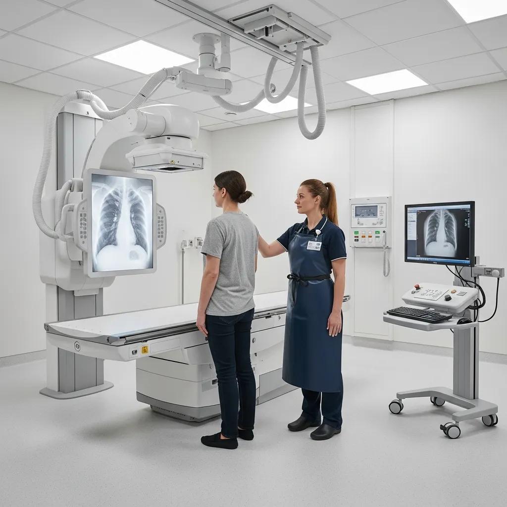
How is a digital chest X‑ray performed?
A digital chest X‑ray is taken with the patient positioned for standard views — usually posteroanterior (PA) and lateral — or anteroposterior (AP) for bedside or supine studies. The radiographer will ask you to remove jewellery and any clothing above the waist that could cause artefact, position your chest against the detector (or have the detector at your side for a lateral), then take a deep breath and hold briefly while the image is captured. The imaging itself takes only a few minutes; the technologist checks centring and motion to ensure diagnostic quality. Portable studies follow the same principles at the bedside, and modern detectors allow quick review so images can be confirmed before you leave.
Why is a chest X‑ray performed? Common uses and benefits
A chest X‑ray is fast, widely available and answers focused clinical questions about the lungs, heart and chest wall. It’s the first‑line test for acute respiratory symptoms, trauma, suspected infection and assessment of cardiac size or devices. Chest X‑rays can detect consolidation, cardiogenic pulmonary oedema, pleural effusion, large masses and lung collapse. Compared with CT, X‑rays use much less radiation, are quicker and easier to organise; CT is used when greater sensitivity or cross‑sectional detail is required. The table below summarises common indications and typical radiographic appearances to help patients and clinicians understand the test’s diagnostic value.
Different chest conditions produce recognisable X‑ray patterns that guide diagnosis and immediate management.
In many cases the chest X‑ray provides the information needed to decide whether further imaging such as CT or ultrasound is required.
Which conditions can a chest X‑ray diagnose?
Chest X‑rays are useful for conditions that change lung density, the pleural space or the cardiac silhouette. Common diagnoses include lobar pneumonia, heart failure with pulmonary oedema, large pleural effusions, pneumothorax and acute rib fractures — each with recognisable radiographic signs. For paediatric patients, chest X‑rays can help identify bronchiolitis, pneumonia or congenital anomalies, with careful justification of radiation. When findings are subtle or don’t match the clinical picture, clinicians often follow up with chest CT or ultrasound for further characterisation and to guide treatment.
How does a chest X‑ray help detect lung and heart issues?
A chest X‑ray evaluates lung aeration, alveolar consolidation and interstitial changes to help distinguish infection, fluid overload, chronic interstitial disease or mass lesions. Cardiac assessment focuses on the size and shape of the cardiac silhouette — an enlarged heart on X‑ray raises suspicion for cardiomyopathy or chronic pressure/volume overload and usually prompts echocardiography or CT. The strength of the X‑ray is rapid triage: it identifies urgent, treatable conditions and points to the next diagnostic step, whether that’s antibiotics, diuretics, advanced imaging or interventional drainage.
How should you prepare for a chest X‑ray scan?

Preparing for a chest X‑ray is straightforward. The aim is to avoid image artefact, confirm clinical details and keep you safe. Wear comfortable, easy‑to‑remove clothing and take off jewellery, piercings or any metal from the neck and upper chest. Let us know if you are pregnant or might be pregnant so the radiographer and referrer can discuss risks and alternatives. Please bring your referral and photo ID, and arrive a few minutes early to complete any check‑in steps — this helps the appointment run smoothly and improves image quality.
- Simple preparation steps that help get clear images and reduce delays:
- Remove jewellery and metal from the chest and neck before the scan.
- Wear comfortable clothing and be ready to change into a gown if requested.
- Tell staff if you are pregnant or could be pregnant so appropriate precautions are taken.
Following these points allows the technologist to obtain clear diagnostic images with minimal need for repeats. Below we explain what happens during the appointment.
What are the key preparation steps before your chest X‑ray?
Key steps are identity verification, removing metal items and informing staff about pregnancy. On arrival our team will check your referral, confirm your identity and ask relevant clinical questions including possible pregnancy in people of childbearing potential. Remove necklaces, bras with metal underwires and clothing with metal fastenings, and follow the radiographer’s instructions if a gown is required. For children, caregivers should bring any previous imaging or clinical notes and follow the centre’s guidance to keep the child calm during positioning.
What to expect during your chest X‑ray appointment?
Your visit usually includes check‑in, verification of clinical details, positioning by the radiographer and a short breath‑hold for exposure. Most appointments take about 10–20 minutes. The radiographer will explain PA, AP or lateral positioning and clear breathing instructions to reduce motion and ensure good lung inflation. Images are reviewed immediately for technical adequacy and a repeat will only be done if necessary, keeping radiation as low as possible. A radiologist interprets the images and sends a report to the referring clinician, who will discuss the results and next steps with you.
Life Medical Imaging Central Coast accepts online appointment requests and recommends you bring your referral and identification to the appointment; see the booking section below for clinic locations and how to request an appointment online.
What are the risks and safety considerations of chest X‑rays?
Chest X‑rays use a low level of ionising radiation and are safe when clinically justified, following the ALARA principle (As Low As Reasonably Achievable) and standard safety protocols. The main concern is cumulative exposure from repeated imaging, so clinicians balance immediate diagnostic benefit against long‑term risk and consider alternatives where appropriate. Techniques such as careful collimation and shielding of sensitive areas reduce dose, and modern digital detectors generally deliver lower effective dose than older systems. For children and pregnant patients we apply additional justification and optimisation to reduce exposure while still obtaining the needed diagnostic information.
- Common safety measures used to reduce radiation and protect sensitive tissues:
- Use of modern digital detectors to minimise dose while keeping image quality high.
- Careful collimation to restrict the X‑ray field and shielding where clinically appropriate.
- Referrer justification so each exposure directly informs patient management.
These measures keep individual exposure low and ensure the diagnostic benefit outweighs the minimal radiation risk. The table below summarises typical dose characteristics and practical notes.
Standardising exposure indices for digital radiography provides an opportunity to optimise dose, especially for paediatric patients.
Standardised Exposure Index for Digital Radiography Optimisation
The exposure index is a method digital radiography manufacturers use to give radiographers feedback on detector exposure as a surrogate for image signal‑to‑noise ratio. Historically there have been many manufacturer‑specific values; an international standard developed with IEC and AAPM aims to harmonise this. The exposure index itself is not a direct measure of patient dose but indicates incident exposure to the detector. Using a standardised exposure index, plus target and deviation index values, should improve radiographer technique and dose uniformity — a particular benefit for optimising paediatric radiography. Radiologists and institutions will also gain consistent terminology and the ability to compare values across dose registries. The Image Gently campaign helps disseminate this standard to support safer paediatric imaging.
The standardized exposure index for digital radiography: an opportunity for optimization of radiation dose to the pediatric population, 2011
How much radiation does a chest X‑ray involve?
A single digital chest X‑ray typically delivers an effective dose of about 0.02 to 0.1 millisievert (mSv), which is small compared with annual natural background radiation. For perspective, this is far lower than a chest CT (which can be tens to hundreds of times higher) and is roughly equivalent to a few days to a few months of background exposure depending on technique. Clinicians weigh this low dose against the value of rapid diagnosis; modern digital systems and strict collimation help keep doses as low as reasonably achievable while preserving diagnostic detail.
Is a chest X‑ray safe during pregnancy?
If you are pregnant or suspect you might be, tell the imaging team so the referrer and radiologist can consider whether the X‑ray is essential or if an alternative like ultrasound is preferable. When imaging is clinically necessary for maternal or fetal care, shielding and careful technique are used to minimise fetal exposure while obtaining vital diagnostic information. Many chest X‑rays are still performed in pregnancy when the benefit outweighs the small risk, especially in urgent situations where missing a diagnosis would be harmful. Open discussion with your referrer and our imaging team ensures documented justification and the safest possible approach.
Life Medical Imaging Central Coast follows recognised safety protocols and highlights NATA accreditation and experienced staff as quality signals to reassure patients that safety and image quality are priorities.
Clinical studies have shown that moving from film‑screen systems to computed radiography (CR) and digital radiography (DR) has enabled optimisation of patient surface doses while maintaining acceptable image quality.
Optimisation de la dose et de la qualité d’image en radiographie numérique
Measurements of patient surface doses for thoracic, abdominal and pelvic radiographs during the introduction of CR and DR systems demonstrated opportunities to adjust technique and reduce dose while keeping image quality acceptable. For CR, algorithm changes and optimisation reduced dose substantially in some cases; for DR, adjustments balanced noise and dose. Final surface doses with optimised CR and DR techniques fell in line with or below published diagnostic reference levels, illustrating the potential for dose optimisation with modern systems.
Optimization of dose and image quality for computed radiography and digital radiography, 2006
How are chest X‑ray images interpreted and what do the results mean?
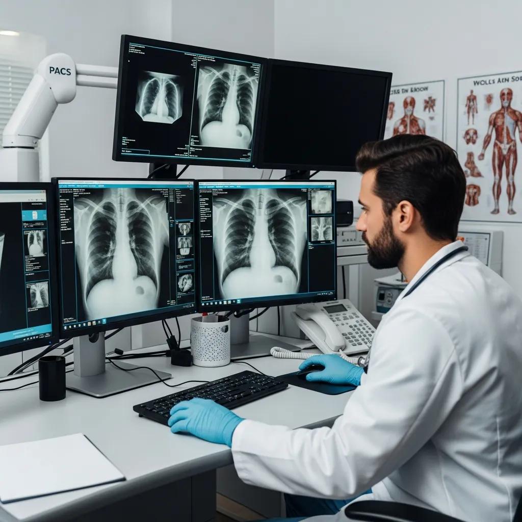
Interpretation follows a simple, reliable workflow: the technologist acquires the images, a radiologist reviews them for diagnostic findings and issues a written report that goes to the referring clinician. Radiologists use a systematic approach — airways, lungs, heart, bones and soft tissues — to reduce the chance of missed findings and to align imaging observations with the clinical question. Reports usually include an impression that summarises key findings and recommends next steps such as follow‑up radiography, chest CT, ultrasound, laboratory tests or clinical review. Knowing this workflow helps you understand when to expect results and why additional tests may sometimes be necessary.
Radiology reports connect what is seen on the image to likely causes and clinical implications, guiding immediate management and further investigation.
What does a normal chest X‑ray look like?
A normal chest X‑ray shows clear lung fields without focal consolidation, a cardiac silhouette within expected size limits, normal diaphragm contours and intact bony structures without acute change. Lung markings should taper toward the periphery and there should be no blunted costophrenic angles or pleural lines to suggest effusion or pneumothorax. Radiologists may note minor, clinically insignificant variations such as scoliosis or surgical clips. When a report reads “no acute cardiopulmonary disease identified,” it generally means no urgent radiographic abnormality was seen, though clinical assessment remains important.
What are common abnormalities found on chest X‑rays?
Common abnormalities include lobar consolidation (often infection), interstitial or alveolar patterns (oedema or chronic lung disease), pleural effusion, and pneumothorax. Rib or sternal fractures can also be seen and, if present, prompt assessment for associated lung injury. Any suspicious mass on X‑ray usually leads to cross‑sectional imaging such as CT for better characterisation and staging where malignancy is a concern. Radiology reports pair these imaging findings with recommended next steps to support the referring clinician’s decisions.
This approach links visible X‑ray patterns to likely causes and helps patients anticipate potential follow‑up investigations.
How to book a chest X‑ray appointment at Life Medical Imaging Central Coast?
Life Medical Imaging Central Coast is an independent, NATA‑accredited radiology provider offering digital X‑ray services across Central Coast locations including Bateau Bay, Killarney Vale, Umina Beach and Erina. We use up‑to‑date equipment, offer sub‑specialist reporting in areas such as Women’s and Cardiac Imaging, and our staff are experienced and qualified to perform and report diagnostic studies. You can request an appointment online via our appointment request form or call the clinic directly — please have a valid referral when booking. Bring your referral and ID to the appointment; a radiologist will report the study and the results will be sent to your referring clinician.
- Simple steps to request a chest X‑ray appointment at our clinic:
- Obtain a valid referral from your doctor that clearly states the clinical question.
- Request an appointment online using our appointment form or call the clinic to schedule.
- Bring photo ID and the referral to your chosen centre on the day of the exam.
These steps make accessing digital chest X‑ray services at local Central Coast sites straightforward and help our team prepare for your visit.
How can you book your chest X‑ray online or by phone?
To book, ask your referring clinician to prepare a written referral that includes the patient’s details and the reason for imaging. With that referral you can use our online appointment request form or call the preferred centre to arrange a time. Online requests usually ask for contact details, preferred location (Bateau Bay, Killarney Vale, Umina Beach or Erina) and the referral upload or confirmation that your referrer will forward it. If the scan is urgent, tell our staff so we can advise on availability or fast‑track scheduling in coordination with your referrer. On the day, bring the referral and ID to ensure a smooth check‑in and exam.
What referral information do doctors need for chest X‑ray requests?
Referrals should include standard patient identifiers, the referring clinician’s details and a clear clinical indication so the imaging team and radiologist understand the purpose of the exam. A concise history — for example acute cough, chest pain, trauma or suspected infection — and any urgency flags help prioritise and tailor imaging. For paediatric or pregnant patients, include age and pregnancy status so the clinic can apply appropriate dose optimisation and positioning. Mentioning prior imaging or known chronic conditions also assists radiologists in comparing studies and interpreting findings.
This practical referral checklist helps ensure timely, high‑quality diagnostic imaging and reporting.
Frequently Asked Questions
What should I expect after my chest X‑ray appointment?
After your X‑ray, a radiologist will review the images and prepare a written report. That report is sent to your referring clinician, who will discuss the findings and any recommended next steps with you. Depending on the result, further tests or treatment may be advised. Always follow up with your healthcare provider to understand what the report means for your care.
How long does it take to receive chest X‑ray results?
Timing varies, but most reports are available to the referring clinician within 24–48 hours. In urgent cases the report can be expedited and you or your doctor will be contacted sooner. If you are concerned about the timing, check with your referring clinician or the clinic.
Can I eat or drink before a chest X‑ray?
There are generally no dietary restrictions for a chest X‑ray, so you may eat and drink as usual. If other imaging is scheduled at the same visit that requires fasting, your clinician will advise you. When in doubt, confirm any special instructions with the referrer before your appointment.
Are there alternatives to chest X‑rays for diagnosing lung conditions?
Yes. Alternatives include computed tomography (CT), ultrasound and magnetic resonance imaging (MRI). CT provides detailed cross‑sectional images and is used when X‑ray findings are inconclusive. Ultrasound is useful for assessing pleural effusions. MRI is less commonly used for routine lung imaging but may be appropriate in select cases. Your clinician will recommend the best modality for your situation.
What happens if my chest X‑ray shows an abnormality?
If an abnormality is seen, your referring clinician will discuss the findings and may request further tests such as CT, ultrasound or laboratory investigations to gather more information. Treatment or specialist referral will depend on the nature of the finding. Keep an open dialogue with your healthcare team to understand the implications and next steps.
Is there a risk of radiation exposure with chest X‑rays?
Chest X‑rays involve a low level of ionising radiation and are generally safe when clinically justified. A single chest X‑ray delivers about 0.02 to 0.1 mSv — far lower than a CT scan. While the risk from a single exam is minimal, cumulative exposure from multiple studies should be monitored. Speak with your clinician if you have concerns about radiation; they can explain risks in the context of your care.
Conclusion
Knowing what to expect from a chest X‑ray — the simple preparation steps, the low level of radiation, and how results are reported — can reduce anxiety and make your appointment smoother. If you’re ready to book, Life Medical Imaging Central Coast offers convenient digital chest X‑ray services across the Central Coast. Request an appointment online or call your chosen centre; bring your referral and ID on the day and our team will take care of the rest.