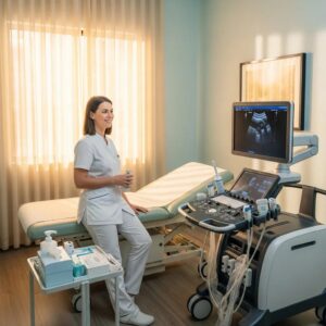
Diagnostic ultrasound is a non-invasive imaging technique that uses high-frequency sound waves to create images of organs and structures inside the body. This method is widely used for various applications, including monitoring fetal development during pregnancy, assessing organ health, and guiding certain medical procedures.
One of the primary advantages of diagnostic ultrasound is its safety, as it does not involve exposure to ionizing radiation. It is commonly used in obstetrics for prenatal scans, but it also plays a crucial role in evaluating conditions related to the abdomen, pelvis, and musculoskeletal system. For instance, it can help diagnose issues such as gallstones, kidney stones, or even tumors.
Preparation for Ultrasound Procedures
Preparation for an ultrasound procedure can vary depending on the type of scan being performed. Generally, patients may be required to fast for several hours before the examination or to drink water to fill their bladder, especially for pelvic ultrasounds. These preparations help improve the clarity of the images obtained during the scan.
It's essential for patients to follow the specific instructions provided by their healthcare provider to ensure optimal results. For example, during an abdominal ultrasound, having a full bladder can help visualize pelvic organs more effectively. Patients should also inform their medical team about any medications they are taking or previous surgeries that may affect the procedure.
Safety Considerations in Medical Imaging
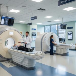
When undergoing medical imaging procedures, safety is a paramount concern. Diagnostic ultrasound is considered one of the safest imaging modalities available, as it does not use ionizing radiation like X-rays or CT scans. However, it is still important to use ultrasound judiciously and only when medically necessary.
Healthcare professionals adhere to strict guidelines to minimize any potential risks associated with medical imaging. For instance, the American Institute of Ultrasound in Medicine recommends that ultrasound exams should be performed only for valid medical reasons, ensuring that the benefits outweigh any risks. Continuous advancements in ultrasound technology further enhance its safety profile.
Types of Ultrasound Scans
There are several types of ultrasound scans, each designed for specific diagnostic purposes. Common types include abdominal ultrasounds, pelvic ultrasounds, and obstetric ultrasounds, each tailored to visualize different organs and structures in the body. For example, abdominal ultrasounds are typically used to examine the liver, gallbladder, and kidneys.
Obstetric ultrasounds are particularly well-known for monitoring fetal development during pregnancy. These scans can be performed at various stages of pregnancy to assess growth, detect abnormalities, and determine the baby's position. Other specialized ultrasound types include Doppler ultrasounds, which evaluate blood flow, and echocardiograms, which assess heart function.
Understanding Diagnostic Ultrasound
Diagnostic ultrasound is a non-invasive imaging technique that uses high-frequency sound waves to create images of organs and structures inside the body. This method is widely used for various applications, including monitoring fetal development during pregnancy, assessing organ health, and guiding certain medical procedures.
One of the primary advantages of diagnostic ultrasound is its safety, as it does not involve exposure to ionizing radiation. It is commonly used in obstetrics for prenatal scans, but it also plays a crucial role in evaluating conditions related to the abdomen, pelvis, and musculoskeletal system. For instance, it can help diagnose issues such as gallstones, kidney stones, or even tumors.
Preparation for Ultrasound Procedures
Preparation for an ultrasound procedure can vary depending on the type of scan being performed. Generally, patients may be required to fast for several hours before the examination or to drink water to fill their bladder, especially for pelvic ultrasounds. These preparations help improve the clarity of the images obtained during the scan.
It's essential for patients to follow the specific instructions provided by their healthcare provider to ensure optimal results. For example, during an abdominal ultrasound, having a full bladder can help visualize pelvic organs more effectively. Patients should also inform their medical team about any medications they are taking or previous surgeries that may affect the procedure.
Safety Considerations in Medical Imaging
When undergoing medical imaging procedures, safety is a paramount concern. Diagnostic ultrasound is considered one of the safest imaging modalities available, as it does not use ionizing radiation like X-rays or CT scans. However, it is still important to use ultrasound judiciously and only when medically necessary.
Healthcare professionals adhere to strict guidelines to minimize any potential risks associated with medical imaging. For instance, the American Institute of Ultrasound in Medicine recommends that ultrasound exams should be performed only for valid medical reasons, ensuring that the benefits outweigh any risks. Continuous advancements in ultrasound technology further enhance its safety profile.
Types of Ultrasound Scans
There are several types of ultrasound scans, each designed for specific diagnostic purposes. Common types include abdominal ultrasounds, pelvic ultrasounds, and obstetric ultrasounds, each tailored to visualize different organs and structures in the body. For example, abdominal ultrasounds are typically used to examine the liver, gallbladder, and kidneys.
Obstetric ultrasounds are particularly well-known for monitoring fetal development during pregnancy. These scans can be performed at various stages of pregnancy to assess growth, detect abnormalities, and determine the baby's position. Other specialized ultrasound types include Doppler ultrasounds, which evaluate blood flow, and echocardiograms, which assess heart function.