Central Coast Brain Scan Services: Costs, Prep & Conditions
Brain scans—most commonly CT and MRI—are imaging tools used to detect, describe and monitor neurological problems such as stroke, tumours and dementia. This guide explains how each modality works, what findings they can show, how to prepare for CT and MRI, common cost factors and Medicare rebate basics, and practical steps for booking a private brain scan on the Central Coast. Many patients come with questions about differences between CT and MRI, contrast agents, result timing and bulk-billing; we address those concerns in clear, evidence-based language. You’ll find plain explanations of CT, MRI and PET mechanisms, compact comparison tables to aid decision-making, step-by-step preparation checklists, and guidance on how results are reported and acted on. Local booking details for Central Coast providers are included where they help clarify referral and billing pathways. Read on for modality-specific advice, Australian cost context and practical steps to get the right scan at the right time.
Brain Imaging Services: Costs, Preparation, and Neurological Conditions — concise summary
Brain imaging—principally CT and MRI—helps detect, characterise and monitor conditions such as stroke, tumours and dementia. This overview outlines how each technique works, what they typically show, how to prepare for CT and MRI scans, typical cost considerations and Medicare rebate rules, and how to book a private brain scan on the Central Coast. It answers common patient questions about modality choice, contrast, reporting times and bulk-billing, and offers practical, evidence-informed guidance for patients and referrers.
Comparison of CT and MRI for brain imaging, 2024
What are the different brain imaging techniques and how do they work?
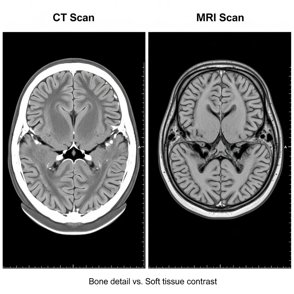
Brain imaging includes several modalities that visualise structure, blood flow and metabolic activity to answer specific clinical questions. Each relies on different physical principles: CT uses X-rays to build fast cross-sectional images, MRI uses magnetic fields and radiofrequency to provide high soft-tissue contrast, and PET uses injected radiotracers to map metabolic or molecular processes. These differences explain why CT is preferred for acute haemorrhage and bone detail, MRI for soft-tissue characterisation and tumour staging, and PET for metabolic or molecular assessment in select cases. The table below summarises key strengths to help match modality to symptom or diagnostic need.
This quick comparison helps clinicians and patients identify an appropriate first-line scan and leads into practical descriptions of CT and MRI.
This table clarifies modality selection by mechanism and clinical application and sets up the more detailed modality descriptions that follow.
What is a CT brain scan and when is it used?
A CT brain scan is a fast X‑ray based exam that acquires thin slices of the head and reconstructs them into cross‑sectional images, producing results quickly for urgent decisions. CT is particularly valuable in emergency settings because it reliably detects acute intracranial haemorrhage, skull fractures and dense lesions within minutes, directly informing immediate treatment. Iodinated contrast may be used for vascular detail or tumour assessment, though many acute scans are performed without contrast. Be aware that CT involves ionising radiation; clinicians weigh diagnostic need against exposure, and modern scanners use dose‑reduction techniques to preserve image quality while limiting radiation.
With CT mechanics and indications clear, the next section explains how MRI differs and when it is preferred for non‑urgent, soft‑tissue questions.
What is an MRI brain scan and how does it differ from CT?
MRI uses magnetic fields and radiofrequency pulses to produce high‑contrast images of brain tissue without ionising radiation, making it ideal for detailed assessment of parenchyma and complex pathology. MRI sequences can be tailored to highlight different tissue properties, detect subtle tumour infiltration, show multiple sclerosis plaques or map structural changes seen in dementia. Scans take longer and are louder than CT and may be unsuitable for patients with certain implants. Gadolinium‑based contrast agents are sometimes used to improve lesion characterisation; clinical teams screen renal function and other contraindications before administration. MRI complements CT rather than replacing it—the choice depends on clinical question and urgency.
This explanation prepares the reader to compare speed, safety and diagnostic yield between CT and MRI in real‑world pathways.
How much does an MRI brain scan cost in Australia and what are bulk‑billing options?
MRI costs in Australia vary by clinic, study complexity, contrast use and whether a Medicare rebate applies. Medicare can partially cover MRI when an eligible referral and appropriate item number are used, but out‑of‑pocket fees depend on clinic billing policy and the specific item applied. On the Central Coast, check whether a clinic offers bulk‑billing for the exact examination and whether there are extra fees for urgent reporting or specialist add‑ons. The table below summarises typical cost drivers, rebate availability and bulk‑billing likelihood to give a practical pricing framework.
This cost matrix shows how rebates, contrast and study complexity influence out‑of‑pocket costs. Always confirm billing policy when booking.
How imaging is billed affects access; some local providers offer bulk‑billing or reduced‑fee pathways for eligible patients to improve affordability.
What are Medicare rebates and private brain scan costs on the Central Coast?
Medicare rebates are government contributions that reduce the fee for some imaging services when a clinician assigns an appropriate item number and the referral meets Medicare criteria. Rebates often cover only part of a private provider’s fee. Private charges reflect clinic operating costs, scanner type, reporting turnaround and contrast use—for instance, studies with intravenous contrast or advanced sequences usually attract higher fees than basic non‑contrast scans. On the Central Coast, confirm whether the referral qualifies for a rebate and whether the imaging centre bulk‑bills the specific procedure. Ask for an itemised estimate before booking so you understand any additional charges such as urgent reporting or specialist review.
Understanding rebate mechanics and typical pricing leads to how local clinics manage affordability and bulk‑billing options.
How we at Life Medical Imaging make brain scans more affordable
Life Medical Imaging Central Coast (LMI) is an independent, NATA‑accredited radiology provider across multiple Central Coast sites. We offer General CT and CT Angiography services and, where clinical criteria and funding allow, advertise bulk‑billing for many examinations. Our clinics use modern, low‑dose CT technology and aim to provide a personalised, community‑focused service with multiple convenient locations. We accept bookings via phone, SMS, email and online enquiry forms to make access simple. If you’re comparing options, contact us to confirm which brain imaging studies we bulk‑bill, whether MRI is available on site, and any conditions for rebate eligibility.
This provider‑specific detail explains local affordability measures and points you to contact us to confirm availability and billing policy before you book.
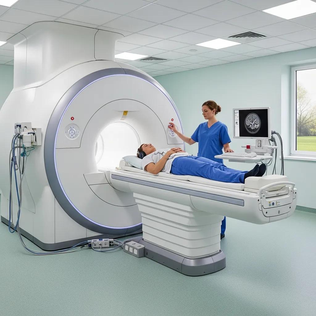
How should you prepare for a brain MRI or CT scan?
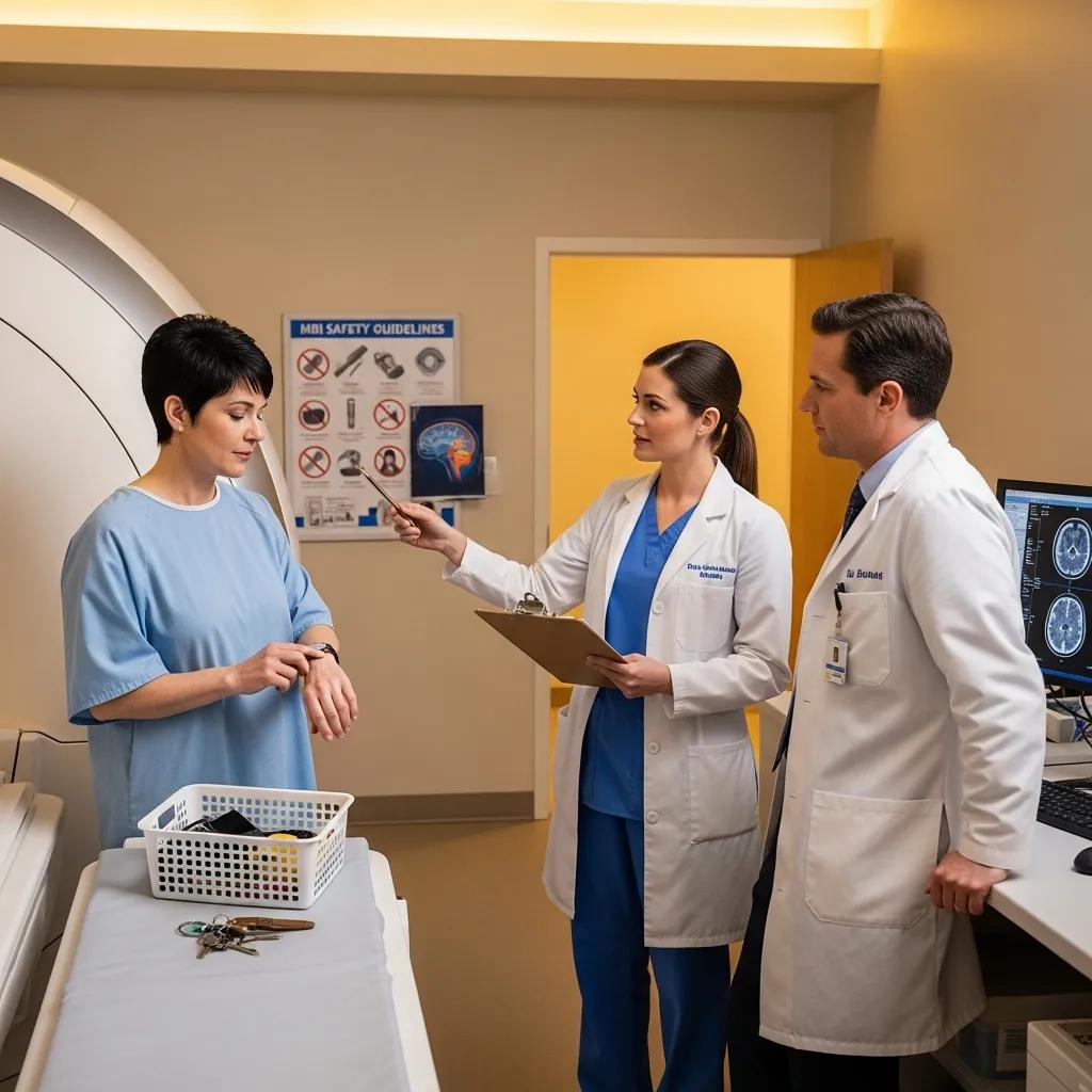
Preparation depends on the modality, contrast use and personal medical history. Good preparation reduces anxiety, improves image quality and lowers risk. Core steps include disclosing implants and relevant medical history, following fasting instructions if contrast is planned, and organising arrival and check‑in details. For MRI, remove metallic items and discuss claustrophobia or sedation options with the clinic; CT preparation focuses on hydration and screening for contrast allergies or renal issues. The table below outlines key preparation steps by procedure with practical notes about contrast, metal precautions and fasting.
This checklist gives a concise view of practical steps to help you be ready on the day.
On arrival, staff will reconfirm your history and screening questions before the scan — a final safety check that ensures the best possible images.
Steps to prepare for an MRI brain scan
Start with accurate screening: tell the clinic about implants, pacemakers, metal fragments, recent surgeries, pregnancy or any implanted device. On the day, remove jewellery, piercings and clothing with metal fastenings, and bring your referral, Medicare card and any previous imaging. Only fast if contrast has been ordered. If you feel anxious or claustrophobic, discuss sedation or comfort measures beforehand—many centres provide headphones, music or other calming options. Allow extra time for administrative checks and screening forms; the radiographer will explain the process and stay in contact during the 20–60 minute scan.
This MRI checklist notes available support and prepares you for the different pace of CT scanning.
How to get ready for a CT brain scan: what to expect
CT brain scans are usually fast, often completed within minutes. Arrive on time with your referral and ID and be ready for a short check‑in. If iodinated contrast is planned, staff will screen for allergies, thyroid issues or renal impairment and may ask you to fast briefly; drinking water before and after contrast is commonly recommended. The scan involves lying still while the gantry rotates around your head; technicians will coach you to hold still for short intervals and will monitor you throughout. After a routine CT you can usually resume normal activities unless sedation or a contrast reaction occurs. Images are reviewed and reported by a radiologist, who sends the report to your referrer.
These CT preparation steps complete practical readiness advice and transition to how imaging is used clinically.
What neurological conditions can brain scans diagnose and monitor?
Brain imaging is central to diagnosing and monitoring many neurological conditions by showing structure, bleeding, ischaemia and, with certain techniques, metabolic changes. Common uses include acute stroke assessment, tumour detection and characterisation, structural imaging for dementia workup, seizure focus localisation in epilepsy and evaluation of traumatic brain injury after concussion. The right modality depends on the clinical question: CT is often first‑line in emergencies, MRI gives detailed soft‑tissue characterisation for chronic or complex problems, and specialised studies like CT angiography or PET are used when vascular or metabolic information is required. Knowing which modality fits a condition helps clinicians and patients select the most informative study while avoiding unnecessary tests.
This overview leads into concise examples showing preferred modalities for specific high‑yield conditions.
How are brain scans used for stroke, tumours and dementia diagnosis?
In acute stroke, rapid non‑contrast CT is used to exclude haemorrhage and identify candidates for reperfusion; CT angiography can show arterial occlusion requiring urgent intervention. MRI may follow to characterise ischaemic injury and uses diffusion‑weighted imaging to detect small infarcts. For tumours, MRI is usually preferred because of superior soft‑tissue contrast and its ability to define lesion extent, oedema and tumour features; CT is helpful when bone involvement or acute haemorrhage is suspected. Dementia workups commonly use MRI to assess atrophy patterns, vascular contributions and structural abnormalities, while cognitive testing and specialist assessment remain central. These roles show how imaging supports immediate management and long‑term monitoring in neurological disease.
These clinical vignettes move into when scans are recommended for common symptoms such as headaches or seizures.
When is a brain scan recommended for headaches, epilepsy or concussion?
Imaging for headaches is recommended when ‘red flag’ features are present—sudden severe onset, new focal neurological signs, progressively worsening pattern, systemic symptoms or signs suggesting a secondary cause; routine imaging for typical primary headache syndromes is usually not necessary. In epilepsy, imaging—preferably MRI—is advised after a first unprovoked seizure to look for structural causes; recurrent or focal‑onset seizures particularly warrant detailed imaging. Concussion imaging depends on severity: patients with worsening neurological signs, focal deficits or prolonged loss of consciousness may need urgent CT to exclude bleed, while uncomplicated concussion without red flags is often managed with observation and deferred specialist review. These indications help balance diagnostic benefit, resource use and patient safety.
This clinical guidance connects directly to what patients can expect after imaging and how results are communicated.
How do you understand and interpret your brain scan results?
A radiology report translates imaging into structured clinical information by summarising the history, technique, findings and an impression that highlights key results and recommended next steps. Reports use standardised language—describing lesion size, location, signal or the presence of haemorrhage—so clinicians can integrate imaging with the clinical picture. Patients benefit from a plain‑language summary of the impression and any suggested referrals. Turnaround varies by urgency: routine studies may be reported in days while urgent or critical findings trigger rapid phone notification to the referring clinician, who will then discuss results and management with you. Knowing what a report contains and typical timelines empowers you to ask focused questions and seek appropriate specialist follow‑up when needed.
This section prepares readers for the structure of a radiologist’s report and the communication pathways between clinics and referrers.
What information will your radiologist provide after a brain scan?
A radiologist’s report usually includes the supplied clinical history, a description of the imaging technique and sequences used, detailed findings and an impression summarising the most important conclusions and recommendations for follow‑up or further testing. The impression is the actionable part of the report—it will state whether the scan is normal, shows an acute problem such as haemorrhage or infarct, or reveals changes consistent with chronic disease or a mass lesion. If technical terms are unclear, ask your referring doctor for a plain‑English explanation—for example, whether a small lesion needs monitoring or immediate intervention. Routine reports are typically available within a few working days, while urgent reports are prioritised for immediate clinical care.
This explanation flows into how findings are communicated to referrers and patients in practice.
How are brain scan findings communicated to referring doctors and patients?
Scan findings are routinely sent electronically to the referring clinician; urgent or unexpected critical results are usually communicated by phone to ensure prompt action. Patients normally receive their results through their referrer, who interprets imaging alongside the clinical assessment. Imaging centres and radiologists can provide expedited reporting for acute cases and coordinate directly with hospital teams for inpatients or emergency presentations. If you’re waiting for results, contact your referring doctor to arrange a follow‑up rather than asking the imaging centre for interpretation—your referrer integrates the imaging findings with your overall care plan. Clear communication between imaging providers and clinicians ensures timely, contextualised explanations and appropriate next steps when abnormalities are identified.
This communication overview leads into practical booking instructions for private brain scans on the Central Coast.
How can you book a private brain scan appointment on the Central Coast?
Booking a private brain scan usually requires a clinician’s referral that states the clinical question and urgency. Prepare the referral, identification and any prior imaging before contacting the imaging centre. Common booking channels include online enquiry forms, phone, SMS and email; each method requires submission of the referral and basic patient details for scheduling and Medicare processing. Some Central Coast providers offer patient‑centred booking and may bulk‑bill eligible studies, but availability varies, so confirm billing and appointment options in advance. The numbered list below summarises practical steps to book a scan and what to have ready for a smooth appointment.
- Obtain a valid referral: Ensure your GP or specialist provides a referral with clinical history and urgency stated.
- Choose a local imaging centre: Pick a provider based on modality availability, billing policy and location convenience.
- Contact the clinic to book: Submit your referral through the clinic’s accepted channel and request an appointment.
- Prepare documentation: Have your referral, Medicare card and any prior imaging ready on the day.
- Confirm billing and contrast requirements: Ask whether the study is bulk‑billed, and whether contrast or fasting is required.
Completing these five steps will help avoid delays and allow the imaging centre to allocate the correct scanner and reporting pathway for your clinical need.
What are the referral requirements for a brain scan at Life Medical Imaging?
Most diagnostic imaging studies require a clinician referral to ensure clinical justification and, where applicable, Medicare rebate eligibility. Referrals should document relevant history, suspected diagnosis and urgency. Life Medical Imaging Central Coast accepts clinician referrals for CT and CT angiography as listed in our services; for MRI availability or bulk‑billing criteria, contact the practice directly to confirm current capabilities. Referrers should include relevant clinical notes and prior imaging summaries to assist interpretation, and urgent referrals should clearly state time‑critical concerns so scheduling can be prioritised. If submission methods are unclear, referrers and patients can use our contact channels to enquire about referral submission and documentation requirements.
This concise referral checklist helps referrers and patients prepare the correct documentation before booking.
What booking methods are available: online, phone, SMS and email?
Most imaging centres offer multiple booking methods to suit patient preferences: online enquiry forms, phone bookings, SMS messaging and email. Each method requires your referral and basic personal details for scheduling and Medicare processing. Life Medical Imaging Central Coast provides convenient booking channels—SMS, email, online form submissions and phone—to make enquiries and appointments across our local sites. When booking, have your referral, Medicare card details and any relevant medical history available, and ask about confirmation times and whether pre‑scan instructions will be sent in writing. Expect the clinic to confirm your appointment and advise arrival time, preparation steps and where to present on the day to streamline check‑in and reduce anxiety.
These practical booking details complete the patient pathway from referral to appointment and close this guide with clear next steps for Central Coast patients seeking brain imaging.
Frequently Asked Questions
What should I expect during a brain scan appointment?
When you arrive, staff will check your referral and medical history. Depending on the scan you may change into a gown and remove metallic items. You’ll lie still on a table while the machine captures images — MRI is louder and takes longer, CT is usually quicker. Staff will guide you through each step and keep you informed to help you stay comfortable and safe.
Are there any risks associated with brain scans?
CT scans use ionising radiation, which carries a small long‑term risk; the immediate diagnostic benefit usually outweighs this risk. MRI does not use radiation but may be unsuitable for people with some implants or severe claustrophobia. Contrast agents carry rare risks such as allergic reactions or renal effects — clinicians screen for these beforehand. Discuss any concerns with your referring doctor to weigh risks and benefits for your situation.
How long does it take to receive brain scan results?
Reporting times vary. Routine scans are typically reported within a few working days; urgent cases are prioritised and may be reported immediately. After images are reported, the radiologist sends the report to your referring clinician, who will discuss the findings and next steps with you.
Can I eat or drink before a brain scan?
Whether you can eat or drink depends on the scan and whether contrast will be used. MRI usually doesn’t require fasting unless contrast is planned. For CT with iodinated contrast you may be asked to fast for a few hours. Follow the specific instructions provided by the imaging centre for the best results.
What should I do if I have anxiety about the scan?
If you’re anxious, tell the clinic ahead of time. Many centres offer calming measures—headphones, guided breathing, or light sedation where appropriate. Bringing a friend or family member for support can help, and knowing what to expect often reduces anxiety.
What happens if a brain scan shows an abnormality?
If an abnormality is found, the radiologist will include it in the report sent to your referring doctor. Your doctor will explain the findings, their implications and recommended next steps, which may include further tests, specialist referral or treatment. Keep open communication with your healthcare team to understand the result and plan follow‑up.
Are brain scans covered by health insurance?
Coverage depends on your health fund and policy. In Australia, Medicare may rebate certain imaging services with a valid referral and qualifying indication, but out‑of‑pocket costs can still apply depending on the clinic’s billing practices. Check with your insurer and the imaging centre before booking to understand likely costs.
Conclusion
Knowing how brain scans work, what to expect and how costs and referrals are handled helps you make informed choices about neurological care. If you think a brain scan is needed, contact your referrer or local imaging provider to discuss options and book an appointment. Taking this step will clarify the next steps in diagnosis and care — we’re here to help you through the process.