
Preventative Body Scans & Health Assessments on the Central Coast
Medical body scanning brings together targeted imaging tests to measure body composition, bone strength and internal organ health — giving clinicians and patients clear, actionable baseline information for early detection and prevention. This guide explains how common diagnostic tools — DEXA for body composition and bone mineral densitometry, plus advanced CT where indicated — work together to answer questions about visceral fat, muscle mass, bone density and cardiac risk. You’ll find what each test measures, how results are interpreted, when to consider testing, and practical steps to prepare for your appointment. The sections that follow cover how body scanning supports preventative care, the mechanics and benefits of DEXA, interpreting bone density, the role of CT in a full assessment, considerations around cost and Medicare, and local booking pathways for people on the Central Coast.
What Is a Body Scan and How Does It Support Preventative Health?
A body scan is a focused set of diagnostic imaging studies designed to detect early, often modifiable changes in body composition, bone density and internal structures that can predict future illness. Different technologies contribute complementary information — DEXA uses dual‑energy X‑ray attenuation for composition and bone metrics, CT provides cross‑sectional organ and vascular detail, and bone densitometry targets skeletal strength — and together they form a multidimensional baseline for risk assessment and monitoring. The main value is timely identification of risk factors you can change, personalised data to guide lifestyle or medical decisions, and objective tracking over time to measure response to treatment or training. Common uses include detecting osteoporosis before fractures occur, estimating visceral fat linked to metabolic disease, and assessing coronary calcification risk.
These scans are performed under clinical oversight with dose optimisation to keep radiation exposure as low as possible while delivering useful diagnostic information. Below are three immediate benefits that explain why clinicians commonly recommend these assessments.
Body scans support preventative health through:
- Early detection: Finding subclinical risk factors — for example, low bone density or elevated visceral fat — before symptoms develop.
- Personalised data: Objective measures of fat, muscle and bone to inform tailored treatment, training or lifestyle plans.
- Preventative action: Results that trigger evidence‑based interventions such as targeted nutrition, exercise programs, medical therapy or further imaging.
With those benefits in mind, the next section outlines what to expect from a typical medical body‑scan pathway.
What Does a Medical Body Scan Involve?
Most body‑scan pathways start with a referral from a GP or specialist, then booking and a short clinical screening on arrival to confirm the indication and address safety concerns. The scan itself depends on the modality: DEXA for composition and bone density usually takes 10–20 minutes, while targeted CT studies can take longer and may require breath‑holding, intravenous contrast or cardiac gating. A trained radiographer performs the acquisition and an imaging specialist reviews the images, issuing a report to the referring clinician with recommended next steps. Patients can expect a clear, interpretive report to be forwarded to their GP or referrer to support follow‑up and management planning.
Understanding this process helps position a body scan as part of ongoing care rather than a one‑off test, and it also clarifies the practical preparation and booking considerations discussed later.
Why Choose Preventative Health Scans on the Central Coast?
Using an accredited local imaging service shortens the pathway from referral to results and improves coordination with your primary care team. Independent, accredited clinics on the Central Coast follow quality‑assured protocols and use equipment calibrated to balance diagnostic accuracy with low radiation exposure — an important consideration for repeat monitoring. Local services also simplify repeat testing, multidisciplinary case review and integrated care with GPs, allied health and specialists across the region. Easier access reduces delays in diagnosis and supports timely lifestyle or medical interventions that improve long‑term outcomes.
For patients and referrers looking for local options, clinics offering body composition DEXA and bone mineral densitometry provide practical, evidence‑based tools to build prevention into routine care on the Central Coast.
Next, we look more closely at the DEXA body composition scan to explain why it’s widely used for assessing fat, muscle and bone — and how modern systems improve accuracy and safety.
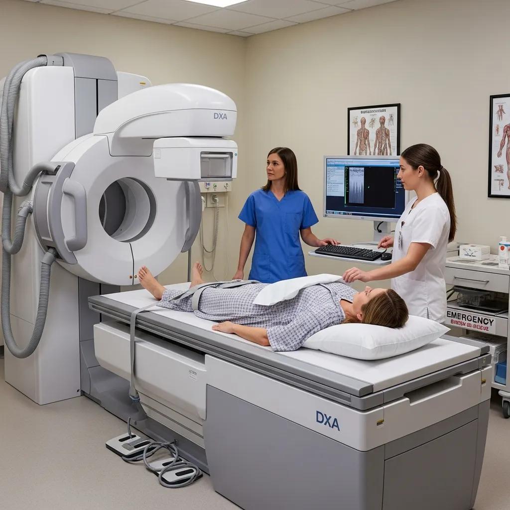
How Does a Dexa Body Composition Scan Work and What Are Its Benefits?
A DEXA (dual‑energy X‑ray absorptiometry) body composition scan measures how tissues attenuate X‑rays at two energy levels to distinguish bone mineral, lean tissue and fat mass. The technique produces precise regional and whole‑body metrics used in clinical practice and performance settings. Because bone and soft tissues absorb X‑rays differently at the two energies, software algorithms can separate compartments with high repeatability and a low radiation dose. The report gives a detailed breakdown of total fat mass, regional distribution (including visceral fat estimates), lean muscle mass and bone mineral density — data used to guide weight management, sarcopenia screening and cardiometabolic risk assessment. DEXA is often seen as a reference method for body composition because it combines accuracy, speed and a minimal dose compared with full CT.
Below is a quick breakdown of common DEXA‑measured metrics, what they look like in the report, and why they matter to patients and clinicians.
The DEXA measurement breakdown:
This summary shows what DEXA quantifies and why those metrics matter. DEXA’s low dose and reproducibility make it suitable for serial monitoring — valuable for athletes and patients on treatment programs.
The study “Reference values of body composition parameters and visceral adipose tissue (VAT) by DXA in adults aged 18–81 years—results from the LEAD cohort, 2020” supplies important age‑ and sex‑related reference values for body composition and VAT mass using Lunar Prodigy DXA.
DXA Reference Values for Body Composition and Visceral Adipose Tissue
Understanding how body composition varies by age and sex is central to interpreting DXA results, because fat and lean mass distribution influence cardiometabolic risk. The LEAD cohort study used GE‑Lunar Prodigy DXA scans from 10,894 adults aged 18–81 years (recruited 2011–2019) to build reference curves for parameters such as fat mass index (FMI), lean mass index (LMI), appendicular LMI, android/gynoid and trunk/limb ratios, and visceral adipose tissue (VAT) mass. Findings show sex differences in central fat accumulation and progressive increases in VAT with age for both sexes. The study provides practical age‑ and sex‑related reference values that help clinicians place an individual’s DXA results in population context.
The groups who benefit from DEXA are varied, and booking choices often depend on local equipment and clinical pathways. Some providers highlight ultra‑low dose, high‑definition systems and accredited reporting to maximise accuracy and safety. For people on the Central Coast seeking DEXA body composition or bone assessments, accredited clinics that use low‑dose equipment and deliver clear reports to referrers make it straightforward to turn results into action; local booking and referral support is usually available from the imaging provider.
What Can a Dexa Scan Measure: Fat, Muscle, and Bone Density?
DEXA separates whole‑body composition into fat mass, lean mass and bone mineral content with regional breakdowns (for example, trunk and limbs) that show distribution patterns important for health and performance. Visceral fat estimates are especially useful because central adiposity correlates strongly with insulin resistance and cardiovascular risk — quantifying it helps guide targeted risk‑reduction strategies beyond BMI alone. Lean mass measures are critical for older adults and athletes, helping clinicians and trainers detect sarcopenia or optimise training and nutrition. BMD values from DEXA allow simultaneous skeletal assessment, so clinicians can track bone health alongside changes in muscle and fat.
Seeing these metrics together supports integrated interpretation: for example, falling lean mass with stable or rising visceral fat suggests a higher metabolic risk and prompts interventions such as resistance training and dietary adjustment.
Who Can Benefit from Dexa Body Composition Analysis?
DEXA is useful for a wide audience: athletes refining training and body‑composition targets, adults monitoring weight loss or fat distribution, and older people where muscle and bone trends guide fracture prevention. Clinicians use DXA to monitor treatment effects — whether pharmacological, surgical or lifestyle — and to quantify changes that don’t show on the scales. Athletes may scan seasonally to inform periodised programs; clinical monitoring typically follows guideline‑based intervals or treatment timelines. Matching the test to the clinical or performance goal ensures DEXA results drive practical next steps rather than producing data without interpretation.
What Is Bone Mineral Densitometry and How Does It Detect Osteoporosis?
Bone mineral densitometry (BMD) quantifies bone mineral content at key skeletal sites — most commonly the lumbar spine and hip — and reports results as T‑scores and Z‑scores used to diagnose osteoporosis and estimate fracture risk. The scan uses X‑ray attenuation to estimate bone density and compares an individual’s values with a young‑adult reference (T‑score) or an age‑matched population (Z‑score); thresholds then classify bone status and inform treatment decisions. Because fractures are the clinical outcome of concern, BMD is integrated with clinical risk calculators and falls history to guide management. Early identification of low bone mass enables timely interventions — both lifestyle measures and medical therapy — to reduce future fracture risk.
Below is a concise table summarising measured sites, the score types you’ll see in a report, and what they mean clinically.
BMD interpretation table:
Interpreting BMD in the broader clinical context ensures appropriate follow‑up; low scores usually prompt discussion of bone‑protective strategies and referral where needed.
How Does a Bone Density Scan Help Assess Bone Health?
A bone density scan provides objective data that, combined with clinical risk factors, helps referrers stratify fracture risk and decide on prevention or treatment. T‑scores compare bone density to a young‑adult reference and classify results as normal (T‑score ≥ −1.0), osteopenia (−1.0 to −2.5) or osteoporosis (≤ −2.5), reflecting increasing fracture risk. Z‑scores, adjusted for age and sex, help identify unexpectedly low bone density in younger patients or those with secondary causes. Results commonly lead to evidence‑based interventions — lifestyle changes, supplements or pharmacotherapy — tailored to the individual’s risk and preferences.
Rapid, clear reporting to the referring clinician is important so bone‑protective strategies can start without unnecessary delay when indicated.
When Should You Consider a Bone Density Scan on the Central Coast?
Bone density testing is commonly recommended for postmenopausal women, men with risk factors, people with prior fragility fractures, long‑term corticosteroid users, or those with conditions that affect bone metabolism; a GP will assess individual timing. Other triggers include unexplained height loss, recurrent falls, or endocrine and metabolic disorders that increase fracture risk. Screening intervals depend on baseline scores and clinical context: low‑normal results may be rechecked every few years, while osteoporotic ranges usually prompt treatment and closer monitoring. Patients should discuss personal risk and possible Medicare rebate eligibility with their GP before referral to ensure testing follows appropriate pathways.
Local accredited imaging services can support referrers with clear reports and recommended follow‑up, streamlining patient management across the Central Coast.
How Are Advanced CT Scans Used in Full Body Health Assessments?
Advanced CT delivers high‑resolution cross‑sectional images that reveal structural changes in organs and blood vessels, complementing composition‑focused tests like DEXA and BMD. CT is excellent for visualising lung nodules, liver lesions, renal stones and vascular calcification; cardiac CT techniques such as coronary calcium scoring and CT angiography directly assess coronary atherosclerosis and refine cardiovascular risk estimates. Modern CT protocols can be dose‑optimised to reduce radiation while preserving diagnostic detail, making CT valuable when anatomical information is required. Because of cost, specificity and radiation, CT is used selectively — typically to answer a targeted question raised by other tests or clinical assessment rather than for indiscriminate whole‑body screening.
In preventative pathways, CT often follows an abnormal finding or clinical concern and provides definitive anatomical detail that guides specialist referral, biopsy or specific treatment.
The paper “Normal organ volume assessment from abdominal CT, JM Boone, 2004” offers normative organ‑volume data from abdominal CT that clinicians can use to compare an individual’s organ size against expected ranges.
Assessment of Normal Organ Volumes Using Abdominal CT Scans
This study measured abdominal organ volumes from 149 adult CT studies and manually outlined 711 organs to calculate normal distributions. Organs studied included kidneys, adrenal glands, spleen, pancreas and liver, with volumes adjusted for height and weight by sex. Results provide mean volumes (for example, the male liver mean was reported as 1710 mL) and cumulative distributions that help clinicians judge whether a particular organ volume lies within expected ranges. These normative data can assist in identifying organ enlargement or atrophy as quantitative indices of disease.
What Internal Organs and Cardiac Health Can CT Scans Screen?
CT can evaluate many organs and structures with high sensitivity for structural disease: chest CT detects lung nodules and interstitial disease, abdominal CT can identify liver lesions and renal masses, and cardiac CT measures coronary calcium and arterial anatomy. Coronary calcium scoring quantifies calcified plaque and refines cardiovascular risk beyond traditional risk factors, helping clinicians personalise prevention. CT angiography visualises vessel lumens and stenoses when an anatomical question exists, while low‑dose chest or abdominal protocols can screen for specific high‑risk features in appropriately selected patients. Choosing CT over other modalities depends on the clinical question, prior imaging and a careful risk–benefit assessment.
Given its strengths, CT’s role in a full assessment is to answer targeted anatomical questions and to provide definitive imaging when structural disease is suspected.
How Do CT Scans Support Early Disease Detection?
CT can reveal structural abnormalities before symptoms appear, enabling earlier intervention that may alter prognosis — for example, identifying potentially resectable lung nodules or quantifying coronary calcification to intensify preventive therapy. CT findings can lead to biopsy, specialist referral or specific treatment plans; when managed appropriately, these downstream actions translate incidental detection into improved outcomes. The sensitivity of CT must be balanced against radiation exposure and the possibility of incidental findings that require follow‑up; for that reason, CT is most useful when guided by clinical indications and evidence‑based pathways. Dose‑optimised protocols and clear reporting help ensure the potential benefits of early detection outweigh harms.
Integrating CT into a body‑scan programme therefore relies on clinical judgement, with imaging chosen to answer precise diagnostic questions rather than used indiscriminately.
What Are the Costs and Medicare Options for Body Scan Services?
Knowing typical cost ranges and how Medicare rebates work helps patients and clinicians plan diagnostic pathways and set expectations for out‑of‑pocket expense. Prices vary with scan type, whether contrast or specialised cardiac protocols are needed, and whether multiple modalities are done in one visit. Medicare rebates apply to certain DEXA and bone density studies when a valid referral and clinical indication are provided, but item numbers and eligibility can change — so always confirm with your GP and the imaging provider. For transparent planning, clinics can provide indicative pricing and booking guidance and explain what to bring to your appointment.
The table below summarises common scan types, typical cost expectations and Medicare/recommended next steps to help patients compare options.
These entries outline typical expectations; final pricing depends on the chosen package and protocols, so contact the imaging provider for an exact quote.
How Much Does a Full Body Health Assessment Cost on the Central Coast?
A combined assessment that includes DEXA, BMD and targeted CT where clinically indicated will cost more than standalone scans, reflecting the additional time, technology and specialist reporting involved. Discuss with your GP which combination of tests is clinically justified, then request an itemised quote from the imaging provider to compare package versus standalone pricing. Transparent clinics will explain which components are likely to attract Medicare rebates and which are private‑billed, allowing patients to weigh comprehensiveness against cost. If budget is a concern, prioritising tests most likely to change management is a practical approach.
For the most accurate pricing and booking details, contact the imaging centre’s enquiry or booking team so they can provide a tailored estimate based on your referral.
What Medicare Rebates Are Available for Dexa and Bone Density Scans?
Medicare rebates for bone density testing usually require a valid clinical referral that meets eligibility criteria — for example, risk factors for osteoporosis or monitoring response to therapy — so patients should confirm entitlement with their GP. DEXA scans performed mainly for body composition may not always be rebateable unless bone density measurement is clinically indicated and correctly itemised on the referral. Because Medicare item numbers and rebate rules change, ask your referring GP to confirm eligibility and check billing details with the imaging provider before booking. Doing so clarifies likely out‑of‑pocket costs and the documentation needed for rebate processing.
Clear pre‑booking communication between patient, GP and clinic minimises surprises and streamlines administration.
How Should You Prepare for Your Body Scan Appointment?
Good preparation improves scan quality, reduces the chance of rescheduling, and helps keep you safe — especially when CT contrast is involved. General steps include bringing your referral and ID, a list of medications and any prior imaging, wearing comfortable clothing without metal, and arriving in plenty of time for administrative checks. CT‑specific instructions may include fasting or hydration guidance and disclosure of contrast allergies or pregnancy; DEXA/BMD usually require minimal preparation but avoid heavy lotions or jewellery that could affect the images. Follow the pre‑scan guidance from your GP and the imaging provider to ensure accurate and timely results.
The two subsections below outline what to expect during DEXA/BMD scans and the preparation needed for CT so you can arrive confident and ready.
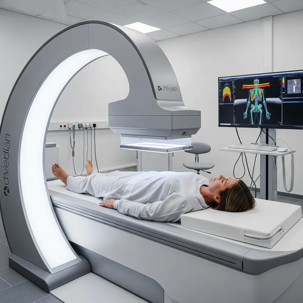
What to Expect During Your Dexa and Bone Density Scans?
DEXA and bone density scans are non‑invasive, quick and generally comfortable. During a DEXA you lie on a padded table while a scanning arm passes over your body; the exam is painless, usually takes 10–20 minutes, and uses a very low radiation dose. Reports are prepared by imaging specialists and sent to the referring GP or clinician within a clinically appropriate timeframe to support discussion and management. Knowing how brief and low‑dose these scans are often reassures patients who are considering serial monitoring for performance or bone health.
Being familiar with the test logistics reduces anxiety and helps ensure an efficient visit, which in turn supports better quality images for interpretation.
How to Get Ready for Advanced CT Scans and Other Imaging Services?
Preparation for advanced CT depends on the protocol: some studies require fasting, others ask you to hydrate or temporarily stop certain medications, and contrast studies require screening for allergies and kidney function where indicated. Tell the imaging team if you are pregnant, breastfeeding, or have allergies or kidney disease so staff can adapt the protocol or offer alternatives. When contrast is used, the team will explain the process, how it might feel and any aftercare; most people tolerate contrast well, but screening prevents avoidable reactions. If you’re unsure, contact the imaging provider before your appointment to confirm requirements and ensure safe, efficient care.
Local centres usually offer practical booking support and referrer guidance to streamline documentation and appointment logistics for clinicians and patients.
- Booking steps for patients and referrers:
Provide a valid GP or specialist referral that states the clinical question and desired scan type.Call the imaging provider’s booking team to schedule an appointment and request a quote if needed.Follow the clinic’s pre‑scan instructions, including fasting or medication adjustments for CT studies. - Referrer support and reporting pathways:
Radiology reports are issued to the referring clinician to support follow‑up and ongoing management.Accredited clinics commonly offer direct communication channels for urgent or complex findings to ensure timely care.
For patients and referrers on the Central Coast seeking accredited, low‑dose imaging with clear reporting and clinician‑focused guidance, Life Medical Imaging Central Coast provides body composition DEXA, bone mineral densitometry and a range of CT and ultrasound services. Our NATA‑accredited clinic uses ultra‑low dose, high‑definition equipment and delivers referrer‑focused reports to support preventative health pathways; patients and referrers can call the clinic to enquire about bookings, quotes and referral requirements.
- Practical reminders before your appointment:
Bring identification, your referral and any prior imaging or treatment summaries.Tell the imaging team about current medications, allergies and pregnancy status.Arrive early to complete administrative checks and consent forms.
Being prepared minimises delays and helps ensure your clinician receives the best possible diagnostic information to act on.
Frequently Asked Questions
What are the risks associated with body scans?
Body scans are generally safe but do involve exposure to ionising radiation, particularly with modalities such as DEXA and CT. With modern, dose‑optimised equipment the risk is low for most patients, though cumulative exposure from repeated studies is a consideration. Discuss any concerns with your GP or the imaging team so you can weigh the potential benefits of the scan against any risks in your individual case.
How often should I get a body scan?
Scan frequency depends on your health goals and risk profile. Athletes might scan seasonally to monitor composition changes; older adults or people at risk of osteoporosis may be advised to repeat scans every 1–2 years depending on baseline results and treatment. Your healthcare provider will recommend the appropriate interval based on your history, recent results and clinical needs.
Can body scans detect diseases other than osteoporosis?
Yes. DEXA can estimate visceral fat, which relates to metabolic risk, while CT can detect structural abnormalities such as lung nodules, liver lesions or vascular calcification. Together, these tools can uncover a range of health issues and help guide prevention or treatment strategies beyond bone health alone.
What should I wear to my body scan appointment?
Wear comfortable clothing without metal fastenings, as metal can affect image quality. Loose‑fitting garments are ideal, and you may be asked to remove jewellery or heavy lotions before the scan. Following these simple guidelines helps ensure accurate results and a smooth appointment.
Are there any specific dietary restrictions before a CT scan?
Some CT studies — especially those using contrast — require fasting for several hours beforehand. Hydration is often recommended unless otherwise instructed. Always follow the specific instructions provided by your imaging centre, as preparation can vary with the type of CT being performed.
How can I access my scan results?
Results are usually sent to your referring clinician, who will discuss the findings with you. Many imaging centres also offer patient portals where you can view reports directly. If you have questions about your report or need clarification, contact your GP or the imaging provider for a detailed explanation and next steps.
What should I do if I have a history of allergies or medical conditions?
If you have a history of allergies — particularly to contrast agents — or medical conditions that could affect the scan, inform the imaging team before your appointment. They can adjust the protocol or offer alternatives to ensure your safety. Clear communication about your medical history helps deliver a safe, effective imaging experience.
Conclusion
Body scans provide practical, objective information that helps detect early risk factors, personalise care and trigger preventative actions tailored to your needs. Understanding the roles of DEXA, bone mineral densitometry and advanced CT lets you and your clinician make informed choices on the best assessments for your situation. On the Central Coast, accredited local imaging services make these tests accessible — if you’d like to explore options or book an assessment, contact us to take the next step towards proactive health management.