Medical Imaging — Central Coast: Clear, Trusted Diagnostic Care
Clear diagnostic answers begin with knowing what tests are available, how to prepare, and how results will guide treatment. This guide explains medical imaging on the Central Coast — the core modalities we offer, simple preparation checklists, dedicated women’s and cardiac pathways, and practical referrer workflows such as eReferral and secure image access. You’ll find plain‑language answers to common safety and results questions, plus guidance on which scan usually fits common symptoms, step‑by‑step prep for MRI, CT and ultrasound, and how to ensure images and reports reach the treating clinician. We also highlight the role of accredited centres and modern equipment in lowering radiation and improving diagnostic confidence, with clear next steps from booking to report.
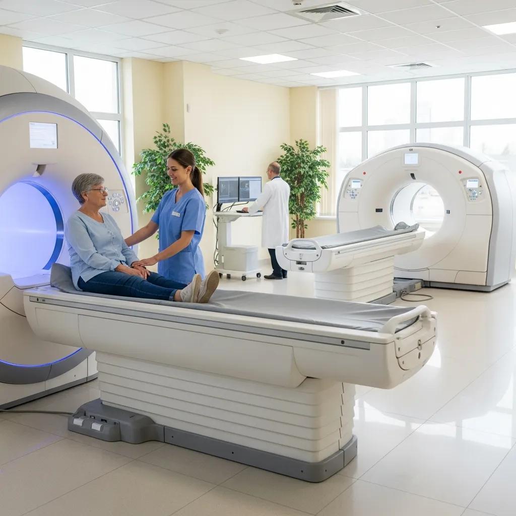
Which medical imaging services are available across the Central Coast?
Medical imaging provides non‑invasive views of the body to detect, monitor and guide treatment. Each modality uses different physics and answers different clinical questions, so understanding the differences helps patients and referrers choose the right test for pain, suspected fracture, pregnancy assessment or cardiac concerns. Core services include CT, MRI, ultrasound, digital X‑ray, dental imaging and DEXA bone densitometry, alongside specialist options such as cardiac CT, CT angiography, paediatric imaging and image‑guided interventional procedures. The sections below list our offered modalities and explain how focused women’s and cardiac imaging pathways improve diagnostic speed and accuracy.
Which diagnostic imaging modalities does Life Medical Imaging offer?
Life Medical Imaging Central Coast provides a wide range of imaging tests tailored to specific clinical questions. CT uses X‑rays and fast detector arrays to produce cross‑sectional images ideal for trauma, chest, abdominal and vascular assessment, including cardiac CT and CT angiography for coronary and vascular evaluation. Ultrasound uses high‑frequency sound to show soft tissues and is the first‑line choice for obstetric, gynaecological, vascular and musculoskeletal issues — it’s safe, radiation‑free and often portable. Digital X‑ray and dental imaging give rapid views for bone and dental concerns, while DEXA measures bone density for osteoporosis assessment. To book, patients and referrers can choose online booking, phone contact or eReferral — options that make scheduling and referral submission straightforward.
Quick comparison: typical uses and simple preparation for common modalities.
Use this table to match common symptoms with the likely imaging test and to check basic preparation steps before booking.
How do specialised services like women’s and cardiac imaging improve care?
Specialised pathways focus protocols, equipment and reporting expertise to answer specific clinical questions more precisely and safely. Women’s imaging brings obstetric and gynaecological ultrasound together with breast ultrasound capability to monitor pregnancy, investigate pelvic pain or assess breast symptoms using dedicated protocols and reporting templates. Cardiac imaging uses tailored cardiac CT protocols and high‑definition scanners to assess coronary anatomy and cardiac structure without an invasive procedure, helping guide decisions about invasive angiography or medical treatment. Specialisation speeds diagnosis by providing targeted information sooner and supports multidisciplinary care where obstetricians, cardiologists and GPs rely on consistent, high‑quality reports.
How can patients prepare for their medical imaging appointments?

Good preparation reduces repeat scans, shortens appointment time and improves image quality. Preparation varies by test and clinical question, but thinking ahead about fasting, hydration, clothing and bringing referral paperwork or prior images will make your visit smoother. The sections below explain how to prepare for MRI, CT, ultrasound and DEXA, and outline the clinic safety checks that protect patients during imaging. Simple steps also ease anxiety and help staff capture diagnostic images on the first attempt.
What are the preparation guidelines for common scans like MRI and ultrasound?
MRI: remove metallic objects, tell us about implants and discuss claustrophobia — some MRI studies use intravenous contrast that may need fasting or hydration instructions. Bring your referral and any previous images; if you have a pacemaker or certain implants, notify staff beforehand. Ultrasound: abdominal scans often require 6–8 hours fasting to reduce bowel gas; pelvic or obstetric scans usually need a full bladder for better views. DEXA: wear light clothing without metal and avoid recent contrast studies where advised. When in doubt, call the imaging team — clear communication ensures a safe, accurate exam.
Immediate checklist: what to bring and do before most imaging appointments.
- Bring your referral, Medicare card if applicable, and any previous imaging studies.
- Wear comfortable, metal‑free clothing and remove jewellery before arrival.
- Follow modality‑specific instructions such as fasting or bladder filling.
- Tell staff about pregnancy, implanted devices or allergies to contrast agents.
These simple steps reduce delays and help the imaging team focus on capturing high‑quality diagnostic images.
How is patient safety managed during imaging procedures?
Patient safety rests on accreditation standards, dose‑optimised technology, trained staff and clear protocols for contrast and sedation when required. Accredited centres follow external quality programs, routine equipment checks and staff credentialing to keep standards high. Modern ultra‑low dose, high‑definition scanners and optimised protocols reduce radiation for CT and X‑ray while preserving image detail. Staff screen for contrast allergies and renal function to manage risk, and where sedation or interventions are needed, consent, monitoring and recovery protocols prioritise patient safety and communication with the referring clinician.
Safety and contraindications summary for common procedures.
This table helps you decide when to seek pre‑appointment advice and identify potential contraindications.
Why choose Life Medical Imaging Central Coast for diagnostic radiology?
Choosing a diagnostic provider should balance technical capability, safety standards and local access so care is timely and communication is clear. Independent, NATA‑accredited centres using ultra‑low dose, high‑definition imaging deliver rigorous quality assurance and dose reduction to improve diagnostic confidence. Multiple local sites across the Central Coast increase access and often reduce travel and wait times, while dedicated women’s and cardiac pathways give focused expertise for complex referrals. Patients and referrers can use online booking, eReferral and the listed contact pathways to arrange appointments, making the referral‑to‑report journey more predictable and efficient.
Feature → patient benefit: how our capabilities translate into practical advantages.
This table explains how technical claims deliver real benefits: safety, clarity and convenience for patients and referrers.
What are the benefits of NATA accreditation and ultra‑low dose technology?
NATA accreditation shows independent verification that a radiology service meets defined quality and safety standards through external audits and ongoing checks. For patients, this means procedures follow documented protocols, equipment is calibrated and staff are trained — all of which reduce variability and build trust in reports. Ultra‑low dose, high‑definition technology uses modern detectors and reconstruction methods to minimise radiation per study while keeping the resolution clinicians need. Together, accreditation and dose optimisation lower procedural risk, improve diagnostic accuracy and can reduce the need for repeat imaging.
How do multiple locations improve access to imaging services?
Multiple clinics across the Central Coast give patients and referrers practical choices: closer locations, flexible appointment times and increased capacity for urgent and routine studies. Local availability supports faster diagnosis when timing matters, and it makes repeat monitoring, paediatric visits and pregnancy scans easier for families. Combined with online booking, eReferral and local referral coordination, multiple sites help integrate imaging smoothly into community healthcare pathways.
What should referring doctors know about eReferral and image access?
Efficient referral and image‑sharing workflows cut administration and speed clinical decisions; modern eReferral systems and secure image access options are central to that efficiency. Referrers should know how to submit complete referral information, what prior imaging formats are accepted, and typical report turnaround times to coordinate follow‑up. The subsections below outline how eReferral streamlines patient management and the technical options for accessing images and reports, giving clear steps to integrate imaging into care pathways with minimal friction.
How does the eReferral process streamline patient care?
eReferral lets referrers send clinical details, attachments and scheduling priorities electronically, reducing paperwork and time to triage. A typical workflow is: complete an electronic referral form, attach relevant clinical notes and prior imaging where available, and indicate urgency or specific protocols required. Using eReferral allows clinics to triage faster, request any missing information, and provide estimated appointment windows back to the referrer. Linking referrals to reports supports continuity of care and quicker clinical decisions.
Steps for referrers to submit an effective eReferral.
- Complete the electronic referral form with the clinical history and reason for imaging.
- Attach prior imaging files or reports in commonly used formats when available.
- Indicate urgency and any required protocols (for example, CT angiography or pregnancy scan).
- Confirm patient contact details so the clinic can arrange scheduling and pre‑scan instructions.
Following these steps helps clinics process referrals efficiently and reduces delays caused by missing information.
What are the options for accessing and sharing diagnostic images?
Images and reports can be shared via secure cloud PACS links, dedicated referrer portals, or physical media (CDs/USB) where needed — each option balances convenience and IT security. Online portals usually offer web‑based viewing and downloadable reports, while PACS links allow integration into hospital systems for multidisciplinary review. For urgent cases, clinics can prioritise reporting or provide direct phone/email notifications to the referrer’s office, subject to local policies. Referrers should confirm the provider’s preferred transfer formats and access procedures to ensure timely receipt and integration into the patient record.
In short: choosing the right image access method supports faster multidisciplinary decisions and better patient outcomes — when available, use eReferral and secure image‑sharing channels.
How are women’s imaging services tailored to health needs on the Central Coast?
Women’s imaging brings together obstetric, gynaecological and breast imaging protocols to manage reproductive health, pregnancy monitoring and symptomatic breast problems with sensitivity and precision. Services use timing and technique matched to gestation or the gynaecological cycle, high‑resolution transducers and structured reporting templates to capture clinically relevant details, and coordinate with referrers on appropriate follow‑up. Below we outline common obstetric and gynaecological scans and explain how breast ultrasound supports screening and symptomatic pathways. Clear, patient‑centred communication and safe imaging practices are central to these specialised services.
What types of obstetric and gynaecological ultrasounds are offered?
Obstetric scans include early pregnancy viability checks, dating scans, nuchal translucency and routine fetal anatomy scans at standard gestations, with targeted growth or follow‑up scans for specific concerns. Gynaecological ultrasound assesses pelvic pain, abnormal bleeding, infertility workups and pelvic masses (for example cysts or fibroids), often using both transabdominal and transvaginal approaches to optimise views. Each scan has timing and preparation considerations — for instance, first‑trimester and anatomy scans occur at defined gestational windows to maximise diagnostic yield — and the imaging team communicates results and recommended next steps to the referring clinician. Patients receive clear instructions on bladder preparation and expected appointment length to ensure efficient, informative exams.
Common obstetric and gynaecological scan types and their primary purposes.
- Early pregnancy (viability) scan: confirms an intrauterine pregnancy and heartbeat.
- Dating scan: establishes gestational age and estimated due date.
- Fetal anatomy scan: detailed assessment of fetal organs and structure.
- Pelvic / gynaecological scan: examines the uterus, ovaries and adnexal regions for pain or bleeding.
These scans support pregnancy care and gynaecological diagnosis; clinicians use findings to plan surveillance, treatment or further referral.
How does breast ultrasound support early detection and diagnosis?
Breast ultrasound complements mammography by better characterising palpable lumps, focal pain and assessments in dense breast tissue. It’s preferred for younger patients and pregnant women when limiting radiation is important. Ultrasound differentiates solid from cystic lesions and guides needle biopsy or aspiration when tissue sampling is needed, improving diagnostic accuracy and avoiding unnecessary procedures. Reports classify findings and advise on follow‑up imaging, biopsy or clinical correlation so referring clinicians can decide on surgery, surveillance or conservative care. Timely ultrasound reduces uncertainty for symptomatic patients and supports early detection within diagnostic pathways.
Key FAQs about imaging procedures and results
Patients and referrers often ask about radiation risk, contrast safety, report timing and how to interpret findings. Clear, concise answers reduce anxiety and help plan next steps. This FAQ section explains risks in plain language, describes how contrast reactions are managed, and clarifies report contents and typical follow‑up. Each answer includes a prompt to arrange imaging or contact the clinic so knowledge leads directly to action. If you’re unsure, speak with your referrer or contact our booking team.
What are the common risks and safety considerations of medical imaging?
Radiation exposure from diagnostic imaging is kept as low as reasonably achievable through protocol optimisation and equipment advances — for example, ultra‑low dose CT reduces exposure while maintaining diagnostic quality. The absolute cancer risk from a single diagnostic CT is small and is carefully weighed against the clinical benefit of accurate diagnosis. Clinicians and radiologists apply dose‑sparing strategies and consider alternatives such as ultrasound or MRI when appropriate. Contrast agents for CT or MRI carry a low risk of allergic reaction; screening questions, pre‑medication and on‑site management minimise that risk. If you’re concerned, discuss risks and alternatives with your referrer, and use online booking or eReferral to arrange the most suitable test.
Advances in multidetector CT (MDCT) have improved coronary imaging and plaque visualisation, often allowing detailed coronary assessment within a single breath‑hold.
Coronary CT Angiography — advanced cardiac imaging
Modern multidetector CT (MDCT) systems, with submillimetre slice capability and high temporal resolution, enable contrast‑enhanced imaging of the coronary arteries and plaque in a single breath‑hold. Achieving diagnostic quality requires appropriate patient preparation, careful recognition of common imaging artefacts (for example beam hardening or motion artefacts) and suitable post‑processing techniques to detect stenosis and plaque. Multiple studies have shown that 64‑slice coronary CT angiography is highly accurate for excluding significant coronary artery stenosis (>50% luminal narrowing), with reported negative predictive values often between 97% and 100% when compared with invasive selective coronary angiography. MDCT can also identify calcified and non‑calcified atherosclerotic plaques, particularly in proximal vessel segments.
Coronary CT angiography, U Hoffmann, 2006
How can patients understand and discuss their imaging results?
Radiology reports list findings, an impression and suggested follow‑up. Common terms include “incidental finding” (an unexpected, often low‑risk observation) and “suspicious” (needs further correlation or investigation). Patients should review reports with their referring clinician, who combines imaging with clinical assessment to recommend further tests, biopsy or specialist referral. Urgent results trigger prioritized notification to the referrer for prompt action; routine reports have variable turnaround times, so check with your clinic or referrer for expected timing. To act on results, patients can book follow‑up appointments online or referrers can submit further eReferral requests for additional tests.
Clear communication between imaging teams, referrers and patients supports safe, timely care. If you have questions, contact the imaging provider through the listed appointment channels or submit an eReferral to discuss next steps.
Frequently Asked Questions
What should patients expect during their first imaging appointment?
At your first appointment you’ll complete basic paperwork and provide your medical history and referral. Our staff will explain the procedure, answer questions and guide you through any preparation. Depending on the exam, you may change into a gown and remove metal objects. The team will ensure you’re comfortable and informed at every step.
How long do imaging results typically take to process?
Turnaround varies with the type of scan and clinic workload. Most routine results are available within 24–48 hours, while urgent cases are prioritised and reported sooner. After a radiologist reviews the images, the report is sent to your referring clinician, who will discuss the findings with you. Check with your clinic for exact timing for your test.
Are there special considerations for paediatric imaging?
Yes. Paediatric imaging is adapted to keep children safe and comfortable, with techniques to minimise radiation and staff trained to reduce anxiety. Parents are welcome to accompany children for reassurance. Specific preparation (for example fasting or a full bladder) may be needed depending on the study — contact the imaging centre for tailored advice.
What should patients do if they have concerns about radiation exposure?
If you’re worried about radiation, talk to your referring clinician or the imaging team. We can explain safety measures such as ultra‑low dose technology and optimised protocols, and discuss alternative tests like ultrasound or MRI where appropriate. Your clinician will help weigh the risks and benefits so you can make an informed choice.
How can patients access their imaging reports?
Report access depends on the clinic’s options. Many centres offer secure online portals for viewing and downloading images and reports, or can send reports to the referring clinician who will discuss results with you. You can also request a printed copy during your visit or afterwards. Check with your imaging provider for the available choices.
What should patients do if they experience discomfort during a scan?
If you feel discomfort during a scan, let the imaging staff know immediately — they will adjust positioning, provide support or pause the scan if needed. If you have known issues such as claustrophobia, tell us in advance so we can plan accommodations to help you through the procedure.
Conclusion
Knowing the medical imaging options on the Central Coast helps patients and referrers make informed, timely decisions. With modern equipment, accredited processes and specialised pathways, Life Medical Imaging aims to deliver accurate, safe and accessible diagnostic care. Explore our services and book online for a smooth appointment experience — take the next step towards clarity in your health journey today.