Understanding ultrasound scans — how they work, what to expect and safety
Ultrasound scans are a common, non-invasive way to see inside the body using high‑frequency sound waves. This guide explains what an ultrasound is, how images are made, and why clinicians choose ultrasound for many diagnoses — especially when avoiding ionising radiation is important. You’ll find plain-language descriptions of common scan types, practical preparation advice, and a clear walkthrough of what happens before, during and after an appointment so you can come prepared and feel confident. We also cover safety questions, new developments like AI-assisted and fusion imaging, and the local services available to Central Coast patients. Each section balances practical tips with enough technical detail to help both patients and referrers make informed decisions. First, we define ultrasound and outline the basic imaging process.
What is an ultrasound scan and how does it work?
An ultrasound scan uses a handheld transducer to send high‑frequency sound waves into the body and listens for the echoes that bounce back. The transducer’s piezoelectric crystals both create the sound waves and convert returning echoes into electrical signals; the machine then processes those signals into real‑time images (sonograms) that show structure, movement and — when Doppler is used — blood flow. Because ultrasound images soft tissues and moving anatomy without ionising radiation, it’s widely used for pregnancy checks, vascular studies and musculoskeletal assessments. The next section describes what you can expect during a routine scan.
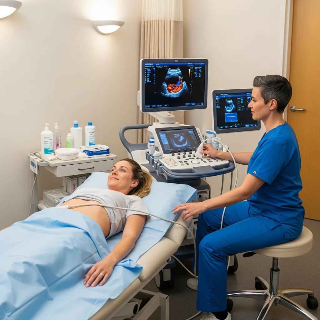
What happens during an ultrasound procedure?
When you arrive we’ll check your referral and identity, then escort you to the exam room. The sonographer will explain the scan, confirm relevant medical details and ask any questions needed for a thorough exam. You’ll lie on the couch while a water‑based gel is applied to the skin to help the transducer make good contact. The sonographer moves the probe to capture the best views and may ask you to hold your breath or change position briefly. Most people feel no pain; you might notice some pressure if the area is tender. Our sonographers talk you through the process and can pause if you need a break — knowing the steps usually helps reduce anxiety and makes the appointment straightforward.
How does ultrasound technology create images?
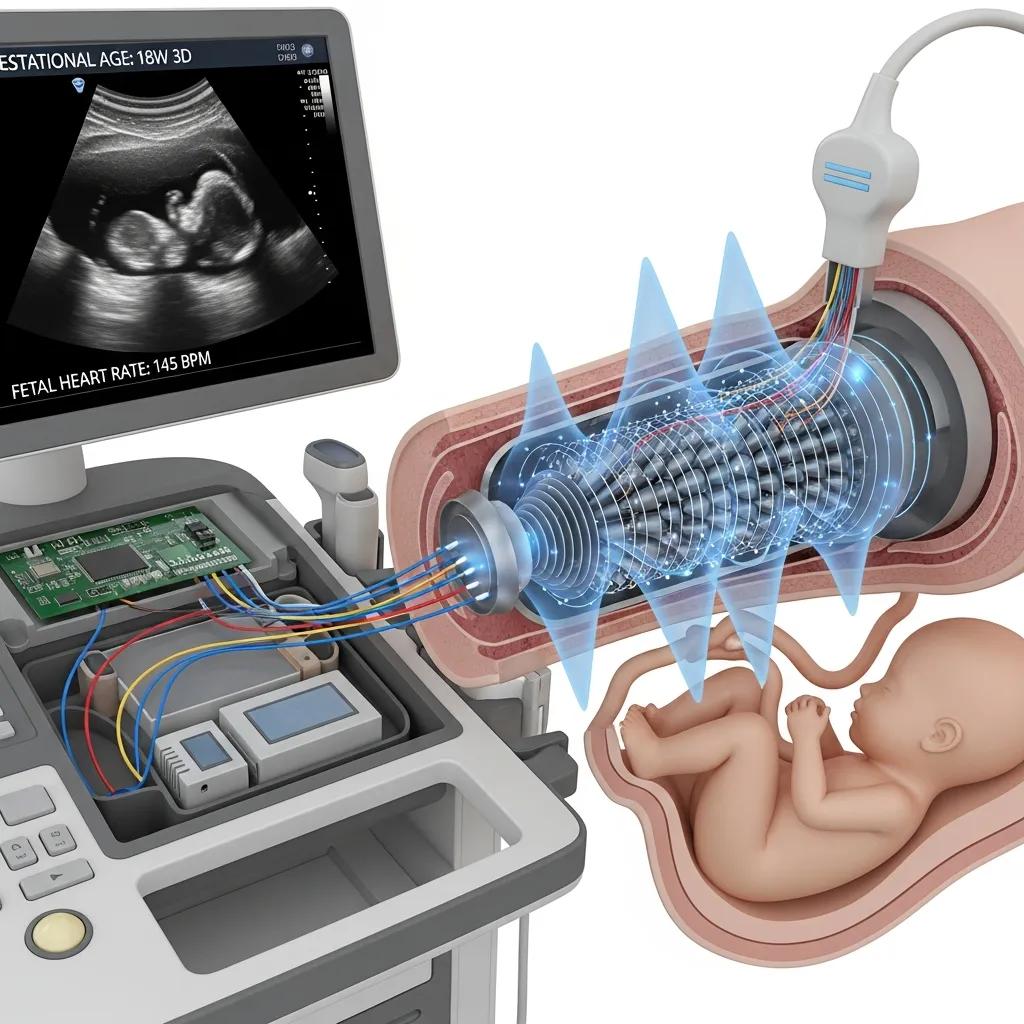
Inside the transducer are piezoelectric crystals that turn electrical energy into sound waves and then back into electrical signals when echoes return. The machine measures the strength and timing of those echoes to calculate distances and build a greyscale image; Doppler modes add colour or spectral displays to show blood flow. Different tissues reflect sound differently because of acoustic impedance — for example, fluid and bone produce distinct echo patterns — and modern systems use image processing (harmonic imaging, speckle reduction and other algorithms) to improve clarity. Next we look at safety and how clinicians keep scans as low exposure as practicable.
Is ultrasound safe? Addressing common patient concerns
Ultrasound is considered safe for diagnostic use because it relies on mechanical sound energy rather than ionising radiation. Professional and regulatory bodies recommend keeping exposure as low as reasonably achievable (ALARA), and modern machines show thermal and mechanical indices so operators can monitor output during the exam. Research shows minimal risk when scans are performed correctly, but sonographers and radiologists remain vigilant about scan time and settings to balance diagnostic benefit with exposure. The sections that follow summarise benefits and any limited risks in plain terms.
What are the risks and benefits of ultrasound scans?
Ultrasound offers several practical benefits: it’s non‑invasive, produces real‑time images, is portable for bedside use and is safe in pregnancy because it does not use ionising radiation. Potential risks are minimal and mainly theoretical — for example, prolonged or unnecessary exposure could produce thermal or mechanical effects — and these are managed by trained operators who monitor output indices and follow ALARA. Ultrasound also has limitations: body habitus or bowel gas can affect image quality, and in some cases complementary imaging (CT or MRI) may be needed for a definitive diagnosis. Understanding these trade‑offs helps clinicians choose the right test for each patient.
- Common patient benefits:
Non‑invasive, real‑time imaging for pregnancy checks and soft tissue assessment.No ionising radiation, reducing long‑term exposure compared with CT or X‑ray.Portable and repeatable, useful for dynamic studies such as Doppler blood‑flow measurements. - Key risks and how we manage them:
Very limited theoretical thermal/mechanical effects — monitored via display indices.Experienced operators and ALARA principles reduce unnecessary exposure.Some scans may be inconclusive and require complementary imaging for clarity.
Those points lead into how our clinic maintains safety through accreditation and staff training.
How does Life Medical Imaging ensure ultrasound safety?
At Life Medical Imaging Central Coast we follow recognised quality and safety practices: regular equipment maintenance, ongoing operator training and adherence to professional safety frameworks such as ALARA. Our practice is NATA accredited — an independent review of our technical and quality systems — and sonographers work closely with reporting radiologists to ensure each exam is clinically justified and performed at appropriate settings. We also run routine quality assurance checks to keep machines performing reliably. If you have specific safety concerns or special requirements, please contact the clinic before your appointment so we can discuss them and make your visit as comfortable as possible.
What are the different types of ultrasound scans?
Ultrasound covers several specialised examinations tailored to clinical needs: abdominal and pelvic scans, vascular Doppler studies, and musculoskeletal imaging that looks at tendons and joints. Modalities include standard 2D anatomical imaging, Doppler for blood flow assessment, and advanced 3D/4D reconstructions when extra spatial detail is useful — for example, in selected obstetric assessments. The table below compares common scan types by typical indications and core benefits to help referrers and patients choose the right test.
This comparison clarifies the usual role of each scan and the outcomes you can expect. The next section outlines the ultrasound services we offer locally at Life Medical Imaging Central Coast.
Which ultrasound scans are offered at Life Medical Imaging Central Coast?
We provide a broad range of ultrasound services for Central Coast patients, including general abdominal scans, vascular Doppler studies, musculoskeletal assessments, obstetric and gynaecological imaging, and breast ultrasound. Our clinic also supports interventional ultrasound guidance for procedures and offers paediatric and cardiac imaging within our diagnostic scope. These services let local referrers and patients access targeted imaging — from pregnancy monitoring to detailed tendon or joint evaluations — without travelling far. If you’re booked for a scan, please note your referral details and follow the preparation guidance in the next section to make your visit smooth.
What are the benefits of 3D and 4D ultrasound imaging?
3D ultrasound builds volumetric views to show anatomy in three dimensions, while 4D adds live motion. Clinically, 3D/4D can improve spatial understanding of fetal features or complex anatomy and assist surgical planning where surface detail matters. For many diagnostic tasks, standard 2D remains the primary tool, but 3D/4D can enhance parental engagement and counselling by producing clearer rendered images. Clinicians weigh the added visual benefit against diagnostic need and exposure principles to decide when 3D/4D is appropriate.
3D and 4D ultrasound have steadily improved and can be a valuable tool in prenatal diagnosis and patient counselling.
Current 3D/4D ultrasound technology in prenatal diagnosis and patient counselling
3D and 4D ultrasound modes extend what conventional 2D imaging can show by reconstructing volumes and displaying real‑time motion. These modes can reveal views not easily obtained with 2D alone, assist in assessing fetal anatomy and movements (including the heart using rapid volume acquisition), and provide rendered images that help parents and clinicians discuss findings. Stored volumes can be reviewed later for detailed analysis or used for education. This technology complements routine imaging where added spatial information or dynamic assessment is clinically useful.
How should you prepare for your ultrasound scan?
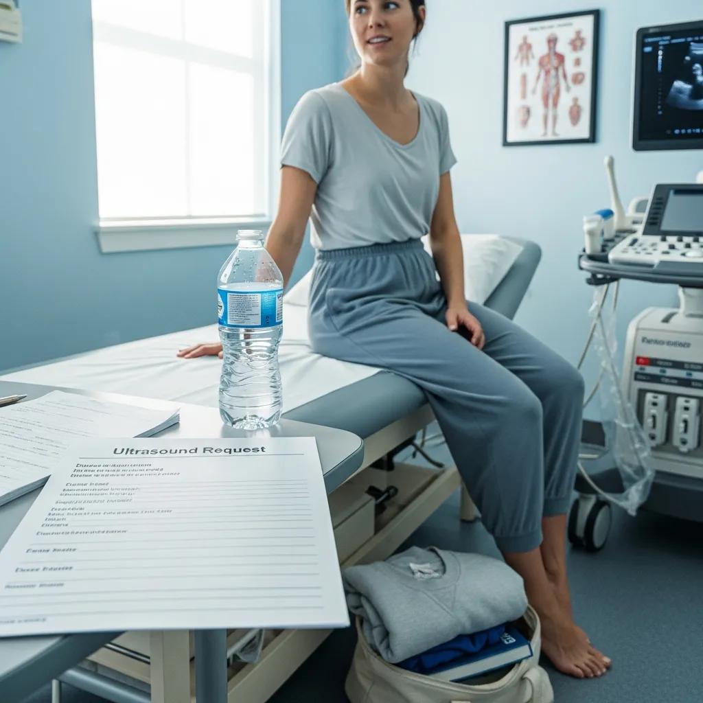
Good preparation helps us get clear diagnostic images and reduces the need for repeat scans. Requirements vary by scan type — common instructions include fasting, having a full bladder or wearing loose clothing — and bringing your referral and any prior imaging always helps. Central Coast patients should allow time for check‑in and bring previous reports if available; the next subsection lists general steps that apply to most appointments.
What are the general preparation guidelines for ultrasound?
Follow these simple steps before most ultrasound appointments to keep your visit efficient and comfortable.
- Bring the referral and identification: Present the referral form and photo ID at check‑in so we can confirm clinical details.
- Wear loose, comfortable clothing: Choose clothes that let the sonographer access the area being scanned and remove jewellery that might interfere.
- Bring relevant medical information: A short list of medications and any past imaging or reports helps with comparison and reporting.
These checks reduce administrative delays and let the imaging team focus on getting the best diagnostic images. The table below summarises scan‑specific preparation rules.
Note that fasting or bladder instructions depend on the body region targeted; follow the specific guidance provided when you book.
These instructions help you arrive ready. For local logistics, allow extra time for parking, bring referral paperwork and call us if you need mobility assistance or have questions about preparation. To change or confirm an appointment, contact the clinic on 02 4326 7000.
What can you expect during and after your ultrasound scan?
A typical appointment follows a clear sequence: check‑in and identity verification, brief pre‑scan questions, the imaging exam, then reporting and communication of results to your referrer. During the exam the sonographer captures the necessary images and may give a short verbal summary when appropriate, but a formal written report is prepared by a reporting radiologist and sent to the referring clinician. Knowing the reporting pathway — sonographer image acquisition → radiologist interpretation → referrer communication — helps you understand when to expect results and any next steps.
What happens in the examination room?
In the room the sonographer confirms your identity and the reason for the study, explains the process and asks for verbal consent. You’ll be positioned to optimise imaging while we protect your privacy and modesty. A clear, water‑based gel is applied and the transducer moved methodically to capture diagnostic images; in some cases internal probes (for example transvaginal or transrectal) are used with full explanation and consent. Staff will communicate during the scan to guide breath holds or position changes and address any discomfort — sonographers aim to balance thorough imaging with your comfort. After the scan images are reviewed and an interpretive report is generated by the radiologist.
How long does an ultrasound take and when will you receive results?
Most scans take 15–45 minutes depending on complexity and whether Doppler or interventional guidance is needed. Targeted studies are usually quicker; comprehensive obstetric or vascular exams take longer. Sonographers may give a brief verbal summary at the end of the scan when appropriate, but the formal report is finalised by a radiologist and sent to the referring clinician. Turnaround times vary with urgency and clinic workflow; if a significant or urgent finding is seen during the exam, the sonographer or radiologist will escalate it to the referrer promptly to ensure timely follow‑up.
Medical imaging technology keeps evolving — artificial intelligence is playing an increasing role in image analysis and workflow support.
AI in ultrasound: improving diagnostic accuracy and workflows
Recent advances in artificial intelligence — especially machine and deep learning — are improving ultrasound image acquisition, quality assessment and automated analysis. AI tools can assist with lesion detection, standardise measurements, flag potential errors and streamline reporting, helping clinicians work more efficiently and consistently. While promising, these technologies are tools to support trained operators and require careful validation and integration into clinical workflow.
Why choose Life Medical Imaging Central Coast for your ultrasound scan?
Life Medical Imaging Central Coast provides NATA‑accredited diagnostic imaging across several Central Coast locations, giving local access to a wide range of ultrasound services with strong quality assurance. Our team supports subspecialist imaging — including women’s and cardiac studies — and we use collaborative reporting by experienced sonographers and radiologists to help ensure accurate, timely results. To book or enquire call 02 4326 7000. We accept referrals for ultrasound at our Bateau Bay, Killarney Vale, Umina Beach and Erina locations so local patients and referrers can access accredited imaging close to home.
What makes Life Medical Imaging’s ultrasound services unique?
Our services combine independent NATA accreditation with a broad clinical scope: general, vascular, musculoskeletal, obstetric and gynaecological, and breast ultrasound, plus interventional guidance and specialist reporting where needed. Standardised quality assurance and close collaboration between sonographers and radiologists help maintain consistent image quality and reporting times. Multiple Central Coast locations improve accessibility, reducing the need to travel for specialist imaging.
How can you book an ultrasound appointment?
To book or enquire about an ultrasound at Life Medical Imaging Central Coast, call 02 4326 7000 and have your referral details ready. Our staff will advise on any preparation required for your specific scan. Bring the referral form, photo identification and any prior imaging or reports to your appointment, and let us know about any mobility or communication needs when you book. Referrers should submit imaging requests through their usual pathways so we can allocate the correct appointment type and urgency.
- Prepare referral and ID: Have paperwork ready when calling to schedule.
- Confirm scan preparation: Ask about fasting or bladder requirements at booking.
- Notify accessibility needs: Tell staff if you need assistance on arrival.
These straightforward steps make attendance easier and help our team provide the most effective diagnostic service.
- Common next steps after reporting:
The referrer reviews the report to plan clinical management or decide on further imaging.If more detail is needed, alternatives such as CT or MRI may be discussed.For urgent findings, we communicate promptly to support timely care.
These practical next steps complete the patient pathway from booking to results and help referrers and patients make informed choices about care.
Frequently asked questions
What should I expect after my ultrasound scan?
After the scan the sonographer may give a brief verbal summary if appropriate, but a formal written report is prepared by a radiologist and sent to your referring clinician. Report turnaround varies by clinic and urgency, though most results are available within a few days. If an urgent issue is found during the exam, the clinic will notify the referrer quickly so follow‑up can happen without delay.
Can ultrasound scans be used for all types of medical conditions?
Ultrasound is versatile and works well for many conditions — especially those involving soft tissues, organs and blood flow (for example obstetric, abdominal and musculoskeletal assessments). However, it’s not ideal for every situation; some problems require CT or MRI for greater detail. Your healthcare provider will recommend the best imaging method based on your clinical needs.
How often can I have an ultrasound scan?
How often you can have an ultrasound depends on the reason for the scan and clinical guidelines. Pregnant patients may have several scans during pregnancy for monitoring, while chronic conditions may require periodic imaging. Follow your clinician’s advice on timing and frequency to ensure appropriate monitoring.
Are there any dietary restrictions before an ultrasound?
Preparation depends on the scan type. For abdominal scans we usually ask you to fast for 6–8 hours to reduce bowel gas and improve images. Pelvic or obstetric scans often require a comfortably full bladder, so you may be asked to drink water beforehand. Always check the specific instructions given when you book.
What should I do if I have concerns about the ultrasound procedure?
If you’re worried about the scan, talk with clinic staff or your healthcare provider before your appointment. We can explain the procedure, address safety questions and accommodate medical or accessibility needs so the scan is performed safely and comfortably. Open communication helps reduce anxiety and ensures a better experience.
Can ultrasound be used for therapeutic purposes?
Yes — therapeutic ultrasound is a different application that uses sound waves to support tissue healing, reduce inflammation and relieve pain, often used in physiotherapy. Diagnostic ultrasound, which is what we describe here, focuses on imaging. If you’re interested in therapeutic ultrasound, discuss it with a healthcare professional to see if it’s appropriate for your condition.
Conclusion
Knowing how ultrasound works and what to expect helps you make informed choices about diagnostic imaging. Ultrasound is a safe, flexible tool for many clinical needs, and Life Medical Imaging Central Coast offers accredited services to support local patients and referrers. If you’d like to book or discuss an ultrasound, contact us to arrange an appointment — we’re here to help.