Comprehensive Life Medical Imaging Services on the Central Coast — your clear guide to advanced diagnostic care
Medical imaging uses specialised equipment to see inside the body so clinicians can diagnose, monitor and plan treatment. This guide outlines the imaging options available on the Central Coast, explains how each test works, describes what to expect at your appointment and gives practical steps for booking and preparing. Local, timely imaging reduces uncertainty and helps clinicians make faster, better-informed decisions. We cover common modalities (x‑ray, CT, MRI, ultrasound, DEXA), compare their uses and safety, describe the patient experience and technology improvements, and explain how to arrange scans and prepare for them locally.
What Medical Imaging Services Are Available on the Central Coast?
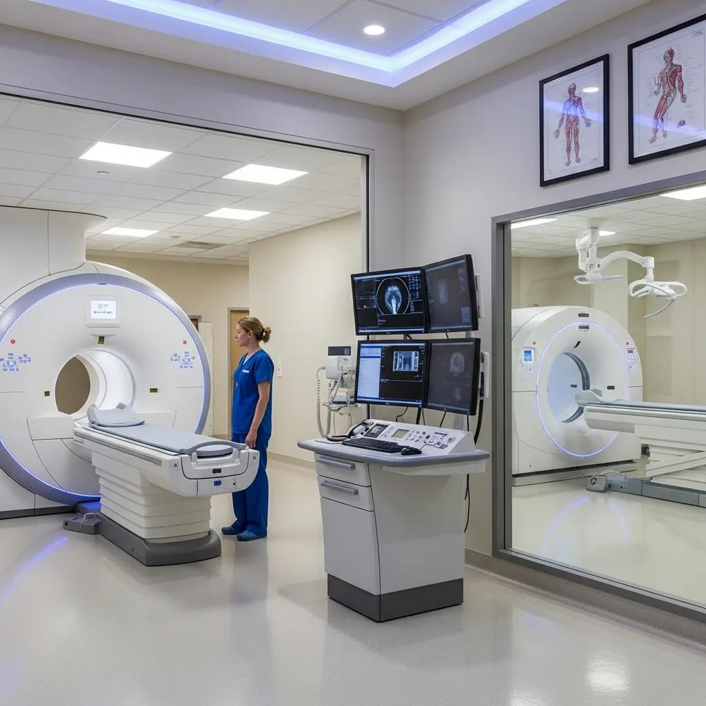
On the Central Coast you can access a full range of diagnostic imaging designed to show bones, soft tissues and physiological activity so clinicians can make accurate diagnoses and treatment plans. Each modality works differently — x‑ray uses ionising radiation to show dense structures, CT combines many x‑rays into cross‑sectional images, MRI uses magnetic fields and radiofrequency to capture soft tissue detail, and ultrasound uses high‑frequency sound waves to show movement and blood flow. Knowing how each test works helps patients and referrers choose the most appropriate scan for symptoms and urgency. The sections below list common scans, when they’re used and what patients should plan for.
Which types of imaging scans does Life Medical offer?
Life Medical Imaging provides x‑ray, CT, MRI, ultrasound and specialist DEXA bone density scans to meet common clinical needs on the Central Coast. X‑ray is ideal for suspected fractures and chest checks; CT is used for complex trauma, lung and urgent abdominal problems; MRI offers detailed soft‑tissue, neurological and joint imaging; ultrasound supports pregnancy, abdominal, vascular and musculoskeletal exams; and DEXA measures bone density for osteoporosis assessment. Most scans require a referral from a GP or specialist, and completed reports are sent to the referring clinician to guide next steps.
Each modality visualises different tissues and pathologies using its own imaging physics and contrast options. X‑ray quickly highlights bone and dense structures and is often the first test for suspected fractures. CT gives rapid, cross‑sectional detail that clarifies complex injuries and acute conditions. MRI provides superior soft‑tissue contrast for ligaments, brain and spinal cord issues. Ultrasound shows real‑time motion and blood flow, useful for vascular studies and guided procedures. Radiographers perform the scans and radiologists interpret the images; the final report links image findings to clinical recommendations and follow‑up.
The table below compares common scan types so patients can match a likely test to their referral and preparation needs. It shows each modality’s best uses and typical preparation.
This comparison makes it easier to see which test best matches a clinical question: x‑ray for suspected fractures, CT for urgent or complex assessment, MRI for detailed soft‑tissue imaging, ultrasound for dynamic or pregnancy exams, and DEXA for bone health. Having these options locally helps Central Coast patients avoid extra travel and speeds diagnosis.
- The table summarises key features of each modality to help you decide quickly.
- Use this as a basis for discussion with your referring doctor.
- If a test requires contrast or fasting, your referrer or imaging team will give clear instructions before the appointment.
How Do Life Medical Imaging Services Benefit Patients on the Central Coast?
Local imaging services improve access, shorten diagnostic pathways and support closer coordination between patients and referring clinicians. Having imaging nearby reduces travel and disruption, so scans and reports can happen sooner — helping clinicians act faster. Imaging bridges symptoms and treatment plans: a clear report gives referrers the information they need to recommend the next step. Below we outline practical patient benefits and how a local provider supports each area.
What are the advantages of local access to advanced imaging?
Central Coast patients benefit from less travel, easier follow‑up and stronger links with local GPs and specialists when imaging is available nearby. Shorter journeys make attending appointments and completing preparations (like fasting or consent for contrast) simpler. Quicker local reporting helps clinicians correlate scan results with clinical findings so care stays connected. For example, someone with an acute ankle injury can get an x‑ray locally, receive the report the same day and start physiotherapy or specialist referral without delay.
How does timely imaging improve treatment outcomes?
Timely imaging often changes clinical management: it enables earlier detection, avoids unnecessary procedures and guides targeted treatment for conditions such as trauma, acute abdominal emergencies or cancer staging. Early diagnosis of appendicitis, displaced fractures or tumour spread allows quicker intervention, which can shorten hospital stays and reduce complications. Imaging also supports monitoring chronic conditions so clinicians can adjust treatment based on objective findings. Speed and accuracy are especially valuable for Central Coast patients who need coordinated local care.
These comparisons show that local diagnostic imaging improves the patient experience, shortens time‑to‑treatment and supports clear communication between imaging teams and local clinicians. If you need help arranging a scan, our team can provide the information you need and assist with bookings.
What Should You Expect During Your Medical Imaging Appointment?
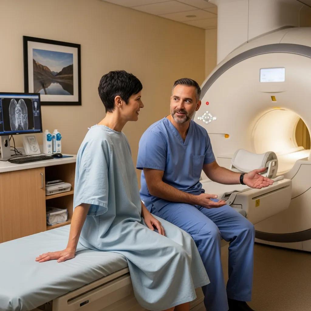
Your appointment will usually follow a simple sequence: arrive and check in, complete any screening, prepare for the exam, have the images taken by a radiographer and receive post‑scan instructions. Staff will confirm your referral and medical history, screen for pregnancy or implants, and explain the procedure to help reduce anxiety. During the scan you’ll be positioned for the best images and can communicate with the radiographer at any time. If contrast is used there may be a short observation period afterwards; reports are prepared and sent to your referring clinician.
How is patient comfort ensured during scans?
Our teams use clear communication, positioning aids and small environmental adjustments to keep you comfortable — cushions, warm blankets and step‑by‑step guidance are common. For MRI, we offer ear protection and music options to reduce noise‑related stress; staff explain breath‑hold instructions and timing to improve cooperation and image quality. Paediatric imaging uses distraction and child‑friendly explanations, and sedation is available in selected cases when stillness is essential. If you have claustrophobia or anxiety, tell the team ahead of time so we can discuss comfort measures or alternatives.
What are the common procedures and safety measures?
Imaging departments follow strict safety practices for radiation exposure, contrast administration and infection control to protect patients and staff. Radiation doses are kept as low as reasonably achievable; x‑ray and CT use ionising radiation while MRI and ultrasound do not. Contrast agents require screening for allergies and kidney function, and staff will ask relevant medical questions before administration. Standard infection control and hygiene protocols keep equipment and patient flow safe. These safety steps are part of routine care and are explained during check‑in and consent.
- Your appointment usually includes check‑in, screening, imaging and report delivery.
- Safety checks focus on pregnancy, implants, allergies and renal function where relevant.
- Comfort measures include positioning aids, ear protection for MRI and clear staff communication.
Knowing these steps can reduce uncertainty before your appointment. If you need reassurance or help arranging a scan, our team is available to answer questions and support bookings.
How Do You Choose the Right Imaging Service for Your Needs?
Picking the right imaging modality depends on the clinical question, the body area involved, urgency and patient factors such as pregnancy or implants. Clinicians consider tissue type (bone versus soft tissue), the need for speed (trauma or acute abdomen), radiation concerns and the likely diagnostic yield. Referrers usually recommend the appropriate test, but a clear decision flow helps patients understand why one scan is preferred. Below is a concise guide to match symptoms or referral notes to likely imaging options.
What factors determine the best imaging modality?
Important factors include the suspected tissue type (bone, soft tissue, vascular), how urgent the diagnosis is, previous imaging and patient considerations such as pregnancy or claustrophobia. For example, swollen painful joints without trauma often start with ultrasound to assess soft tissue and fluid, while suspected complex fractures or internal bleeding typically require CT for rapid, detailed cross‑sectional imaging. MRI is preferred for neurological symptoms or when superior soft‑tissue contrast is needed, and DEXA is specific for bone density checks. Availability and local scheduling can also influence the final choice.
When should you consult a specialist for imaging advice?
Specialist imaging input is useful for complex diagnostic uncertainty, pre‑operative planning, cancer staging and when previous scans are inconclusive. Multidisciplinary discussion between surgeons, oncologists and radiologists helps plan advanced studies — for example, contrast‑enhanced protocols, targeted MRI sequences or PET‑CT in specialised settings. If your GP is unsure which test will best answer the clinical question, they can request radiologist advice or suggest specialist referral to tailor imaging. Specialist input helps avoid unnecessary tests and focuses resources on the most informative studies.
- Key considerations: tissue type, urgency, patient safety and prior imaging.
- If unsure, ask your referrer to consult a radiologist for targeted guidance.
- Specialist input matters for surgical planning and complex oncology cases.
These decision points help patients and referrers choose the most informative, least invasive test for each clinical scenario.
What Are the Latest Technologies Used in Life Medical Imaging on the Central Coast?
Modern imaging equipment improves diagnostic accuracy with higher resolution, faster scans and advanced contrast techniques that make pathology easier to see. High‑field MRI systems deliver better soft‑tissue detail and shorter scan times, multislice CT scanners capture fine anatomy quickly, and digital x‑ray provides clearer images with lower dose. Ultrasound systems with Doppler and 3D capabilities enhance vascular and structural assessment. These upgrades reduce repeat scans and give clinicians more confidence in their decisions.
How do advanced imaging machines improve diagnostic accuracy?
Advanced systems offer higher spatial and contrast resolution, faster acquisition and more powerful post‑processing, all of which improve lesion detection and characterisation. Higher‑resolution MRI can show subtle soft‑tissue changes, while multislice CT produces thin‑slice reconstructions that reveal complex bone anatomy or small lung nodules. Digital workflows and PACS speed up report distribution and support multidisciplinary review. Together, these capabilities reduce diagnostic uncertainty and help plan precise treatment.
What innovations set Life Medical apart from other providers?
Life Medical focuses on timely reporting, patient comfort and integration with local clinical workflows. Improvements typically include streamlined digital reporting that delivers images and radiology reports directly to referring clinicians and patient‑centred scheduling that minimises wait times. Emphasising modern equipment and efficient processes helps local providers deliver prompt, high‑quality diagnostic information to support patient care.
- Advanced imaging raises accuracy through better resolution, speed and post‑processing.
- Efficient workflows and digital reporting shorten the time from scan to clinical decision.
- Local access to modern equipment supports better outcomes for Central Coast patients.
How Can You Book and Prepare for Your Medical Imaging on the Central Coast?
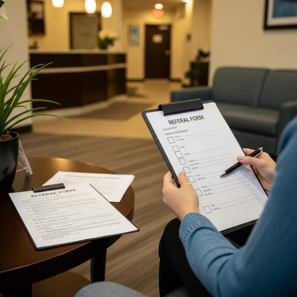
Booking usually begins with a referral from your GP or specialist; the referral gives clinical context so we can schedule the right modality and protocol. When booking, have your referral, any previous imaging reports and a list of current medications ready. Instructions may include fasting for certain scans, screening questions about contrast or implants, and guidance on clothing and personal items. Use the practical booking steps and preparation checklist below to arrive ready and reduce the chance of a rescheduled appointment.
What is the booking process for Life Medical imaging services?
To book with Life Medical Imaging you’ll generally need a referral from your GP or specialist and to provide relevant clinical information when you call or book online. Appointments are prioritised by clinical urgency and you’ll receive clear instructions about fasting, medication adjustments and consent for contrast where required. Bring your referral and any prior imaging to the appointment, and try to arrive a little early for check‑in and screening. For enquiries or to arrange scans, our team can guide you through the process and answer questions.
How should patients prepare for different types of scans?
Preparation varies by test: x‑ray usually needs minimal preparation and comfortable clothing; CT may require fasting and kidney function checks for contrast studies; MRI requires removal of metal and implant screening; and some ultrasound exams need a full bladder for pelvic views. Paediatric patients may need special fasting or sedation instructions, and claustrophobic adults should discuss options with the imaging team before arrival. The table below summarises common preparation points to help you plan for your Central Coast appointment.
Follow the specific instructions you receive with your appointment to ensure the best image quality and safety. Good preparation reduces delays and increases the chance the scan will answer the clinical question on the first attempt. If you need help or have questions about preparation, contact our team — we’re here to help with bookings and advice.
- Bring your GP or specialist referral and any prior imaging to the appointment.
- Follow fasting or medication instructions given at booking to avoid rescheduling.
- Tell staff about pregnancy, implants or allergies during screening.
Following these steps makes booking and preparation straightforward and helps you get the most from your imaging appointment on the Central Coast.
Frequently Asked Questions
What should I do if I have concerns about radiation exposure during imaging?
If you’re concerned about radiation, talk with your referrer or the imaging team. We follow the ALARA principle (As Low As Reasonably Achievable) to minimise dose and can explain why a test is necessary. In many cases, a non‑radiation alternative such as MRI or ultrasound may be suitable — your clinician can help decide the best option for your situation.
Can I bring someone with me to my imaging appointment?
Yes — you can usually bring a friend or family member for support. Some procedures have restrictions, especially those involving radiation or a sterile environment, so check with the facility beforehand. Having someone with you can ease anxiety and help with practical arrangements after the scan.
What happens if I need a follow-up scan after my initial appointment?
If a follow‑up scan is needed, your referring clinician will discuss the reasons with you and provide a new referral if required. Our team will help schedule the follow‑up so it fits your treatment plan. Keep open communication with your healthcare provider to understand the next steps in your care.
Are there any specific instructions for paediatric patients undergoing imaging?
Yes. Children often require special preparation. Parents or guardians should discuss instructions with the imaging team before the appointment. This may include fasting, sedation options or distraction techniques to help children stay still. Staff use child‑friendly language and strategies to make the experience as comfortable as possible.
How can I access my imaging results after the appointment?
Results are normally sent to your referring clinician, who will discuss them with you at a follow‑up. Some centres also offer a patient portal where you can view reports and images. If you need results sooner or have questions, contact the imaging facility and they’ll advise how to obtain them securely.
What should I do if I feel anxious about my imaging procedure?
If you feel anxious, tell the imaging staff when you arrive. They can explain the process, offer reassurance and suggest comfort measures such as music or relaxation techniques. For severe anxiety or claustrophobia, discuss sedation options with your healthcare provider before the appointment. Our team’s priority is your safety and comfort.
Conclusion
Advanced medical imaging on the Central Coast delivers real benefits: less travel, faster diagnosis and better continuity of care. Understanding the available modalities helps you make informed choices that match your clinical needs. If you have questions or want to book a scan, reach out to Life Medical Imaging — we’re here to provide clear information and convenient local care that puts your health first.