Understanding Ultrasound Scans: What to Know About Technology, Safety, Types, Preparation and Results
Ultrasound scans are safe, non-invasive tests that use high-frequency sound waves to produce real‑time images of organs, tissues and blood flow. This guide explains how ultrasound works, why it’s often chosen for soft-tissue and pregnancy imaging, and how you can prepare to get the clearest results. Many people want straightforward answers about safety, what happens during an appointment, and how results are reported; this article covers the basic physics in plain language, the common scan types, practical preparation checklists and the key safety concepts such as the ALARA principle, the Thermal Index and the Mechanical Index. You’ll also find what to expect during and after a scan and how accredited local clinics on the Central Coast maintain quality imaging and reporting. Read on to understand how ultrasound helps diagnosis and monitoring, how to prepare, and where to find trusted services when you need them.
What is an ultrasound scan and how does the technology work?
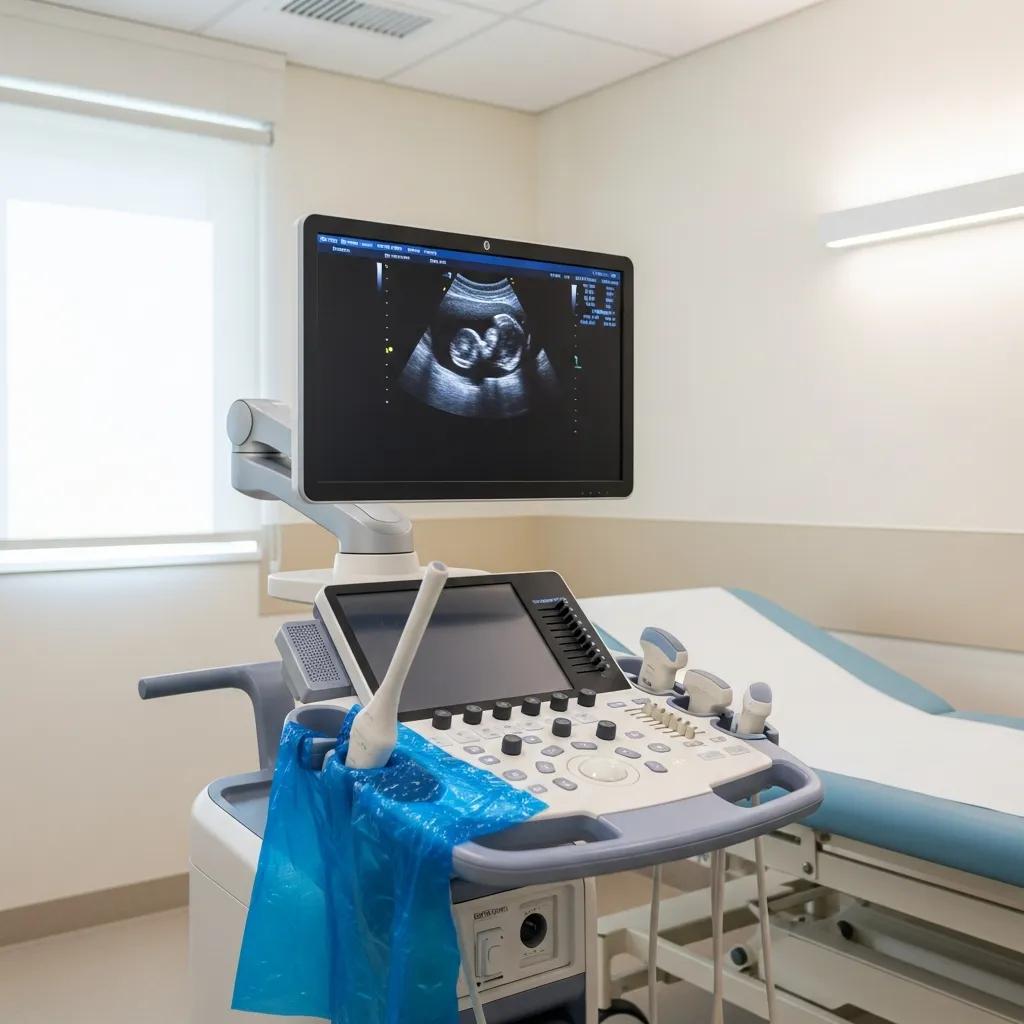
An ultrasound scan (also called sonography or ultrasonography) sends brief pulses of high-frequency sound into the body and records returning echoes to build an image. A handheld transducer contains piezoelectric crystals that turn electrical energy into sound waves and then convert returning echoes back into electrical signals, producing live sonograms. This method gives dynamic views of soft tissues, fluid collections, fetal growth and blood flow with immediate feedback for the operator and referrer. Because ultrasound uses non‑ionising sound rather than X‑rays, it’s especially useful for repeated monitoring and for imaging during pregnancy. Knowing how the transducer and echo principles work also makes terms like echogenicity and Doppler flow easier to understand—these are described below.
What is an ultrasound scan? Explaining sonography and ultrasonography
An ultrasound scan is a targeted imaging test that visualises structures beneath the skin using reflected sound waves and produces images called sonograms. Clinicians commonly request sonography for abdominal concerns, musculoskeletal injuries, breast lumps, vascular checks and pregnancy monitoring. Referrals usually state the clinical question—pain, suspected fluid, pregnancy dating or vascular assessment—and the sonographer tailors the exam to that question. Trained sonographers perform the scan; radiologists interpret the images and issue the report that guides diagnosis and treatment.
How do ultrasound sound waves and transducers create images?
The transducer emits ultrasound pulses that travel through tissue and reflect back at boundaries where properties change. The machine times and analyses those echoes to map internal structures. Piezoelectric crystals in the transducer deform under an electric current to create sound, then generate electrical signals when echoes return—this is how images form. Echogenicity describes appearance on the image: fluid is anechoic (black), soft tissue ranges from hypoechoic to hyperechoic, and calcifications are typically very bright. Doppler mode detects frequency shifts in returning waves to assess blood flow and velocity, which helps evaluate vessel patency and flow direction—linking the physics directly to clinical assessment.
What are the different types of medical ultrasound scans?
Ultrasound covers a range of targeted exams tailored to the body area and clinical question—from general abdominal scans to specialised vascular and musculoskeletal studies. Each type uses the same basic physics but adjusts transducer frequency, patient position and Doppler settings to answer specific diagnostic needs. Knowing which scan is appropriate helps you follow the right preparation and reduces the chance of returning for repeat imaging. Below we outline the major categories, common uses and simple prep notes so you can see which study matches your referral.
Which ultrasound types are used for general, obstetric and gynaecological imaging?
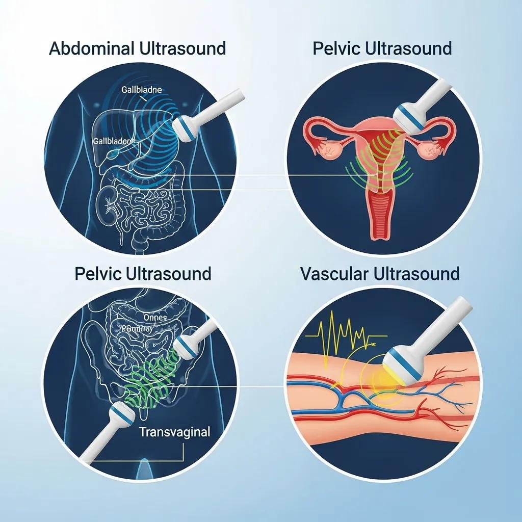
Abdominal scans look at the liver, gallbladder, pancreas, spleen and kidneys for causes of pain, abnormal liver tests or suspected stones; these exams often require fasting to reduce bowel gas. Pelvic ultrasound assesses the bladder, uterus and ovaries and can be transabdominal (usually with a full bladder) or transvaginal for higher resolution of pelvic structures; gynaecological reasons include ovarian cysts and uterine fibroids. Obstetric scans range from dating and growth checks to anatomy surveys and optional 3D/4D imaging for improved fetal surface detail—3D/4D adds volumetric views that can aid assessment while giving expectant parents clearer images. Knowing these differences explains why appointment instructions vary.
What are specialised ultrasounds like vascular, musculoskeletal, breast and Doppler scans?
Specialised studies adapt ultrasound to specific tissues or functions. Vascular duplex combines Doppler and B‑mode imaging to check blood flow, detect clots or stenoses. Musculoskeletal ultrasound visualises tendons, ligaments and joint fluid for injuries. Breast ultrasound helps characterise palpable lumps or findings seen on mammography. Doppler measurements quantify flow velocity and direction to diagnose conditions such as deep vein thrombosis or arterial disease, while elastography (where available) estimates tissue stiffness to help describe lesions. These high-resolution, dynamic studies often complement other imaging and are suitable for follow-up because they do not use ionising radiation.
Different ultrasound types and typical preparation are summarised below to help you know what to expect before your appointment.
If you need local access to these services, Life Medical Imaging Central Coast offers a wide range of ultrasound exams at multiple Central Coast locations. Services include general, vascular, musculoskeletal, obstetric and gynaecological imaging, breast ultrasound and 3D/4D obstetric options. You can request an appointment or book online to arrange the correct study and receive clear pre‑scan instructions. Local availability helps match your referral to the right test quickly and conveniently.
How safe is ultrasound? Understanding safety guidelines and common concerns

Ultrasound is regarded as a safe imaging method because it uses non‑ionising sound energy rather than ionising radiation. Scans follow the ALARA (As Low As Reasonably Achievable) principle and are monitored using indices like the Thermal Index (TI) and Mechanical Index (MI). Clinical governance and scanning protocols ensure operators limit exposure time, use appropriate settings and perform scans only when clinically indicated. For pregnancy, ultrasound is the preferred imaging choice for fetal assessment because diagnostic‑level ultrasound, when used responsibly, has no known harmful effects. The short table below explains safety indices and what they mean in plain language.
This table shows how safety is measured and managed during diagnostic ultrasound.
Safety of Ultrasonic Examinations: Mechanical and Thermal Indices Explained
ABSTRACT: This review summarises the biophysical effects relevant to ultrasonography and explains the reasoning behind the Mechanical Index (MI) and Thermal Index (TI) as part of the Output Display Standard (ODS). Safe diagnostic ultrasound doses follow specific rules and are reported using MI and TI. These indices consider the physical interactions between ultrasound fields and biological tissue, which depend on temporal and spatial parameters of the acoustic field produced by transducers. Predicted temperature rises are modelled using homogeneous, layered and bone/tissue interface scenarios.
Safety of ultrasonic examinations; thermal and mechanical indices, A Nowicki, 2020
Why clinicians often choose ultrasound:
- It uses non‑ionising sound, avoiding the radiation exposure associated with X‑rays and CT scans.
- It delivers real‑time imaging, enabling dynamic assessment without increasing patient risk.
- Operators follow ALARA and monitor TI and MI to keep exposures within accepted diagnostic practice.
These safety safeguards explain why ultrasound is commonly used for pregnancy care and serial follow‑up. Understanding indices like TI and MI helps patients see why sonographers may adjust settings or limit scan time to answer the clinical question safely and efficiently.
Why is ultrasound considered safer than X‑rays and some other imaging?
Unlike ionising modalities, ultrasound relies on reflected sound waves rather than photon radiation, so there’s no cumulative radiation dose from repeat exams. That makes ultrasound the preferred first‑line test for fetal imaging, pelvic assessments and many soft‑tissue problems where radiation would be unnecessary. Practically, this means scans can be repeated for monitoring without the radiation risks that come with CT or radiography—which is why ultrasound is often the initial imaging choice for common clinical presentations.
What are the Thermal and Mechanical Indices and their role in safety?
The Thermal Index (TI) estimates potential tissue heating from ultrasound energy; the Mechanical Index (MI) estimates the chance of non‑thermal mechanical effects such as cavitation. Sonographers keep both indices low and will reduce power or shorten scan time when imaging sensitive tissues, for example in early pregnancy or neonatal imaging. In simple terms, monitoring TI and MI ensures we capture the necessary diagnostic information while minimising any theoretical risk—consistent with ALARA and best clinical practice.
How should you prepare for an ultrasound scan? Essential tips and checklists
Good preparation helps improve image quality, shortens appointment time and lowers the chance of needing repeat imaging. General preparation focuses on clothing, arrival logistics and paperwork, plus simple adjustments such as fasting or bladder filling depending on the scan. Below are actions to take before most ultrasound appointments, followed by scan‑specific guidance for abdominal, pelvic and other specialised studies.
What general preparation steps should patients follow before any ultrasound?
Before your scan, bring your referral, any relevant medical history and previous imaging reports—these help the sonographer and radiologist interpret findings. Wear loose, comfortable clothing so the area being scanned is easy to access, and remove jewellery from the region of interest; you may be asked to change into a gown for some exams. Arrive a few minutes early to complete registration and tell staff about mobility needs, pregnancy or implanted devices; this keeps appointments running smoothly and ensures you get the right assistance. These simple steps reduce stress on the day and lead into the specific preparation needed for particular scan types.
General checklist before an ultrasound:
- Bring your referral and any previous imaging or reports.
- Wear loose clothing and remove jewellery from the scan area.
- Arrive early and inform staff of mobility, language or other needs.
How do preparation requirements differ for abdominal, pelvic and other specific ultrasounds?
Preparation varies because bowel gas, bladder volume and diet affect image clarity. Abdominal scans usually require fasting for 6–8 hours to reduce bowel gas and better visualise the gallbladder and pancreas. Pelvic or obstetric transabdominal scans generally need a comfortably full bladder to provide an acoustic window; transvaginal exams require an empty bladder and give higher pelvic resolution. Musculoskeletal and vascular studies typically only require comfortable clothing and easy limb access, though your referral may include other instructions.
If you’re unsure about instructions or need to change your appointment, contact Life Medical Imaging Central Coast—clinic staff can confirm the correct preparation for your referral. Clear pre‑scan preparation reduces the likelihood of repeat imaging and helps ensure the diagnostic question is answered on your first visit.
What can you expect during and after your ultrasound scan? A practical overview
A typical ultrasound visit follows a predictable sequence: check‑in and paperwork, a short clinical discussion with the sonographer, the scan itself (usually 15–45 minutes depending on the study) and then image interpretation by a radiologist. During the exam you may feel light pressure as the sonographer moves the transducer and brief cold from the gel; most people describe the experience as comfortable and painless. After the scan, images are reviewed and a formal report is issued to your referring clinician, who will discuss results and next steps with you. The step‑by‑step outline below sets expectations and can help reduce anxiety about the process.
What happens during the appointment:
- Registration and check‑in; confirmation of identity and referral details.
- Brief clinical review with the sonographer to confirm symptoms and the area of concern.
- The scan—sonographer acquires images and may ask you to change position or hold your breath.
- Post‑scan: images sent for radiologist review and report generation.
This sequence clarifies timing and responsibilities and leads into how reports are structured and explained to patients.
What happens during an ultrasound scan? Step‑by‑step procedure overview
When you enter the room, the sonographer will explain the procedure and confirm relevant history to focus the exam. You’ll lie on an examination couch and ultrasound gel will be applied to the skin; the transducer is then moved over the target area while the sonographer captures still images and video loops. For vascular or Doppler studies you may be asked to remain still and breathe gently while flow measurements are taken. For obstetric scans the sonographer will measure fetal structures and assess growth. Routine scans generally take 15–30 minutes, though detailed studies or body habitus can extend that time; images are then forwarded for reporting.
How are ultrasound images interpreted and what do your results mean?
Sonographers obtain diagnostic images but do not issue the final report. A radiologist with specialist training reviews the images, correlates findings with clinical history, and issues a structured report to the referring clinician. Reports commonly use terms such as “anechoic” (fluid) or “hyperechoic” (bright) while a plain‑language summary may accompany formal findings to guide next steps. If further investigation or referral is recommended, your clinician will explain options—follow‑up ultrasound, complementary imaging or specialist consultation. Clear communication between sonographer, radiologist and referrer helps translate imaging findings into appropriate clinical care.
Why choose Life Medical Imaging Central Coast for your ultrasound scan? Expertise, technology and patient care
Life Medical Imaging Central Coast is an accredited, independent radiology provider with multiple sites across the Central Coast, including Bateau Bay, Killarney Vale, Umina Beach and Erina. We deliver a full suite of diagnostic ultrasound services with particular strengths in women’s and cardiac imaging, alongside general, vascular, musculoskeletal, obstetric and gynaecological scans, breast ultrasound and 3D/4D obstetric imaging. Accreditation and specialist oversight help ensure consistent quality in image acquisition and reporting. Our booking process is designed to be straightforward for referrers and patients—request an appointment or book online to arrange the right study and receive clear pre‑scan instructions.
How do our skilled sonographers and radiologists ensure accurate ultrasound diagnoses?
Experienced sonographers perform targeted exams using standardised protocols and capture the images that radiologists review for diagnostic interpretation—this two‑step process verifies image quality and findings. Radiologist oversight and structured reporting translate sonographic observations into clinically meaningful conclusions to support patient management. Our emphasis on women’s and cardiac imaging brings sub‑specialist expertise where complex protocols or detailed assessment is needed; quality assurance practices support reliable, consistent results for referrers and patients alike.
What advanced ultrasound technologies and patient facilities do we offer?
Life Medical Imaging Central Coast offers advanced capabilities such as Doppler flow assessment and 3D/4D obstetric imaging in addition to high‑resolution B‑mode ultrasound, covering a wide range of diagnostic needs. Our facilities are modern and patient‑centred, with comfortable scanning rooms and staff who assist with preparation and appointment logistics. For convenience you can request an appointment or book online, and clinic staff are available to clarify any preparation instructions or accommodate specific needs before your scan.
Key advantages of local accredited services:
- Specialist expertise in women’s and cardiac imaging for accurate interpretation.
- Advanced ultrasound capabilities (Doppler, 3D/4D) to answer complex clinical questions.
- Multiple Central Coast locations to improve access and reduce travel time.
These local strengths make it simple to access high‑quality ultrasound imaging when referred by your clinician or when follow‑up is required.
Frequently Asked Questions
What should I expect during my first ultrasound appointment?
On arrival you’ll check in and complete any necessary paperwork. A sonographer will briefly review your medical history and the reason for the scan. You’ll lie on a couch, ultrasound gel will be applied and the sonographer will move the transducer to capture images. Most scans take 15–45 minutes depending on the type. Afterward the images are reviewed by a radiologist who prepares a report for your referring clinician.
How long does it take to receive ultrasound results?
Images are sent to a radiologist for interpretation after the scan. Depending on clinic workflow and scan complexity, this usually takes a few hours to a couple of days. The radiologist’s report is sent to your referring clinician, who will discuss results with you—often at a follow‑up appointment. If urgent findings are found, your clinician may contact you sooner to arrange next steps.
Are there any risks associated with ultrasound scans?
Diagnostic ultrasound is considered very safe because it uses non‑ionising sound waves rather than ionising radiation. Operators follow the ALARA principle to minimise exposure, and safety indices such as the Thermal Index (TI) and Mechanical Index (MI) are monitored during the exam. While no known risks exist from standard diagnostic use, discuss any concerns with your healthcare provider before the scan.
Can I eat or drink before my ultrasound?
Preparation depends on the type of scan. For abdominal scans you’ll typically fast for 6–8 hours to reduce bowel gas and improve image quality. Pelvic or obstetric scans may require you to drink water beforehand to fill your bladder. Always follow the specific instructions provided by your clinician or imaging centre to ensure the best results.
What should I wear to my ultrasound appointment?
Wear loose, comfortable clothing to make access to the scanned area easy. You may be asked to change into a gown for some exams, especially if the area is difficult to access. Remove jewellery from the scan area to avoid interference with imaging.
How can I prepare my child for an ultrasound scan?
Explain the procedure in simple terms and reassure them it is painless and quick. Role‑playing the process can help, and bringing a favourite toy or book can comfort younger children. If the scan requires fasting or a full bladder, explain those steps clearly beforehand to avoid confusion on the day.
What happens if the ultrasound results are abnormal?
If results are abnormal, your referring clinician will discuss the findings and explain what they mean. They may recommend further tests, additional imaging, or specialist referral for more detailed evaluation. Ask questions and request clear next steps—early follow‑up often leads to better outcomes.
Conclusion
Knowing what an ultrasound can and can’t do helps you make informed choices about your care. Ultrasound is a non‑invasive, low‑risk tool that provides valuable diagnostic information without radiation. By following preparation instructions and understanding how scans are performed and reported, you can help ensure a smooth appointment and the best possible images. For more information or to schedule your ultrasound, visit Life Medical Imaging Central Coast or book online today.