Comprehensive Radiology & Imaging Services — Benefits, Procedures and Innovations
Diagnostic imaging combines specialised scanners, experienced clinicians and digital reporting to give clear, non‑invasive views of the body. This article explains how imaging informs clinical decisions and describes the strengths of CT, MRI, ultrasound, digital X‑ray and DEXA. You’ll find practical guidance for Central Coast patients on booking and preparing for scans, an overview of safety considerations like radiation and contrast, and a look at recent advances such as ultra‑low dose CT and AI‑assisted workflows. We also outline image‑guided interventional options and local access routes so clinicians and patients can act on results promptly. Below we cover what imaging services do, CT benefits and risks, ultrasound uses, technology trends including AI and teleradiology, how to book private MRI and other scans, and the interventional procedures available locally.
What Are Diagnostic Imaging Services and How Do They Support Medical Care?
Diagnostic imaging creates internal images of the body using technologies such as CT, MRI, ultrasound, digital X‑ray and bone densitometry (DEXA). Each technique converts physical signals — X‑rays, sound waves or magnetic resonance — into images radiologists interpret to confirm diagnoses, stage disease and monitor treatment. Imaging reduces uncertainty, often avoids invasive procedures and plays a vital role in emergency assessment, chronic disease follow‑up, surgical planning and targeted interventions. The list below summarises the main modalities and their typical clinical uses for quick reference.
Our clinics offer a practical mix of modalities to meet most diagnostic needs:
- CT (Computed Tomography): Fast cross‑sectional imaging for trauma, chest, abdomen and bone detail.
- MRI (Magnetic Resonance Imaging): Detailed soft‑tissue characterisation for neurological and musculoskeletal conditions.
- Ultrasound: Real‑time imaging for soft tissue and blood flow, plus obstetric and gynaecological studies.
- Digital X‑ray: Quick, high‑resolution imaging for bones and chest examinations.
- DEXA (Bone Densitometry): Precise measurement of bone density to assess metabolic bone disease and fracture risk.
Together, these modalities provide the evidence clinicians need to diagnose conditions, guide biopsies or injections, and track treatment response. The next section explains the specific services we offer locally and how accreditation and specialist staff maintain quality.
Which Types of Diagnostic Imaging Are Offered at Life Medical Imaging Central Coast?
Life Medical Imaging Central Coast provides a wide range of services across multiple sites on the Central Coast. Our offerings include general CT, cardiac CT/CT angiography, dental imaging, digital X‑ray, comprehensive ultrasound services, DEXA bone densitometry and paediatric imaging. We also deliver image‑guided procedures such as spinal and joint injections, biopsies and PRP therapy. As a NATA‑accredited independent radiology provider, we prioritise equipment calibration, staff credentials and consistent reporting to ensure reliable image quality and timely, clinically useful reports. Patients benefit from dedicated women’s and cardiac pathways, up‑to‑date scanner technology and easy online booking for local appointments.
Robust quality assurance and accreditation support accurate diagnosis and patient safety, while multiple Central Coast locations make scheduling more convenient for referrers and patients.
How Do Radiologists and Imaging Technologists Ensure Accurate Diagnoses?
Radiologists provide the diagnostic interpretation and reports; sonographers and radiographers acquire the images using standardised protocols to ensure reproducible, high‑quality studies. Our workflows include patient screening, contrast safety checks, routine equipment calibration and peer review to reduce errors and keep standards high. Reporting may use structured templates and, where appropriate, teleradiology to secure subspecialist input and fast turnaround for urgent cases. These layers of quality control help imaging findings reliably inform clinical decisions and treatment planning.
Good coordination between referring clinicians and imaging teams ensures the right study is requested and that critical results are communicated promptly, improving patient care and reducing delays.
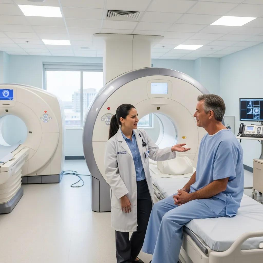
What Are the Benefits and Risks of CT Scans in Modern Radiology?

CT delivers rapid, high‑resolution cross‑sectional images that are particularly good for visualising bone, lungs, acute abdominal conditions and vascular anatomy. The scanner rotates an X‑ray source and detectors around the patient to reconstruct detailed axial images and 3D reformats useful for surgical planning and vascular assessment. The main advantages of CT are speed, broad clinical utility and excellent spatial resolution; the principal risks relate to ionising radiation and, when used, intravenous contrast. Modern dose‑reduction techniques and careful clinical justification keep risk low while preserving diagnostic value. The table below compares common CT types so patients know what to expect.
Key clinical reasons for selecting particular CT studies include:
- General CT: Fast assessment for trauma and emergency chest or abdominal conditions.
- Cardiac CT / CT Angiography: Non‑invasive coronary anatomy and vascular assessment.
- Dental CT (OPG / Cone Beam): Detailed imaging for oral surgery planning and implant assessment.
The table that follows contrasts CT types by typical use, dose considerations and basic preparation.
How Does Ultra-Low Dose, High Definition CT Technology Improve Patient Safety?
Ultra‑low dose, high‑definition CT reduces ionising radiation exposure through combined hardware and software advances while keeping image quality suitable for diagnosis. Techniques include iterative reconstruction, more sensitive detectors and tailored protocols matched to patient size and the clinical question. For follow‑up scans, younger patients or screening situations, low‑dose protocols cut cumulative exposure without sacrificing the detail clinicians need. Life Medical Imaging Central Coast highlights dose optimisation as a core patient‑safety strength — offering clinicians imaging that balances diagnostic confidence with lower exposure.
That focus on dose optimisation leads into what patients should expect when preparing for and having a CT scan.
What Should Patients Expect When Preparing for and Undergoing a CT Scan?
Preparation depends on the study and whether intravenous contrast is used. Typical steps include arrival and registration, safety screening for allergies and implants, and brief positioning in the scanner for the scan itself. You may be asked to fast for a few hours if contrast is planned; technologists will review medication history and any prior contrast reactions as part of safety checks. During the scan you’ll be in contact with the technologist via intercom. The actual image acquisition often takes only minutes, though the appointment includes consent and preparation time. A radiologist interprets the images and the results are shared with your referring clinician according to clinical urgency and local reporting pathways.
Clear pre‑visit instructions and efficient reporting help reduce anxiety and ensure each study is safe and diagnostically useful.
How Is Ultrasound Imaging Used and What Are Its Key Advantages?
Ultrasound uses high‑frequency sound waves from a transducer to create real‑time images of soft tissues, vessels and moving structures — all without ionising radiation. This makes ultrasound ideal for pregnancy care, vascular assessments and musculoskeletal scans. Doppler techniques show blood flow and direction, offering dynamic information that static imaging cannot. Portable systems also allow bedside assessment in acute settings. Key benefits include safety in pregnancy, dynamic and comparative scanning, and accurate guidance for interventional procedures. Below we list common ultrasound types with brief preparation notes for patients and referrers.
Common ultrasound variants and their uses include:
- Obstetric Ultrasound: Dating scans, fetal anatomy and selected 3D/4D views; early pregnancy scans may require a full bladder.
- Vascular (Doppler) Ultrasound: Assessment of arterial and venous flow for DVT, carotid disease and peripheral vascular conditions; usually no fasting required.
- Musculoskeletal Ultrasound: Evaluation of tendons, ligaments and soft tissues with dynamic testing; may include comparative scanning of the opposite side.
- Abdominal / Pelvic Ultrasound: Organ and pelvic assessment; fasting or a full bladder may be requested depending on the exam.
The table below compares common ultrasound subtypes by typical use, preparation and main advantages to help guide selection.
What Are the Different Types of Ultrasound Scans Available?
Ultrasound exams are tailored to the body area and clinical question using specific probes, presets and scanning approaches. Obstetric scans focus on fetal measurements and anatomy (with optional 3D/4D where indicated). Vascular Doppler evaluates flow velocity and direction to detect stenosis or thrombosis. Musculoskeletal studies visualise tendons, ligaments and fluid collections, often with dynamic provocation to reproduce symptoms. Preparation varies by test — pelvic and early obstetric scans commonly need a full bladder; abdominal scans may ask you to fast to reduce bowel gas. Knowing the scan type helps referrers request the correct exam and prepares patients for the right pre‑visit actions.
Choosing the appropriate ultrasound ensures the clinical question is answered efficiently and reduces the need for further tests.
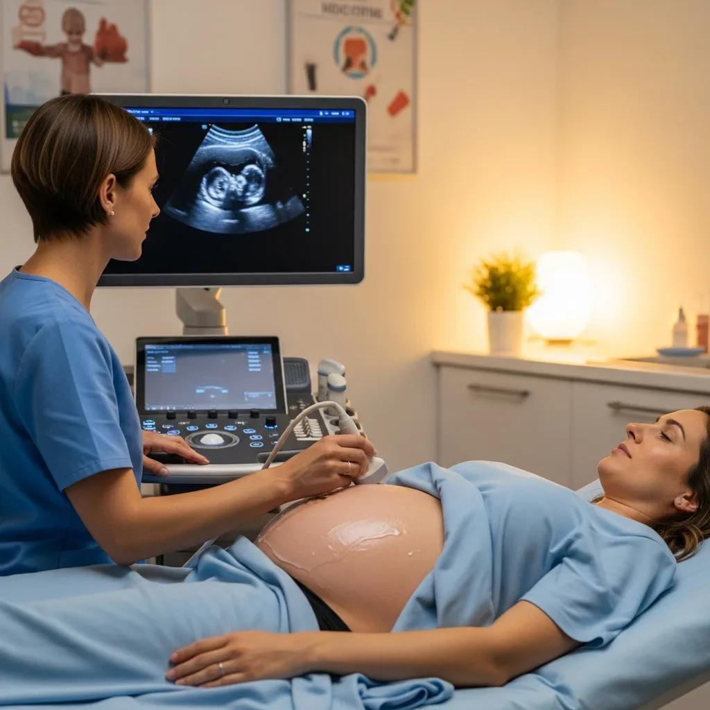
Why Is Ultrasound Considered Safe and Effective for Pregnancy and Women’s Imaging?
Ultrasound is the preferred modality in pregnancy because it uses sound waves rather than ionising radiation, posing no known radiation risk to the fetus. It provides clear information on fetal growth, placental position and anatomy, and Doppler techniques assess blood flow where needed. For gynaecological concerns, transabdominal and transvaginal approaches give complementary views of the uterus and adnexa without radiation exposure. The ability to perform repeat examinations safely makes ultrasound central to antenatal care and many women’s health pathways.
These safety and effectiveness features explain why ultrasound is a first‑line tool for pregnancy and gynaecological imaging.
What Advances in Medical Imaging Technology Are Shaping Radiology Today?
Recent developments — AI‑assisted image analysis, expanded teleradiology, iterative reconstruction for dose reduction and improved detector technology — are changing how imaging is delivered. AI can automate detection, quantify imaging biomarkers and help prioritise urgent studies, acting as a clinical support tool rather than replacing radiologists. Teleradiology provides access to subspecialist reporting and overnight coverage, which is especially helpful for regional services. Hardware improvements such as lower‑dose CT with high‑definition reconstruction and progressive MRI hardware upgrades also boost image quality and patient safety.
Together, these trends shorten diagnostic timelines and allow more personalised imaging protocols, benefiting both patients and referring clinicians.
How Is Artificial Intelligence Enhancing Diagnostic Accuracy and Workflow?
AI enhances imaging by automating routine tasks, flagging urgent findings and quantifying features that support early detection and treatment monitoring. Examples include lung‑nodule detection, coronary calcium scoring and volumetric liver assessment — objective measures that complement radiologist interpretation. AI can triage worklists to reduce reporting backlog and provide on‑scanner quality checks to identify acquisition issues early. Importantly, radiologists review and validate AI outputs, keeping clinical judgement central to diagnosis.
Practical AI adoption delivers faster, more consistent reporting while preserving radiologist oversight and responsibility for final interpretation.
AI in Medical Imaging: Optimising Workflows and Enhancing Diagnostics
Recent advances in artificial intelligence are being applied across the medical imaging pathway — before, during and after examination. AI tools now help optimise image acquisition, automate lesion detection and support structured reporting, improving efficiency and consistency. Research into large language models and specialised imaging AI continues to expand, offering new ways to streamline workflows and assist clinicians. While AI shows strong potential to speed reporting and enhance image quality, its role remains one of clinical support alongside human experts.
What Role Does Teleradiology Play in Improving Access to Specialist Reporting?
Teleradiology links local imaging sites with remote radiologists so subspecialist interpretation and after‑hours coverage are available. Secure data transfer, standardised reports and quality assurance processes maintain consistency and privacy across networks. For patients and referrers, teleradiology shortens wait times for expert reads and supports continuity of care where local specialist access is limited. It complements on‑site services and helps clinics deliver timely results that guide acute management.
Expanded teleradiology capacity strengthens service resilience and access without replacing local clinical pathways.
How Can Patients Book and Prepare for Private MRI and Other Imaging Scans on the Central Coast?
To book private MRI and most other imaging tests you normally need a referral from a medical practitioner, completion of a safety screen and adherence to modality‑specific preparation such as fasting or implant disclosure. Private imaging often offers shorter wait times, flexible appointments and online booking with digital pre‑visit forms to speed check‑in. For MRI, check implant compatibility, tell staff about claustrophobia and follow fasting instructions if contrast is planned. The checklist below sets out simple, practical steps to prepare for your visit.
- Why these steps matter: Proper booking and preparation reduce repeat scans, improve diagnostic accuracy and help ensure safe contrast use when required.
Follow these steps for a smooth appointment:
- Obtain a referral: Ask your clinician for a formal referral that specifies the body area and clinical question.
- Check implant information: Notify us of any implants, pacemakers or metalwork so MRI safety can be assessed.
- Complete pre‑visit forms: Provide allergy history and current medications, particularly if contrast might be used.
- Bring required items: Bring your referral, ID and any prior imaging on disc or USB if available.
What Are the Key Considerations for Private MRI Scans in Central Coast Clinics?
Important points for private MRI include referral requirements for funding and clinical justification, private billing and insurance options, and comfort measures such as open MRI alternatives or sedation for claustrophobic patients. Implant safety screening and a current clinical history determine MRI eligibility and whether special protocols are needed. Private scans typically offer quicker appointment availability and options to improve patient comfort; reporting can also be expedited for urgent clinical needs via established workflows. Confirm costs, referral validity and any pre‑scan instructions when booking to avoid delays on the day.
Clear communication between the referrer, patient and imaging team improves safety and the overall experience.
How Does Online Appointment Booking Simplify Access to Radiology Services?
Online booking shows available time slots instantly, lets patients complete intake forms before arrival and reduces time spent on the phone. Digital systems provide confirmations, reminders and tailored pre‑visit instructions (for fasting or bladder requirements), helping patients arrive prepared. For referrers, portals can simplify referrals and upload of prior imaging, easing administrative workload. Strong security and privacy safeguards protect patient data submitted electronically.
These efficiencies create smoother patient journeys and faster access to diagnostic answers.
If you need help scheduling a private scan or want to check modality availability across Central Coast sites, call the clinic’s main number for appointment support and triage.
What Interventional Radiology Procedures Are Available and How Do They Aid Treatment?
Interventional radiology uses imaging guidance (CT, ultrasound or fluoroscopy) to perform minimally invasive diagnostic and therapeutic procedures such as spinal and joint injections, image‑guided biopsies and aspirations, and regenerative therapies like PRP. These targeted procedures reach the source of symptoms while reducing recovery time compared with open surgery, provide tissue diagnosis when required, and deliver treatments directly to problem areas for faster symptom relief. Indications range from chronic musculoskeletal pain to suspicious masses that require biopsy. The table below summarises common image‑guided procedures, their usual indications, guidance method and expected recovery.
Image guidance improves accuracy, reduces collateral tissue injury and supports outpatient care pathways that lower procedural risk while maintaining diagnostic value.
How Do Spinal and Joint Injections Help Manage Pain?
Spinal and joint injections place medication (local anaesthetic, often with corticosteroid) directly into inflamed or compressed structures under imaging guidance to reduce inflammation, interrupt pain pathways and enable rehabilitation. Guidance by CT, fluoroscopy or ultrasound ensures accurate placement, improving effectiveness and reducing complications. Typical candidates include patients with radicular pain, inflammatory joint flares or degenerative conditions who haven’t responded to conservative care and who want symptom relief to participate in physiotherapy or defer surgery. Benefits can range from short diagnostic relief to weeks or months of improved function as part of a broader pain management plan.
Clear expectations and post‑procedure instructions support safe recovery and better outcomes.
What Are Biopsies, Aspirations, and Platelet-Rich Plasma Therapy in Imaging-Guided Treatments?
Image‑guided biopsies and aspirations permit precise sampling or fluid drainage using a needle under real‑time imaging, providing tissue for histology or microbiology that informs oncology, infection or inflammatory management. Platelet‑rich plasma (PRP) concentrates a patient’s own platelets and is injected under imaging guidance into tendons or ligaments to stimulate a healing response for selected chronic injuries. Each procedure involves informed consent, local anaesthesia and brief recovery advice; risks are generally low and include bleeding, infection and temporary discomfort. These targeted, image‑guided therapies expand non‑surgical options and often clarify diagnoses when uncertainty remains.
Image guidance increases procedural safety and effectiveness, enabling clinicians to tailor treatment with clear procedural records.
AI Revolutionises Ultrasound: Enhancing Accuracy and Workflows
The application of artificial intelligence to ultrasound is accelerating improvements in image acquisition, automated analysis and workflow efficiency. Machine‑learning models help with lesion detection, image quality checks and decision support, reducing operator variability and clinician workload. As AI tools mature, they are expected to support standardisation, personalised assessment and smarter clinical workflows while remaining tools that augment — not replace — clinician judgement.
Frequently Asked Questions
What is the difference between CT and MRI scans?
CT (Computed Tomography) uses X‑rays to produce fast, high‑resolution images — it’s especially useful for bone injuries, lung and emergency abdominal problems. MRI (Magnetic Resonance Imaging) uses magnetic fields and radio waves to give superior detail of soft tissues such as brain, spinal cord, muscles and ligaments. CT is usually quicker and common in emergency settings; MRI gives more contrast for soft‑tissue issues and does not use ionising radiation.
How can patients ensure they are well-prepared for their imaging appointment?
Follow the preparation instructions on your appointment confirmation. This may include fasting if contrast is required, completing safety forms and disclosing allergies or pregnancy. Bring your referral, ID and any previous imaging if available. Arrive a little early to finalise paperwork — being prepared helps the exam run smoothly and improves diagnostic quality.
What should patients know about the use of contrast agents in imaging?
Contrast agents improve the visibility of blood vessels and tissues in some CT and MRI studies. They are generally safe but can cause allergic reactions in a small number of people. Tell us about prior contrast reactions, allergies (including to iodine), kidney disease or medications that may affect contrast safety. Staying hydrated before and after the exam helps clear contrast from the body when advised.
Are there any specific risks associated with ultrasound imaging?
Ultrasound is one of the safest imaging methods since it uses sound waves rather than ionising radiation. Discomfort can occur with transvaginal or transrectal probes, and in rare cases prolonged or improper use could cause thermal or mechanical effects. Having trained sonographers perform the scan minimises risk and ensures accurate results.
How does teleradiology improve patient care?
Teleradiology allows radiologists to report studies remotely, giving faster access to specialist reads — especially out of hours or in regions with limited local expertise. This reduces waiting times for results and supports prompt treatment decisions while maintaining secure, quality‑assured reporting standards.
What advancements in imaging technology should patients be aware of?
Key recent advances include AI‑assisted analysis to speed and support reporting, and ultra‑low dose CT techniques that substantially reduce radiation while keeping image quality high. These innovations contribute to safer, faster and more accurate imaging — improving care and patient experience.
Conclusion
Diagnostic imaging is central to modern patient care, offering timely, accurate insights through technologies like CT, MRI and ultrasound. These services support informed clinical decisions while prioritising safety and comfort. By understanding the benefits, risks and simple preparation steps, patients can approach imaging with confidence. For help booking or preparing for your next scan, explore our resources or contact your local Life Medical Imaging Central Coast clinic.