Your Practical Guide to Radiography and Diagnostic Imaging
Radiography covers the diagnostic imaging tools clinicians use to see inside the body and make timely decisions about care. This guide walks you through how X‑rays, CT, ultrasound and bone densitometry (DEXA) work, when each test is preferred, and how recent device and software advances keep scans safer and more accurate. Many patients and referrers want clear comparisons, easy preparation steps and practical checklists — this article offers plain‑language explanations, comparison tables and usable checklists to close that gap. You’ll learn the basics of each modality, common clinical uses, what to expect before and after a scan, and how to access services locally. The next section explains radiography technology in simple terms and outlines the main modalities used in everyday diagnostic care.
What Is Radiography Technology and How Does It Work?
Radiography refers to imaging techniques that use energy — most often ionising radiation or sound waves — to create images of internal anatomy and disease. X‑rays are produced in a tube when accelerated electrons hit a metal target; detectors capture transmitted X‑rays and convert them into digital data for image reconstruction. Ultrasound sends high‑frequency sound into the body and uses the returning echoes to build real‑time images without ionising radiation. DEXA and CT use specialised detectors and reconstruction algorithms to measure bone density or produce detailed cross‑sectional anatomy. These technologies let clinicians quickly see fractures, lung disease, soft‑tissue issues and bone health, improving diagnostic confidence and guiding treatment. With that foundation, the following sections break down the main modalities and how they’re used in practice.
What Are the Main Types of Radiography Technology?
The core diagnostic tools you’ll encounter are plain radiography (digital X‑ray), computed tomography (CT), ultrasound and bone densitometry (DEXA). Each uses different detectors and serves distinct clinical roles: digital X‑ray is fast and ideal for bones and chest checks; CT gives high‑resolution cross‑sectional detail useful in trauma and complex organ assessment; ultrasound provides real‑time views of soft tissues and blood flow without radiation; and DEXA measures bone mineral density for osteoporosis assessment. Choice of modality depends on the suspected condition, body region and whether the clinician needs functional or structural information. Knowing their complementary strengths helps referrers choose the right imaging pathway for each patient.
How Do Radiography Technologies Ensure Patient Safety?
Safety is central to modern imaging and rests on three pillars: justification (ensuring the test is needed), optimisation (ALARA — as low as reasonably achievable) and quality assurance through calibrated equipment and accredited practice. Dose‑reduction methods include automatic exposure control, iterative reconstruction for CT and sensitivity tuning in digital X‑ray systems. Ultrasound and DEXA use little or no ionising radiation by design. Clinics also follow standard procedures such as pregnancy screening, appropriate shielding and regular servicing and accreditation to keep performance consistent. These measures minimise unnecessary exposure while preserving diagnostic quality, helping patients and referrers balance benefit and risk.
How Are Digital X-Ray Imaging Services Used in Medical Diagnosis?
Digital X‑ray (digital radiography) uses flat‑panel detectors to capture images that are available instantly for review and electronic archiving. X‑ray photons are converted to an electronic signal, allowing image post‑processing (contrast tweaks, magnification) that can reveal subtle findings more easily than film. Typical uses include detecting fractures, chest imaging for infection or chronic lung disease, joint assessments and focused scans for localised pain. Its speed and portability make digital X‑ray essential in emergency care and outpatient clinics. Below we compare digital systems with traditional film and outline what patients can expect during a routine procedure.
The main benefits of digital radiography compared with traditional film are practical and patient‑facing:
- Lower or comparable radiation dose: Modern detectors obtain diagnostic images with reduced exposure versus older film systems.
- Faster turnaround: Images are available immediately and can be shared electronically with specialists.
- Improved storage and sharing: Digital files enable long‑term archiving, easy comparison with prior studies and secure transfer for multidisciplinary care.
These advantages mean quicker diagnoses, fewer repeat exposures and smoother workflows for treating teams. The next section explains what patients should expect during a digital X‑ray.
What Are the Benefits of Digital X-Ray Compared to Traditional X-Ray?
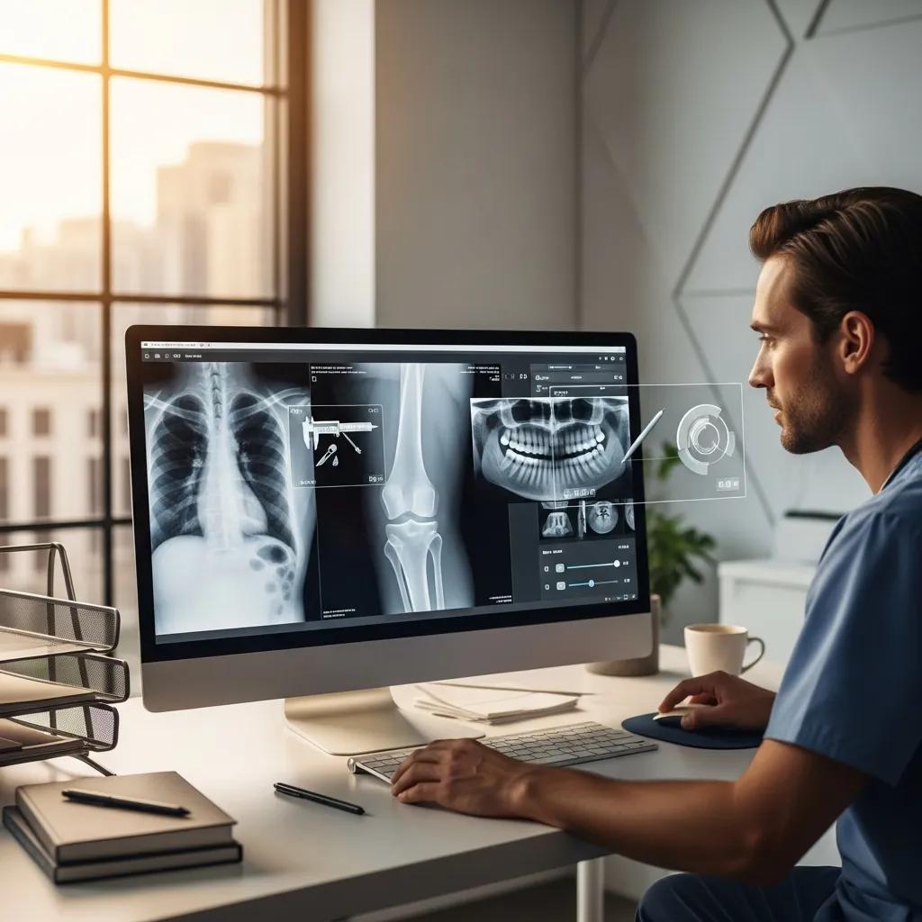
Digital X‑ray shortens the time from exposure to diagnostic review and offers post‑processing tools that can enhance contrast and highlight subtle abnormalities. Modern detectors often achieve the same or better image quality at lower doses than film. Immediate availability of images reduces waiting times and supports faster clinical decisions, while electronic storage makes it easy to compare with previous studies and consult sub‑specialist radiologists when needed. These technical and workflow gains improve the patient journey and help busy diagnostic services run more efficiently.
What Should Patients Expect During a Digital X-Ray Procedure?
When you arrive, staff check your referral and medical details and confirm any safety information such as pregnancy. The scan itself usually takes only a few minutes: a technologist will position you, take brief exposures and may ask you to hold still or breathe in a particular way. Images are checked by the radiographer and then sent to a radiologist for formal reporting; many clinics offer rapid electronic delivery of results to the referring clinician. Understanding this workflow helps you plan your appointment and know when to expect results and any follow‑up.
Local accredited clinics focus on low dose and timely reporting. You can book online, use eReferral systems or phone Life Medical Imaging Central Coast on 02 4326 7000 to arrange studies and ask about preparations — this helps ensure appointments and referrals are processed efficiently.
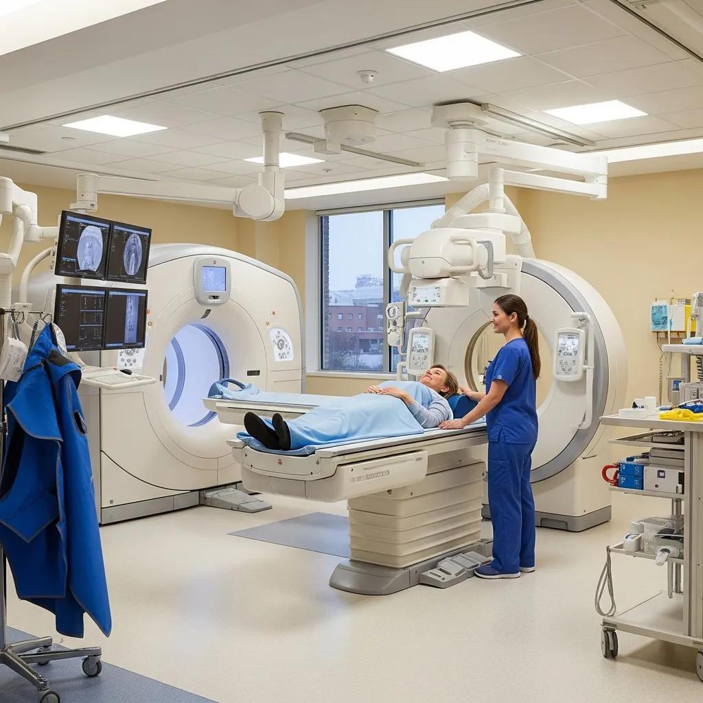
What Are CT Scan Procedures and Their Diagnostic Advantages?
Computed tomography (CT) takes multiple X‑ray measurements around the body and reconstructs cross‑sectional images using advanced algorithms. Helical acquisition and iterative reconstruction produce high‑resolution slices that can be viewed in multiple planes, making CT ideal for trauma, complex abdominal conditions, pulmonary disease and vascular assessment. Modern CT scanners include dose‑saving hardware and software that maintain image quality while reducing exposure, so CT is powerful for urgent diagnostics and elective studies that demand detailed anatomy. The following sections cover ultra‑low dose, high‑definition CT and common CT subtypes, including cardiac techniques.
CT improves diagnostic confidence through a combination of safety and quality strategies that reduce radiation burden without compromising image fidelity. Iterative and deep‑learning reconstruction, more efficient detectors and tailored scanning protocols optimise the trade‑off between image noise and diagnostic information. These improvements make CT safer for follow‑up and screening, while preserving the spatial and contrast resolution needed for trauma assessment, pulmonary embolism evaluation and complex abdominal disease. The table below summarizes common CT types to help select the right study.
Research into advanced reconstruction techniques highlights significant progress in reducing dose while preserving image quality.
Deep Learning Reconstruction for Improved Chest CT Imaging
Deep learning reconstruction (DLR) has been introduced by major vendors and tested across CT examinations. This study compared DLR with hybrid iterative reconstruction (IR) for standard, reduced and ultra‑low‑dose chest CTs using high‑definition CT systems and multiple section thicknesses and matrix sizes in patients with various pulmonary diseases.
Effectiveness of deep learning reconstruction on standard to ultra‑low‑dose high‑definition chest CT images, 2023
How Does Ultra-Low Dose, High Definition CT Technology Improve Safety?
Ultra‑low dose, high‑definition CT combines more sensitive detectors with advanced software — iterative reconstruction and AI denoising — to preserve diagnostic detail from fewer photons. The reconstruction and noise‑reduction techniques keep edges and fine structures visible while significantly lowering radiation per scan. For patients this reduces cumulative exposure from repeat studies and makes serial imaging safer for vulnerable groups, while clinicians still get the high spatial resolution needed for surgical planning and detailed assessment. Understanding these strategies helps referrers pick CT protocols appropriate for higher‑risk patients.
What Are Common CT Scan Types Including Cardiac Imaging Techniques?
CT protocols are tailored to the clinical question: non‑contrast CT is fast and useful for acute haemorrhage or renal colic; contrast‑enhanced CT improves soft‑tissue characterisation for abdominal or pelvic disease; CT angiography visualises vessels and lumen; and dedicated cardiac CT assesses coronary anatomy and calcium scoring. Preparation varies — contrast studies need renal function checks and fasting in some cases, and cardiac CT may require heart‑rate control for sharp images. Clinicians should match CT type to the diagnostic need, balancing preparation, radiation and diagnostic yield.
Many local centres now offer ultra‑low dose, high‑definition CT as a patient‑centred option. Life Medical Imaging Central Coast promotes this technology and provides online booking and eReferral pathways for cardiac and general CT studies. To discuss protocols or schedule a CT, phone the clinic on 02 4326 7000 to confirm the best approach and available times.
How Do Ultrasound Diagnostic Services Support Medical Imaging?
Ultrasound uses reflected sound waves to create real‑time images of soft tissues, organs and blood flow — a radiation‑free option for obstetrics, vascular assessment and musculoskeletal problems. Sound emitted from a probe reflects at tissue interfaces and the returning echoes are processed into moving images that show anatomy, function and dynamic relationships (for example, tendon motion or valve opening). Ultrasound is portable, repeatable and can include Doppler to assess blood flow; it’s often selected when avoiding radiation is important or when scans guide procedures in real time. The next sections list common ultrasound types and explain how patients should prepare.
What Are the Different Ultrasound Types Offered?
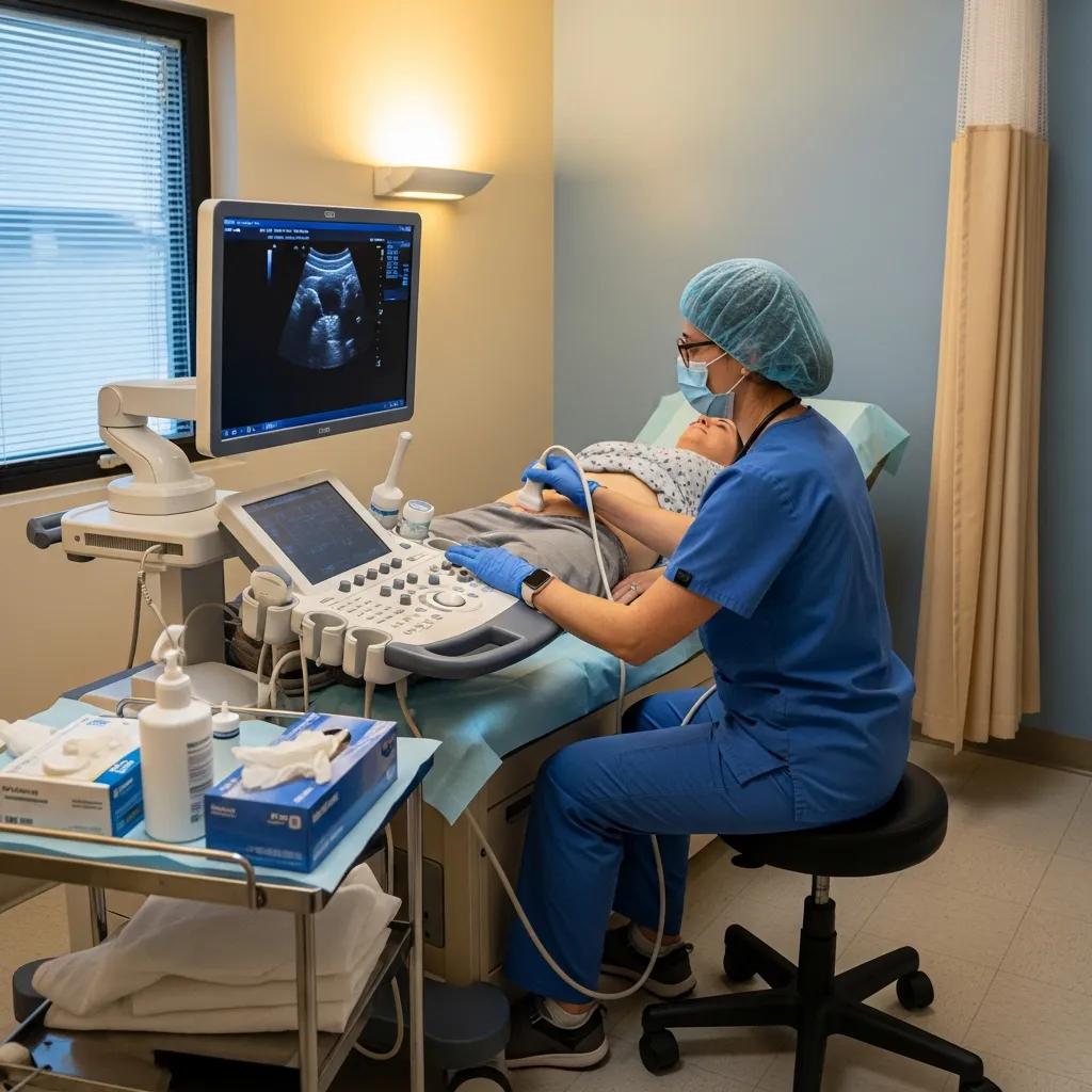
Common ultrasound exams include obstetric scans (2D/3D/4D), vascular duplex studies, musculoskeletal scans and gynaecological pelvic imaging — each focused on specific regions and clinical needs. Obstetric ultrasound monitors fetal growth and anatomy and can include 3D/4D surface views; vascular duplex assesses arterial and venous flow for clots or stenosis; musculoskeletal scans evaluate tendons, ligaments and superficial joints, often providing dynamic information complementary to MRI; and pelvic/gynaecological scans assess the uterus and ovaries for masses, cysts or pelvic pain. Appropriate pre‑scan preparation improves image quality and diagnostic yield.
How Should Patients Prepare for an Ultrasound Scan?
Preparation depends on the study and can meaningfully affect image quality. For abdominal ultrasound you’ll usually be asked to fast for several hours to reduce bowel gas; pelvic and obstetric scans often require a comfortably full bladder to improve views of the pelvis. Wear loose clothing for easy access to the area being scanned and bring any prior imaging or referral paperwork for comparison. These simple steps often avoid repeat scans and speed up reporting, helping you arrive prepared and reassured.
Preparation checklist for common ultrasound scans:
- Abdominal: Fast for 6–8 hours before the scan to reduce bowel gas.
- Pelvic/Obstetric: Arrive with a comfortably full bladder unless told otherwise to improve pelvic visualisation.
- MSK/Vascular: Wear clothing that allows access to the limb or joint being examined.
Following these guidelines usually produces higher‑quality images and faster reports. The next section explains bone densitometry and local access on the Central Coast.
What Is Bone Densitometry and How Is It Used on the Central Coast?
Bone densitometry — DEXA (dual‑energy X‑ray absorptiometry) — measures bone mineral density to assess fracture risk and diagnose osteoporosis using a very low‑dose X‑ray technique. Results compare a patient’s bone density with young‑adult and age‑matched reference groups and report T‑scores and Z‑scores that help guide prevention and treatment decisions. On the Central Coast, DEXA services give clinicians objective baseline measures, track treatment response and help stratify fracture risk in older adults or patients on long‑term therapies affecting bone health. The following sections explain how results are interpreted and the patient benefits of BMD testing.
How Does Bone Mineral Density Testing Diagnose Conditions Like Osteoporosis?
DEXA reports T‑scores and Z‑scores: a T‑score compares your BMD to a healthy young adult and a Z‑score compares to age‑matched peers. Clinical thresholds guide diagnosis: a T‑score between −1.0 and −2.5 indicates low bone mass (osteopenia), while a T‑score of −2.5 or lower is diagnostic for osteoporosis and signals higher fracture risk. Clinicians interpret these numbers together with clinical risk factors to form a management plan. Lumbar spine and hip scans are standard because they reliably predict fracture risk and guide treatment initiation or monitoring.
What Are the Patient Benefits of Bone Densitometry?
DEXA provides early detection of reduced bone density, a way to monitor response to bone‑strengthening treatments, and objective data to guide fracture‑prevention strategies. The test is quick, non‑invasive and uses very low radiation compared with CT‑based approaches, making it suitable for baseline and serial monitoring. Detecting osteoporosis early allows clinicians to start lifestyle measures, fall‑prevention plans and medication when appropriate, reducing the chance of future fractures. Local DEXA access on the Central Coast shortens the path from risk identification to treatment, improving long‑term bone health outcomes.
Key patient benefits of DEXA scanning:
- Early detection: Identifies reduced bone density before fractures occur.
- Treatment monitoring: Tracks how bone‑strengthening therapies are working over time.
- Low radiation: Provides reliable results with minimal exposure.
These benefits support preventive care and clinical management aimed at reducing fractures and related morbidity.
How Can Patients and Referrers Access Radiography Technology Services?
Access typically starts with a valid referral from a clinician. After that, patients can book appointments via online systems, eReferral platforms or by phone — knowing what documents and preparations are required helps avoid delays. Referrers should state the clinical question to guide protocol choice and urgency; imaging centres then schedule based on clinical priority and available technology. Many practices offer multiple locations and NATA‑accredited services with sub‑specialist reporting for women’s and cardiac imaging, which is valuable for complex cases needing specialist interpretation. The following sections describe how booking systems help and which resources are available to patients and clinicians.
How Does Online Appointment Booking and eReferral Streamline Access?
Online booking and eReferral speed up the referral‑to‑imaging process by securely transmitting clinical details, cutting phone time and enabling automated scheduling and confirmations. Referrers can attach clinical notes and prior images, helping radiology teams triage urgency and choose the best protocol before the patient arrives. Patients get clear appointment times and pre‑scan instructions. While timelines vary by urgency, eReferral workflows often reduce administrative steps and shorten waits for elective studies. These systems also make it easier to transfer images between providers, improving multidisciplinary care.
To arrange imaging with Life Medical Imaging Central Coast, clinicians can use eReferral pathways and patients may book online or phone 02 4326 7000 to confirm availability and preparation instructions. The clinic operates multiple Central Coast locations and offers NATA‑accredited imaging with sub‑specialist reporting in women’s and cardiac imaging to support complex diagnostic needs.
What Resources Are Available for Patients and Referring Doctors?
Imaging centres typically provide patient preparation guides, referral templates, clinician liaisons and secure image portals to simplify result access and multidisciplinary review. Patients should bring their referral, medical history and any prior imaging to appointments; referrers can include clear clinical questions to speed protocol selection. Many centres publish FAQs and study‑specific checklists covering CT contrast considerations, ultrasound fasting rules and DEXA positioning. These resources help reduce delays and ensure imaging yields diagnostically useful information.
Resources commonly available from diagnostic imaging providers:
- Patient preparation guides and FAQs that explain fasting, bladder needs and clothing.
- Referrer materials including eReferral templates and clinical guidance for choosing the right modality.
- Image access portals and clinician liaisons for report queries and multidisciplinary discussion.
Frequently Asked Questions
What is the role of radiography technology in preventive healthcare?
Radiography plays an important role in prevention by detecting conditions early. Tests like DEXA for osteoporosis and screening mammograms identify issues before symptoms appear, allowing timely intervention. Early detection supports lifestyle changes, monitoring or treatment that can reduce long‑term risk and improve quality of life.
How do advancements in radiography technology impact patient outcomes?
New imaging techniques and dose‑reduction technologies improve diagnostic accuracy and patient safety. Better image quality helps clinicians make more precise diagnoses and treatment plans, while innovations such as AI‑assisted analysis and ultra‑low dose CT reduce radiation exposure and speed up decision‑making. Together, these advances support faster, more effective care and better patient outcomes.
What should patients know about the costs associated with radiography services?
Costs vary by study type, location and insurance coverage. Many clinics provide clear pricing and may offer payment options for those without coverage. It’s sensible to check with your insurer about benefits for specific imaging studies and to ask the clinic about expected out‑of‑pocket costs before booking. Knowing costs upfront helps you plan and avoid surprises.
Are there any risks associated with radiography procedures?
Imaging procedures are generally low risk. The main concern for X‑ray and CT is exposure to ionising radiation, which carries a small lifetime risk that increases with repeated scans. Modern protocols focus on minimising dose through validated strategies. Always discuss any concerns with your referrer or the imaging team so they can weigh the benefits and risks and choose the most appropriate test.
How can patients ensure they receive the most accurate imaging results?
Follow the pre‑scan instructions provided by the clinic — fasting, hydration and clothing guidance all matter. Bring prior imaging and a clear referral or medical history. Tell staff about relevant symptoms or implants, and ask questions if anything is unclear. Good preparation and communication help radiologists produce the most accurate reports.
What are the latest trends in radiography technology?
Recent trends include wider use of AI for image analysis, ultra‑low dose imaging protocols that preserve quality while reducing exposure, and more portable devices for bedside imaging. These developments improve access, speed and safety in diagnostic imaging and support better integration into clinical workflows.
Conclusion
Knowing how radiography technologies work and when each modality is used helps patients and referrers make informed choices about diagnostic imaging. Modern imaging offers improved safety, faster diagnosis and clearer results that support better care. To explore local services, booking options or next steps, contact Life Medical Imaging Central Coast for advice and appointments.