Comprehensive Medical Imaging Services Central Coast: Your practical guide to scans, safety and booking
Medical imaging gives clinicians the visual evidence they need to diagnose, stage and monitor a wide range of conditions — from sudden injuries to chronic disease. This guide walks you through the main services available across the Central Coast (CT, ultrasound, digital X‑ray and DEXA), how each test works, what to expect as a patient, and how referrers and patients can book. You’ll find clear preparation advice, safety notes and condition‑to‑scan guidance designed to reduce delays and support accurate diagnosis. We also explain service types, typical workflows, common clinical indications, equipment and safety standards, plus practical steps for referrals and bookings tailored to local needs. Where relevant, we reference current practice so you can decide when CT, ultrasound or DEXA is the right choice and what happens after imaging is completed.
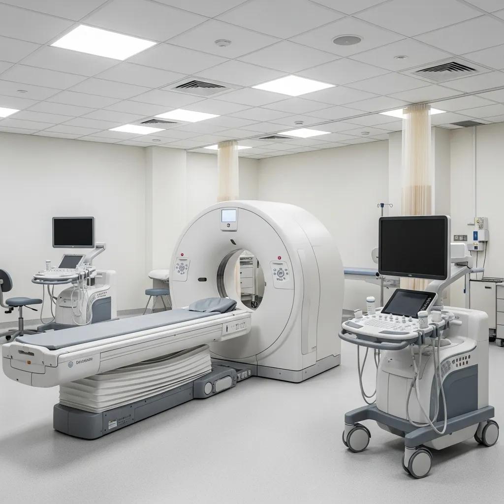
What medical imaging services are available at Life Medical Imaging Central Coast?
Medical imaging includes a suite of diagnostic tools that produce images of the body to inform treatment and diagnosis. CT (computed tomography) uses X‑rays and computer reconstruction to quickly show bones, organs and vessels — which makes CT a common choice for acute head, chest and abdominal problems. Ultrasound uses high‑frequency sound to provide real‑time images of soft tissues and blood flow; it’s radiation‑free and preferred for pregnancy and many abdominal, vascular and musculoskeletal questions. Digital X‑ray captures high‑resolution skeletal and chest images quickly, while DEXA (dual‑energy X‑ray absorptiometry) measures bone density and body composition for osteoporosis assessment. We also offer interventional image‑guided procedures to obtain biopsies or deliver targeted injections with minimally invasive techniques.
Choosing the right modality depends on the clinical question and patient factors. The quick reference below summarises typical uses and the main body areas assessed to help with referrals.
This table summarises common modalities, where they’re typically used and the main body regions assessed.
This comparison helps clinicians and patients select the appropriate test. Below we describe CT subtypes and ultrasound services in practical terms, including brief preparation notes that influence booking and patient instructions.
Which CT scan services does Life Medical Imaging offer?
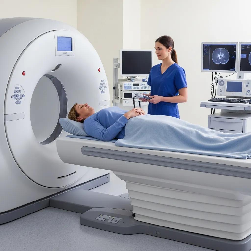
Our CT service includes general CT for trauma and acute abdominal pain, cardiac CT for non‑invasive coronary assessment, and CT angiography for detailed vascular mapping. Referrals for CT usually specify the region and clinical urgency because CT is fast and sensitive for acute pathology and helps clinicians triage care. Some CT protocols require contrast — patients are screened for renal function and allergies when needed, and instructions on hydration or fasting depend on the protocol. Modern scanners use dose‑reduction technology to limit radiation while preserving image quality, which is particularly important when follow‑up scans may be required.
These CT subtypes are valuable for suspected pulmonary embolism, appendicitis, complex fractures and coronary artery disease evaluation. Cardiac CT on the Central Coast offers non‑invasive coronary imaging that can complement functional testing and, in selected patients, reduce the need for invasive angiography.
What types of ultrasound services can patients access?
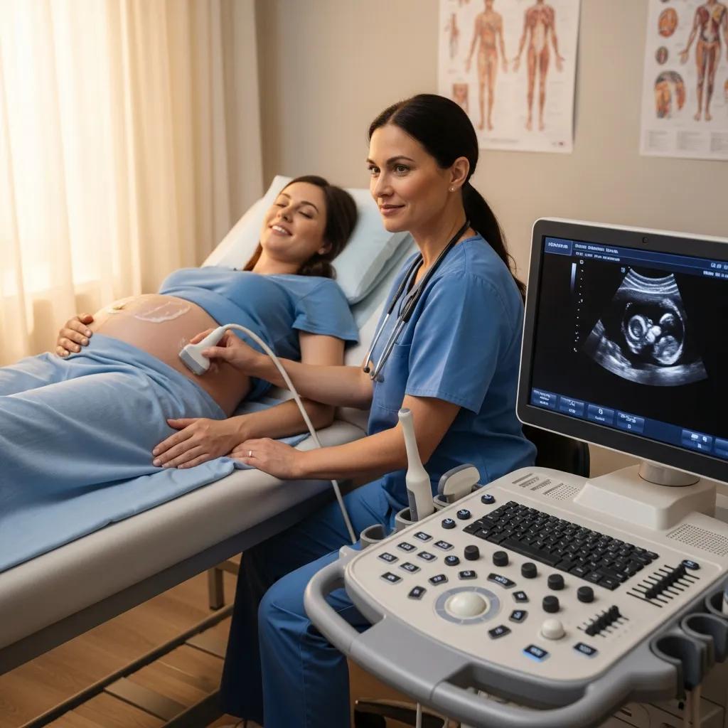
Our ultrasound services include general abdominal scans, obstetric and gynaecological imaging, vascular duplex studies, musculoskeletal (MSK) ultrasound and paediatric ultrasound tailored for children. Ultrasound on the Central Coast is radiation‑free and commonly used for pregnancy checks, gallbladder or liver assessment, venous duplex for suspected deep vein thrombosis, and dynamic assessment of tendons and joints. Most targeted scans take around 20–40 minutes; obstetric and detailed MSK studies may take longer and can require specific bladder or fasting instructions. Because ultrasound is operator‑dependent, experienced sonographers and sub‑specialist radiologists maximise diagnostic yield and produce clinically useful reports.
Please bring your referral and any prior imaging, and wear comfortable clothing. For pregnancy scans, details about gestation and obstetric history help us tailor the exam. The safety and immediacy of ultrasound make it an excellent first‑line option for many presentations on the Central Coast.
How do diagnostic imaging procedures work and what should patients expect?
Imaging procedures follow a clear, safety‑focused workflow to ensure image quality and timely reporting. On arrival we confirm identity and referral details and check any pre‑scan requirements. Technologists perform the study using modality‑specific positioning and protocols, while radiologists oversee complex or interventional cases. Radiologists interpret images and produce structured reports; findings and recommendations go to the referring clinician, with urgent results communicated by phone when needed.
Knowing this pathway helps patients plan their visit and reduces uncertainty about what happens after the scan.
Below is a step‑by‑step outline of a typical CT or ultrasound appointment so patients and referrers can prepare and plan accordingly.
- Arrival and registration: Staff confirm identity, check the referral and screen for contrast risks.
- Pre‑scan consent and preparation: Technologists explain the procedure, position the patient and administer contrast if required.
- Image acquisition: CT or ultrasound images are captured while the patient follows breathing or positioning instructions.
- Post‑scan review and aftercare: Patients receive any immediate instructions and information about how results will be delivered.
- Reporting and results delivery: Radiologists prepare structured reports that are sent to the referring clinician for management discussion.
This workflow clarifies each stage from check‑in to report distribution and explains how urgent findings are escalated and routine results managed.
Many studies require specific preparation to reduce artefact and improve diagnostic yield. The checklist below summarises common instructions and what patients should bring to their appointment.
Which conditions can be diagnosed using radiology services on the Central Coast?
Different imaging modalities suit different clinical questions; selecting the right test reduces delays and unnecessary procedures. CT provides fast cross‑sectional detail useful for acute intracranial haemorrhage, pulmonary embolism and abdominal emergencies. Ultrasound is ideal for pregnancy monitoring, gallstones, ovarian pathology and venous thrombosis because it provides real‑time assessment without ionising radiation. DEXA quantifies bone density to diagnose osteoporosis and estimate fracture risk, while digital X‑ray quickly confirms fractures and assesses chest pathology. Mapping conditions to the appropriate test helps clinicians choose the safest, most effective pathway for diagnosis.
The practical table below pairs modalities with typical indications and explains why each scan is chosen, helping with quick referral decisions and patient education.
What cardiac and women’s health conditions are detected by specialised imaging?
Cardiac imaging — including cardiac CT and CT angiography — can reveal coronary calcification, stenosis and other structural heart findings that affect management. Cardiac CT is useful in selected patients with chest pain and for non‑invasive coronary mapping that complements functional testing. In women’s health, obstetric and gynaecological ultrasound assesses fetal growth, placental position, ovarian cysts and uterine pathology without radiation; sub‑specialist reporting improves detection of subtle abnormalities. Specialist interpretation increases diagnostic confidence and ensures findings are correlated with the clinical picture to guide follow‑up.
Our clinical teams apply these specialised services when the results will influence management, reducing unnecessary tests and focusing care where it matters most.
How does paediatric and musculoskeletal imaging support diagnosis?
Paediatric imaging prioritises radiation minimisation, child‑friendly communication and fast, accurate studies to reduce distress while meeting diagnostic goals. For children, ultrasound and targeted X‑ray are often first‑line; CT is reserved for cases where its detail will change management. Musculoskeletal (MSK) ultrasound provides dynamic assessment of tendons, ligaments and soft‑tissue lesions and complements X‑ray for fractures and alignment. MSK imaging supports sports medicine referrals, guides injection therapies and monitors healing without repeated radiation. Clear paediatric protocols and experienced staff improve image quality while keeping children safe and comfortable.
These approaches blend technical adjustments with communication strategies to make imaging effective and child‑centred.
What advanced technologies and safety measures are used in our imaging services?
Contemporary imaging equipment and structured safety protocols enhance diagnostic accuracy and patient outcomes. Modern CT scanners offer high spatial and temporal resolution useful for cardiac and body imaging, while digital X‑ray systems deliver high‑quality images with lower doses than older film systems. Improved ultrasound transducers increase resolution for obstetric and MSK work. Together, equipment upgrades and specialist reporting create a reliable pathway from image acquisition to an actionable clinical report.
Mentioning advanced technology reassures patients and referrers that examinations are performed to current standards.
- Modern multi‑slice CT scanners that shorten exam time and improve image detail for cardiac and body imaging.
- High‑resolution ultrasound transducers suited to obstetric, vascular and musculoskeletal studies.
- Digital X‑ray detectors and DEXA machines optimised for dose efficiency and reproducible measurements.
How does Life Medical Imaging use cutting‑edge equipment for accurate diagnosis?
Life Medical Imaging Central Coast combines contemporary scanners with sub‑specialist reporting to deliver clinically meaningful results. Faster scanners and higher‑resolution transducers shorten exam times and improve visualisation of small lesions, coronary arteries and soft‑tissue pathology. Sub‑specialist women’s and cardiac reporting further refines interpretation for complex cases. The mix of modern hardware and experienced reporting increases the chance that imaging will directly inform clinical decisions and reduce repeat studies.
Clearer images and specialist interpretation shorten diagnostic pathways and support more timely, targeted treatment.
What patient comfort and radiation safety protocols are in place?
Patient comfort and radiation safety are central to our practice. We apply dose‑reduction protocols, shielding where appropriate and paediatric dose adjustments to minimise unnecessary exposure while maintaining diagnostic quality. For anxious or claustrophobic patients, technologists use clear communication, positioning aids and shorter protocols to improve tolerance. We maintain strict infection control and hygiene standards, and staff training ensures consistent application of safety measures. Explaining these protocols before the scan reduces anxiety and helps patients cooperate during image acquisition.
These safety and comfort measures produce better images, fewer repeats and a more positive patient experience from appointment to report.
How can patients and referring doctors book appointments and access radiology services?
Booking imaging and submitting referrals on the Central Coast is straightforward when the necessary documents and clinical details are ready. Referrals should state the clinical question, relevant history and any prior imaging so the radiology team can choose the correct protocol. Patients can book by phone or use online booking where available; for urgent imaging, calling in speeds triage and allows staff to advise on immediate preparation. Clear booking steps help reduce no‑shows and ensure correct pre‑scan instructions (fasting, medications, contrast information) are given in advance.
The numbered steps below explain the usual referral and booking workflow for referrers and patients so appointments are scheduled with the right priority and preparation.
- Obtain a referral from a GP or specialist that states the clinical question and urgency.
- Contact the imaging provider by phone to schedule the requested modality and confirm pre‑scan preparation.
- Attend the appointment with the referral, photo ID and any prior imaging, following the provider’s preparation instructions.
These steps make the process predictable and efficient for patients and referrers, helping to shorten time to diagnosis and treatment planning.
What is the referral process for diagnostic imaging on the Central Coast?
Referrals usually come from GPs, specialists or hospital teams and should state the clinical indication, relevant history and urgency to guide imaging selection. Electronic referrals speed submission and reduce admin delays, though paper referrals remain acceptable where e‑referral is not possible. Including prior imaging reports avoids unnecessary duplication and enables radiologists to compare current and previous findings. Clear clinical questions such as “rule out appendicitis” or “assess coronary calcium” help staff apply the correct protocol and deliver an actionable report.
A complete referral expedites booking and ensures the imaging study addresses the diagnostic need, reducing repeat studies and shortening the pathway to treatment.
How can patients book imaging appointments online or by phone?
Patients can book via the provider’s published booking channels or by telephone for direct help with preparation and scheduling. When you call, have the referral details, Medicare or private health information if relevant, and any prior imaging ready; staff will advise on fasting or medication adjustments. If online booking is available, the portal usually prompts for the referral and pre‑scan instructions. For urgent cases, calling provides faster triage and clarifies immediate requirements. Always confirm arrival time, identification needs and any special instructions to avoid delays on the day.
To enquire or schedule an appointment at Life Medical Imaging Central Coast, please call 02 4326 7000 to confirm locations and receive tailored guidance for your referral and preparation. This direct contact helps ensure the correct modality is booked and that pre‑scan steps are clearly understood.
What are the most common questions about medical imaging services on the Central Coast?
Common questions include: which test is best for a given problem, how to prepare, whether imaging is safe in pregnancy or for children, and whether bulk billing is available. Short, direct answers help people act: ultrasound is preferred in pregnancy, CT is preferred for acute trauma, and DEXA is used for osteoporosis screening. Reporting times vary by urgency — most routine reports reach the referring clinician within a clinically appropriate timeframe, while urgent findings are communicated immediately. Clear guidance on these questions reduces anxiety and speeds appropriate referral.
The list below summarises primary modality benefits and common use cases to support quick decision‑making for patients and clinicians.
- CT scans: Fast, detailed cross‑sectional images for trauma, chest and abdominal emergencies.
- Ultrasound: Real‑time, radiation‑free evaluation ideal for pregnancy, vascular and soft‑tissue assessment.
- DEXA scans: Measure bone density to diagnose osteoporosis and estimate fracture risk.
What are the benefits and uses of CT scans and ultrasounds?
CT and ultrasound are complementary because they image the body using different physics: CT uses ionising radiation and excels at bone detail, complex anatomy and acute pathology, while ultrasound uses sound waves for real‑time motion and blood‑flow imaging without radiation. CT is typically requested for suspected internal bleeding, complex fractures and detailed chest or abdominal assessment. Ultrasound is preferred for pregnancy surveillance, gallbladder disease and vascular studies. Choice depends on the clinical question, availability and radiation considerations: when radiation is a concern, ultrasound is often first‑line; when depth and cross‑sectional anatomy are essential, CT provides superior detail.
Choosing the right test reduces unnecessary investigations, improving patient safety and diagnostic efficiency.
X‑ray and DEXA play a key role in assessing bone health and identifying fractures.
X‑ray and DEXA for bone density and fracture detection
Thoracolumbar radiography helps identify vertebral fractures, while DEXA is the preferred method for measuring bone mineral density. Hip total‑density predicts hip fracture risk best, and the lumbar spine is often used to monitor treatment response.
Bone mineral density: testing for osteoporosis, A Sheu, 2016
Is bulk billing available for radiology services at Life Medical Imaging?
Bulk billing depends on the service, patient eligibility and the referral. Some studies may be bulk‑billed under Medicare for eligible patients, while other services may incur private fees or co‑payments depending on the examination. Ask about bulk billing and any out‑of‑pocket costs when you book to avoid surprises. For definitive information on eligibility and current billing, contact the imaging provider at the time of booking.
Checking billing at booking lets patients make informed decisions about timing and location of imaging.
In emergencies, rapid initial imaging can be critical for trauma patients.
CT and X‑ray for initial trauma assessment
Objective studies have examined the role of initial rapid imaging (chest and pelvic X‑ray, abdominal ultrasound) in severely traumatised patients before whole‑body CT. These assessments can guide immediate care while definitive imaging is arranged.
Initial imaging assessment of severe blunt trauma, O Langeron, 2001
For further information or to book an appointment and discuss preparation, patients and referrers may call Life Medical Imaging Central Coast on 02 4326 7000 to confirm available locations (including multiple Central Coast sites) and receive tailored guidance for their referral and clinical needs. Calling helps secure timely bookings and clarifies preparation instructions to ensure the best possible imaging results.
Frequently Asked Questions
What should I bring to my imaging appointment?
Bring your referral from the GP or specialist, any previous imaging reports or CDs, and photo identification. Wear comfortable, loose clothing — for DEXA, avoid metal fastenings. Having these items ready helps speed check‑in and keeps your appointment on time.
How long will it take to receive my imaging results?
Turnaround depends on the study and urgency. Routine reports are usually available to the referring clinician within a clinically appropriate timeframe (often a few days). Urgent findings are communicated immediately. If you need a specific timeframe, ask your referrer or the imaging centre when you book.
Are there any risks associated with medical imaging procedures?
Imaging is generally safe. Modalities that use ionising radiation, like CT and X‑ray, carry a small radiation risk that we minimise with modern protocols. Ultrasound is radiation‑free and considered very safe, including in pregnancy. Discuss any concerns with your clinician or the imaging team so the benefits and risks for your situation are clear.
Can I eat or drink before my imaging appointment?
Preparation depends on the exam. Some scans (for example CT abdomen/pelvis) require fasting, while others (certain ultrasound exams) may ask you to have a full bladder. Always follow the specific instructions provided by the imaging centre; if unsure, call ahead for clarification.
What should I do if I have anxiety about the imaging procedure?
If you feel anxious, tell the staff when you arrive or when booking. Our technologists are experienced in helping patients feel comfortable and can explain the process, suggest breathing techniques, allow a support person where appropriate, or use shorter protocols for claustrophobic patients. Let us know so we can support you.
What happens if I need a follow‑up imaging study?
If further imaging is required, your referring clinician will discuss why and provide a new referral if needed. Follow‑up timing will be based on the clinical indication. Keep the imaging centre informed of any change in symptoms and schedule follow‑up promptly as advised by your clinician.
Conclusion
Comprehensive imaging services on the Central Coast help patients and clinicians reach timely, well‑informed decisions about diagnosis and treatment. With CT, ultrasound, DEXA and digital X‑ray available, each test is chosen to match the clinical need. If you have a referral, call us to book an appointment — experience the benefits of modern imaging technology and specialist interpretation at Life Medical Imaging Central Coast.