
Medical Imaging — Central Coast NSW: Complete Diagnostic and Specialist Radiology Services
Medical imaging brings together non‑invasive technologies used to diagnose, monitor and guide treatment across a wide range of health concerns. On the Central Coast, that means local access to comprehensive diagnostic and specialist radiology services. This guide explains the modalities we offer, how each works and what patients and referrers can expect from referral to results. We emphasise safety, low‑dose techniques and specialist women’s and cardiac imaging, and provide practical preparation steps, modality comparisons and referral guidance. You’ll learn which test best suits common clinical scenarios, how to prepare for CT, MRI, ultrasound and DEXA, and how paediatric and cardiac imaging differ in protocol and comfort measures. The sections below cover services, quick comparisons, women’s imaging, booking and preparation, advanced technology, paediatric and cardiac care, and how patients and referrers can access services on the Central Coast.
What Medical Imaging Services Are Available at Life Medical Imaging Central Coast?
Across the Central Coast we provide the core imaging modalities: CT, ultrasound, X‑ray/OPG, MRI and DEXA — each using different physical principles to show anatomy and disease. CT uses X‑rays and detectors to create cross‑sectional images for trauma, chest, abdominal and cardiac assessment; ultrasound uses sound waves for dynamic soft tissue, obstetric and vascular exams; MRI uses magnetic fields and radio waves to produce detailed soft tissue images without ionising radiation; digital X‑ray and OPG give fast skeletal and dental views; and DEXA measures bone mineral density to assess fracture risk. These services are supported by image‑guided interventional procedures (for biopsies and injections) and sub‑specialist reporting in women’s and cardiac imaging to ensure clinical nuance. Our Central Coast network follows NATA‑accredited workflows to maintain safety and quality. The following subsections describe CT subtypes and ultrasound offerings, then explain how to book specific services.
The table below summarises the core modalities — their main uses, common preparation and key safety notes — so clinicians and patients can match tests to symptoms and needs.
This quick comparison highlights the main differences between tests and leads into a closer look at the CT options available locally.
Which Types of CT Scans Does Life Medical Imaging Offer?

Our CT services on the Central Coast include general diagnostic CT, cardiac CT and CT angiography. Where clinically appropriate we use low‑dose protocols to minimise radiation while preserving diagnostic quality. General CT is quick and effective for head, chest, abdomen and trauma; cardiac CT and CT angiography assess coronary anatomy and vascular patency for suspected vascular disease. Preparation depends on the scan: abdominal CT often requires a short fast, and contrast studies need screening for kidney function and allergies via a pre‑scan questionnaire. Advanced reconstruction techniques and NATA‑accredited safety checks help reduce dose and maintain consistency. Knowing the available CT options helps referrers pick the right study and prepares patients for the appointment.
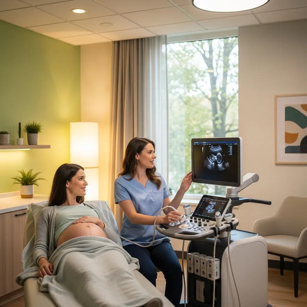
What Ultrasound Services Are Provided at Erina and Other Locations?
Ultrasound across our Central Coast clinics covers general abdominal, vascular, musculoskeletal, obstetric and gynaecological studies. Erina is noted for dedicated women’s imaging services. General ultrasound evaluates liver, kidneys and gallbladder; vascular studies assess blood flow and detect clots; musculoskeletal scans visualise tendons and joints dynamically; obstetric and gynaecological ultrasound monitor fetal growth and pelvic pathology. Preparation differs by exam — pelvic and obstetric scans often require a full bladder, while some abdominal scans benefit from fasting — and staff will give clear, appointment‑specific instructions. Because ultrasound uses no ionising radiation it’s preferred in pregnancy and for paediatric assessments where reducing exposure is important. These location‑specific services help patients choose the site best suited to their needs.
How Does Women’s Imaging Central Coast Support Women’s Health?
Women’s imaging brings together obstetric, gynaecological and breast ultrasound with sub‑specialist input to support diagnosis, monitoring and early detection. 3D/4D obstetric scanning provides enhanced anatomical views for detailed fetal assessment, gynaecological ultrasound evaluates uterine and adnexal conditions, and breast ultrasound complements mammography — particularly in dense breast tissue or for symptomatic assessment. We coordinate imaging with referrers so studies fit into antenatal and gynaecological care pathways, and specialist reports highlight findings most relevant to obstetricians and gynaecologists. By combining advanced techniques with sub‑specialist interpretation, women’s imaging improves diagnostic confidence and helps guide timely management. The sections below describe 3D/4D benefits and the clinical role of breast ultrasound.
What Are the Benefits of 3D/4D Obstetric and Gynaecological Ultrasound?
3D/4D obstetric ultrasound captures volumetric data that improves visualisation of fetal anatomy and structural relationships, aiding detection of certain anomalies and supporting detailed counselling for parents. 3D helps assess facial, limb and complex cardiac structures when 2D views are limited, while 4D (real‑time 3D) adds motion assessment for functional evaluation. Ultrasound remains the preferred imaging method in pregnancy because it uses no ionising radiation; exposure is kept to diagnostic levels. Beyond clinical value, 3D/4D imaging can also help parents connect with their baby by providing clearer images. These scans complement routine antenatal care and help referrers request the most appropriate study.
How Is Breast Ultrasound Used for Early Detection and Diagnosis?
Breast ultrasound is central to assessing lumps, focal pain and palpable changes. It helps distinguish cystic from solid lesions, guides needle biopsies when tissue is needed and characterises lesion features such as vascularity and margins to inform next steps. In screening roles, ultrasound can be an adjunct to mammography for those with dense breasts or higher risk; in diagnostic settings it’s standard for urgent referrals with clinical signs. Structured reporting communicates relevant findings to surgical and oncology teams and helps determine whether biopsy or short‑interval follow‑up is needed. Clear radiologist–referrer communication ensures results are acted on promptly.
What Should Patients Expect When Booking and Preparing for Medical Imaging?
Good preparation helps produce high‑quality diagnostic images, reduces delays and improves patient comfort. Most imaging requires a referral and appointments can be made online or via eReferral systems that triage urgency; modality‑specific preparation instructions are provided at booking and may include fasting, hydration or clothing advice. On arrival you’ll complete a safety screen for implants, allergies and pregnancy where relevant. Technologists will explain the procedure, how long it takes and whether contrast is required. Reports are sent to the referring clinician through standard reporting pathways. Below we give practical checklists for preparation and explain how referrals and appointments are handled across the Central Coast.
Use the checklist below to help your scan go smoothly and produce the best possible images.
- Bring your referral and any recent clinical notes or previous imaging reports.
- Tell staff about implants, pacemakers, allergies, pregnancy or recent illnesses.
- Follow fasting or hydration advice given for CT, MRI or abdominal ultrasound.
- Wear loose, metal‑free clothing and remove jewellery before the scan.
- Arrive early to allow time for paperwork and safety screening.
Following these steps helps with image quality and safety. After the scan, reporting timelines and next steps are communicated to both patient and referrer.
We support booking via online appointment tools and eReferral for clinicians, and our staff can advise on preparation or manage urgent requests. Please check preparation instructions when you book and let us know about any health issues before your appointment.
How Do You Prepare for a CT Scan or MRI at Central Coast Clinics?
Preparation for CT or MRI is condition‑specific and important for safety and image quality, especially when contrast is used. For CT with contrast you may be asked to fast for a few hours, provide a history of allergies or kidney disease and bring recent blood tests showing renal function. The imaging team screens for contraindications and discusses contrast risks and benefits. For MRI, screening covers metallic implants, pacemakers, neurostimulators and claustrophobia; some patients may receive mild anxiolytics under clinical guidance, and alternatives such as open MRI or sedation can be considered. Both modalities include guidance about medications and pregnancy status, and technologists explain what sensations to expect during the scan to help reduce anxiety. Clear pre‑scan communication reduces cancellations and ensures the correct protocol is used.
What Is the Appointment and Referral Process for Medical Imaging Services?
Referrals usually come from GPs, specialists and hospital teams. A clear clinical history and a focused question help triage urgency and choose the right modality and protocol. Many local referrers use eReferral systems for secure information transfer, though paper referrals remain acceptable where electronic pathways aren’t available. Including previous imaging and relevant labs speeds decision‑making. Urgent or same‑day appointments are triaged by clinical need and sub‑specialist radiologists can advise on complex cases; reporting turnaround times vary by modality and urgency and are communicated to referrers. Patients should follow referral instructions and contact the clinic if they need help booking or preparing, which prevents delays and supports coordinated care.
What Are the Advantages of Advanced Imaging Technologies at Life Medical Imaging?
Modern imaging tech improves diagnostic confidence, patient safety and workflow. Examples include low‑dose CT protocols and AI‑assisted tools that support radiologist decision‑making. Low‑dose CT maintains image quality while reducing radiation through iterative reconstruction and dose modulation, useful for screening or repeat studies. AI can help triage studies, flag urgent findings and enhance image clarity for subtle pathologies, speeding time to diagnosis while leaving final interpretation to radiologists. Integrated into accredited workflows, these technologies shorten turnaround times and support better outcomes. Below are the main advantages and a simple comparison of key technologies and their clinical benefits.
Key advantages of modern imaging technology include:
- Reduced radiation exposure while preserving diagnostic detail.
- Faster, more consistent reporting supported by AI triage and prioritisation.
- Improved detection of subtle findings through image‑enhancement algorithms.
These benefits explain why equipment and software choices matter for patient care; the sections that follow look more closely at low‑dose CT and AI features.
How Does Low‑Dose CT Technology Improve Patient Safety?
Low‑dose CT reduces radiation by optimising acquisition settings and using advanced reconstruction algorithms that retain diagnostic detail at lower doses. Clinically, low‑dose protocols are valuable for follow‑up scans, lung screening and where repeated imaging is expected, because they lower cumulative exposure without compromising sensitivity. Safety also depends on thorough pre‑scan screening, appropriate clinical justification and adherence to accreditation standards (such as NATA) that govern dose monitoring and quality assurance. Radiologists choose low‑dose protocols when evidence shows they answer the clinical question while minimising risk. Transparent communication about dose reduction helps patients give informed consent and feel reassured.
What Role Does AI Play in Enhancing Diagnostic Accuracy?
AI is an assistive tool in diagnostic imaging — it highlights patterns, flags possible abnormalities and prioritises studies for radiologist review rather than replacing human interpretation. Examples include automated detection of lung nodules, vertebral fractures or intracranial haemorrhage on CT, helping flag critical findings faster so clinicians can act sooner. AI also denoises low‑dose datasets, improving the signal radiologists use to make diagnoses. Robust governance is essential: algorithms are validated, used with radiologist oversight and applied within the appropriate clinical context to avoid overreliance. When used responsibly, AI speeds workflows and supports more accurate, timely care.
Which Specialist Imaging Services Are Offered for Paediatric and Cardiac Patients?
Our specialist services adapt protocols to the patient group: paediatric imaging focuses on radiation minimisation, child‑centred communication and comfort, while cardiac imaging targets coronary and vascular assessment with Cardiac CT and CT angiography. Paediatric protocols use age‑ and size‑adjusted exposures, distraction techniques and, when needed, non‑sedative calming measures to avoid unnecessary sedation. Cardiac studies use ECG gating and heart‑rate optimisation where appropriate and provide specialised reporting for cardiologists. These services are coordinated with referrers so test selection, preparation and follow‑up are clinically useful. The table below summarises key differences and the safety and comfort measures offered.
How Is Paediatric Imaging Tailored for Children’s Safety and Comfort?
Paediatric imaging follows ALARA (as low as reasonably achievable) principles to minimise radiation, with technologists using size‑adjusted protocols and child‑focused communication. Staff employ distraction aids and calming techniques and, when clinically necessary, sedation is carefully managed. We prioritise non‑ionising modalities like ultrasound when appropriate and apply shielding and low‑dose CT protocols only when required. Our team’s paediatric training and close coordination with referrers help achieve diagnostic images while keeping the child comfortable and safe.
What Cardiac Imaging Procedures Are Available on the Central Coast?
Cardiac services include Cardiac CT and CT angiography to non‑invasively assess coronary arteries, cardiac anatomy and selected vascular conditions. These studies commonly use ECG gating to synchronise acquisition with the cardiac cycle and may involve heart‑rate control to improve image quality. Contrast is sometimes required and is administered after screening for allergies and renal risk. Cardiac CT is useful for excluding significant coronary disease in selected patients, planning structural procedures and assessing complex anatomy without invasive catheterisation in many cases. Structured reports support cardiology decision‑making and we coordinate closely with cardiology referrers to ensure results inform timely treatment.
How Can Referrers and Patients Access Resources and Support at Life Medical Imaging?
We offer resources for both patients and referrers, including downloadable preparation guides, eReferral pathways and online appointment booking to streamline scheduling and triage. Referrers should include relevant clinical history and prior imaging to help select the correct protocol; specialist radiologist consultation is available for complex or urgent cases. Patients can access modality‑specific preparation instructions and should notify the clinic of implants, allergies or pregnancy before arrival to reduce delays. The checklist below helps referrers submit complete referrals, and the following section lists our Central Coast locations and their primary services so patients can choose the most appropriate site.
Referrers should follow this concise checklist when ordering imaging.
- Provide a clear clinical question and relevant history to guide protocol choice.
- Include prior imaging reports or CDs where available for comparison.
- Indicate urgency and any special requirements such as contrast or sedation.
- Use eReferral channels when available for faster triage and scheduling.
What Referral Guidelines and Resources Are Available for Doctors?
Referral guidance focuses on clinical justification, a targeted history and a clear question so the imaging study answers the referrer’s diagnostic or management need. Protocols for common indications help standardise practice. We recommend eReferral systems where available to securely transfer patient information and speed triage; for complex or urgent referrals radiologist consultation can help choose the optimal protocol and expedite reporting. Please include prior imaging and relevant labs (especially if contrast is planned) to support safety screening and scheduling. Specialist reporting and subspecialty reads (women’s and cardiac imaging) are available when advanced interpretation is needed, promoting collaborative care between radiologists and referring clinicians.
Where Are Life Medical Imaging Locations on the Central Coast?
Life Medical Imaging Central Coast operates at multiple sites including Bateau Bay, Killarney Vale, Umina Beach and Erina. Each location offers a selected range of services tailored to local needs — some focus on core diagnostics like CT, X‑ray and ultrasound, while others provide dedicated women’s imaging or interventional procedures. When booking, choose the site that offers the required test and follow the preparation instructions provided. We supply accessibility and parking details and modality‑specific preparation guides with appointment confirmations to make attendance easier. For bookings and enquiries, use our online appointment system or eReferral options, and our staff will advise on the best site for your needs.
Frequently Asked Questions
What should I do if I have a fear of enclosed spaces before an MRI?
If you experience claustrophobia, tell us before your MRI. We can discuss options such as mild sedatives, an open MRI where available, or coping strategies and distraction techniques used by our staff. Preparing ahead and sharing your concerns helps us make the scan as comfortable as possible.
Are there any specific imaging services for children?
Yes. Our paediatric imaging services are designed for children’s safety and comfort. We use low‑dose protocols, child‑friendly communication and distraction techniques to reduce anxiety and movement. We prioritise non‑ionising modalities like ultrasound where clinically appropriate and provide guidance to parents on how best to prepare their child.
How can I access my imaging results after the procedure?
Results are usually sent to the referring clinician, who will discuss them with you. If you prefer direct access, ask the clinic about patient portals or online systems — availability varies by site. Check the clinic’s policy on direct access and what identification is required for privacy compliance.
What should I do if I have allergies to contrast agents?
If you have a known contrast allergy, tell the imaging team before your appointment. We will assess your history and may recommend alternative imaging, premedication (antihistamines or steroids) or additional precautions to reduce risk. Provide a full medical history so we can plan the safest approach.
Can I bring someone with me to my imaging appointment?
Yes — you may bring a support person to most appointments. A companion can help reduce anxiety, but please check with the clinic beforehand as some procedures limit the number of people in the examination room for safety or space reasons.
What are the typical turnaround times for imaging results?
Turnaround times vary by modality and clinical urgency. Routine studies are commonly reported within 24–48 hours, while urgent cases are prioritised for faster reporting. Your referring clinician will usually receive the report first and will discuss results with you. If timing is critical, let the clinic know at booking.
Are there any costs associated with medical imaging services?
Costs depend on the procedure, your private health cover and the clinic. Many clinics provide fee information up front — check with your insurer about coverage for diagnostic imaging. If cost is a concern, speak with clinic staff about payment options or financial assistance that may be available.
Conclusion
Comprehensive medical imaging on the Central Coast helps patients and clinicians achieve accurate diagnoses and personalised care using advanced technology and specialist interpretation. Knowing the available modalities and how to prepare improves the experience and supports better outcomes. Explore our services and book an appointment online or contact your chosen clinic for guidance — Life Medical Imaging is here to deliver safe, reliable imaging when you need it.